Anesthetic Management of Nasopharyngeal Angiofibroma Resection with Carotid I...
IJCCM_2015_chest wall hematoma
1.
2. Indian Journal of Critical Care Medicine April 2015 Vol 19 Issue 4246246
given to prevent further bleed. But the hematoma
continued to increase in size requiring an emergency
angio-embolization of a large branch of left internal
mammary artery. INR came down to 0.8 next day.
Hemoglobin remained stable, and she went home in
3 days. A significant resorption of the hematoma was
observed on review, and anti-coagulant therapy was
re-started after 10 days.
Hematoma in pectoral muscle has been rarely
reported in the literature. Kocer et al. described cases of
spontaneous intra-pectoral bleeding.[3]
It was considered
that in our patient, interaction between levofloxacin and
warfarin could have possibly potentiated the action
of warfarin, and even mild physiotherapy could have
triggered a bleed.[4,5]
This case is rare as it occurred over a very short
duration and involved an arterial bleed requiring
interventional management. The drop in hemoglobin
was a very significant 8%.
The interaction of warfarin with various medications
and food substances is well-known. Its life-threatening
complication is something that should be kept in
mind considering the fact that levofloxacin is a
very common antibiotic prescribed by community
physicians for common cold, urinary tract, and upper
respiratory tract infections. As noted here, the trigger
for a torrential bleed could be very trivial as mild
physiotherapy.[4,5]
To conclude, a careful screening for interactions is
required before prescribing medicines for patients on
anticoagulation therapy.
Pradeep Rangappa,
Tejaswini Arunachala Murthy, Ipe Jacob
Intensive Care Unit, Columbia Asia Referral Hospital, Yeshwantpur,
Bengaluru, Karnataka, India
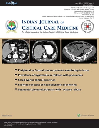
Figure 1: (a) Gross appearance. (b) Chest X-ray. (c) Computed tomography
(CT) image coronal section. (d) CT image transverse section. (e) CT image,
sagittal section
d
cba
e
Drug interaction
resulting in massive
chest wall hematoma in
a patient on therapeutic
anticoagulation
Sir,
The incidence of spontaneous large hematomas is rare
compared to several case reports of major intracranial
bleed in patients on therapeutic anticoagulation. The
most commonly involved anticoagulant is warfarin,
used in the prevention of thrombo-embolism in
deep vein thrombosis, prosthetic valves, atrial
fibrillation, etc.[1]
The incidence of hematomas requiring
invasive intervention is rarely encountered. There is
a case of pectoral hematoma reported by Cinar, et al.,
in a case series, which was managed conservatively.[2]
We report a case of an elderly woman, on therapeutic
anticoagulation for deep-vein thrombosis, who
presented with a large left chest wall hematoma
requiring embolization of the bleeding vessel.
An82-year-oldfemalepatientwasadmittedtoIntensive
CareUnit(ICU)withahistoryofdisorientationfor2days.
She was on therapeutic anticoagulation with warfarin
2 mg OD since 1-year after presenting with chronic deep
vein thrombosis due to hyperhomocysteinemia and
protein S deficiency and required inferior vena cava filter
insertion earlier. Her platelet count and serum creatinine
levels were within normal range.
At present, she was found to have urinary tract
infection for which ertapenem was started. International
normalized ratio (INR) at time of admission was 1.8 and
was monitored daily and antibiotic was de-escalated
to levofloxacin 500 mg intravenous (IV) OD as per the
urine culture report and she was shifted to wards. She
was back in ICU 3 days later, with a painful swelling
in left breast. Ultrasonogram [Figure 1a], chest
X-ray [Figure 1b], and CT chest [Figure 1c-e] showed
a large left anterior chest wall hematoma, inseparable
from pectoralis major, measuring 15 cm × 8.2 cm axially
and 16 cm craniocaudally.
Her hemoglobin had dropped to 4.5, and INR had
shot up to 6.22 over 3 days. She was resuscitated
with IV fluids and packed red blood cells. Fresh
frozen plasma and injection Vitamin K 10 mg were
3. 247247Indian Journal of Critical Care Medicine April 2015 Vol 19 Issue 4
Correspondence:
Dr. Pradeep Rangappa,
Intensive Care Unit, Columbia Asia Referral Hospital,
Yeshwantpur, Bengaluru, Karnataka, India.
E-mail: drpradeepr@aol.com
References
1. Gurwitz JH, Avorn J, Ross-Degnan D, Choodnovskiy I, Ansell J. Aging
and the anticoagulant response to warfarin therapy. Ann Intern Med
1992;116:901-4.
2. Cinar N, Sahin S, Karaoglan A, Karsidag S, et al. Intramuscular
hematomas caused by anticoagulant therapy: Is advanced age a risk
factor. Archives of neuropsychiatry. The Free Library by Farlex, Galen
Publishing. New Jersey: Galen Publishing House, Freehold; 2010.
Available from: http://www.thefreelibrary.com/_/print/cite. 2010-09-01.
3. Kocer B, Ozturk O, Duman T. Pectoral muscle hematoma caused by
enoxaparin. Gazi Med J 2003;14:185-8.
4. Baillargeon J, Holmes HM, Lin YL, Raji MA, Sharma G, Kuo YF.
Concurrent use of warfarin and antibiotics and the risk of bleeding in
older adults. Am J Med 2012;125:183-9.
5. Glasheen JJ, Fugit RV, Prochazka AV. Effect of levofloxacin
coadministration on the international normalized ratios during warfarin
therapy – A comment. Pharmacotherapy 2003;23:1079-80.
Access this article online
Quick Response Code:
Website:
www.ijccm.org
DOI: 10.4103/0972-5229.154590
![Indian Journal of Critical Care Medicine April 2015 Vol 19 Issue 4246246
given to prevent further bleed. But the hematoma
continued to increase in size requiring an emergency
angio-embolization of a large branch of left internal
mammary artery. INR came down to 0.8 next day.
Hemoglobin remained stable, and she went home in
3 days. A significant resorption of the hematoma was
observed on review, and anti-coagulant therapy was
re-started after 10 days.
Hematoma in pectoral muscle has been rarely
reported in the literature. Kocer et al. described cases of
spontaneous intra-pectoral bleeding.[3]
It was considered
that in our patient, interaction between levofloxacin and
warfarin could have possibly potentiated the action
of warfarin, and even mild physiotherapy could have
triggered a bleed.[4,5]
This case is rare as it occurred over a very short
duration and involved an arterial bleed requiring
interventional management. The drop in hemoglobin
was a very significant 8%.
The interaction of warfarin with various medications
and food substances is well-known. Its life-threatening
complication is something that should be kept in
mind considering the fact that levofloxacin is a
very common antibiotic prescribed by community
physicians for common cold, urinary tract, and upper
respiratory tract infections. As noted here, the trigger
for a torrential bleed could be very trivial as mild
physiotherapy.[4,5]
To conclude, a careful screening for interactions is
required before prescribing medicines for patients on
anticoagulation therapy.
Pradeep Rangappa,
Tejaswini Arunachala Murthy, Ipe Jacob
Intensive Care Unit, Columbia Asia Referral Hospital, Yeshwantpur,
Bengaluru, Karnataka, India
Figure 1: (a) Gross appearance. (b) Chest X-ray. (c) Computed tomography
(CT) image coronal section. (d) CT image transverse section. (e) CT image,
sagittal section
d
cba
e
Drug interaction
resulting in massive
chest wall hematoma in
a patient on therapeutic
anticoagulation
Sir,
The incidence of spontaneous large hematomas is rare
compared to several case reports of major intracranial
bleed in patients on therapeutic anticoagulation. The
most commonly involved anticoagulant is warfarin,
used in the prevention of thrombo-embolism in
deep vein thrombosis, prosthetic valves, atrial
fibrillation, etc.[1]
The incidence of hematomas requiring
invasive intervention is rarely encountered. There is
a case of pectoral hematoma reported by Cinar, et al.,
in a case series, which was managed conservatively.[2]
We report a case of an elderly woman, on therapeutic
anticoagulation for deep-vein thrombosis, who
presented with a large left chest wall hematoma
requiring embolization of the bleeding vessel.
An82-year-oldfemalepatientwasadmittedtoIntensive
CareUnit(ICU)withahistoryofdisorientationfor2days.
She was on therapeutic anticoagulation with warfarin
2 mg OD since 1-year after presenting with chronic deep
vein thrombosis due to hyperhomocysteinemia and
protein S deficiency and required inferior vena cava filter
insertion earlier. Her platelet count and serum creatinine
levels were within normal range.
At present, she was found to have urinary tract
infection for which ertapenem was started. International
normalized ratio (INR) at time of admission was 1.8 and
was monitored daily and antibiotic was de-escalated
to levofloxacin 500 mg intravenous (IV) OD as per the
urine culture report and she was shifted to wards. She
was back in ICU 3 days later, with a painful swelling
in left breast. Ultrasonogram [Figure 1a], chest
X-ray [Figure 1b], and CT chest [Figure 1c-e] showed
a large left anterior chest wall hematoma, inseparable
from pectoralis major, measuring 15 cm × 8.2 cm axially
and 16 cm craniocaudally.
Her hemoglobin had dropped to 4.5, and INR had
shot up to 6.22 over 3 days. She was resuscitated
with IV fluids and packed red blood cells. Fresh
frozen plasma and injection Vitamin K 10 mg were](data:image/gif;base64,R0lGODlhAQABAIAAAAAAAP///yH5BAEAAAAALAAAAAABAAEAAAIBRAA7)