SlideShare uma empresa Scribd logo
1 de 39
Granulosa cell tumor
Ovarian tumor withDifferentiation towards
folliculargranulosa cells.Any age after 15 years age;
75% associated with hyperestrogenism,
10 year survival > 90%; tends to recur
locally, - 25% risk of malignancy, cannot predict from
histology
M/S: Small, bland, cuboidal to polygonal cells in
various patterns, including Call-Exner bodies (small
follicle-like structures filled with acidophilic material),
macrofollicular, trabecular, solid and insular patterns
Malignant Theca tumor
• Malignant thecomas are extremly rare. Thecomas
having mitosis more than 4/10 hpf and one ormore
of atypia, necrosis, rupture or adhesions is regarded
as having malignant potential . In this case, there
are sheets of round to spindle cells with ill-defined
pale often vacuolated cytoplasm. The nuclei vary
from round to spindle shape with moderate atypia
and mitotic activity (arrow) is more than 4 per high
power field. Hence, it has been classified as a
malignant thecoma.
Benign serous tumors
• Includes cystadenoma, cystadenofibroma,
adenofibroma, papillary cystadenoma, papillary
cystadenofibroma, papillary adenofibroma
• The term used depends on the relative amount
of fibrous stroma, but distinctions are often
arbitrary
• Benign; 10-20% bilateral; most are partially or
completely cystic; > 1 cm in size (< 1 cm signifies
a cortical inclusion cyst or endosalpingiosis)
Simple serous cyst
• Usually small, uni- to multi-locular cysts lined
by a single layer of tall, columnar, ciliated cells
resembling normal tubal epithelium or
cuboidal non-ciliated epithelium resembling
ovarian surface epithelium
• Stroma contains spindly fibroblasts
Paillary serous cystadenoma
• A case of papillary Serous cystadenoma Ovary
showing papillary processes lined by a single
layer of tubal type columnar epithelium with
cilia and rounded nuclei. The stroma is
edematous to fibrous. There is no atypia or
mitotic activity.
Serous cystadenofibroma
• Serous Cystadenofibroma ovary showing a
lining of a single layer of cuboidal cells with
uniform basal nuclei. The stroma is myxoid to
fibrous. There is no nuclear atypia or epithelial
stratification. Few hypocellular edematous
papillary projections can be seen in the lumen.
adenofibroma
• Adenofibroma: entirely solid appearing; if
surface papillary projections are prominent,
designate as surface papillary serous
adenofibroma
Borderline serous papillary tumor
Serous tumors of low malignant potential, characterized by
broad, branching papillae (hierarchical branching) focally
covered by stratified epithelium with mild to moderate
atypia with few mitoses
Younger women, often pregnant.10% have
microinvasion, but rarely has malignant behavior.
Bilateral in 25%.patiets with borderline tumors may have
peritoneal implants. Which may be noninvasive and have
no influence on prognosis or implants may be invasive
which are associated with tumor recurrence or transition
to invasive carcinoma.
Borderline serous papillary tumor ovary showing
branching papillae of multiple lengths with edematous
connective tissue cores. The papillae are lined by ciliated
columnar epithelium with eosinophilic cytoplasm, round
or oval nuclei showing mild to moderate atypia and
cellular stratification. Mitotic figures are easily seen.
There is no stromal invasion.
Serous cystadenocarcinoma
• Papillary serous carcinoma, Ovary- Low Grade
malignant tumors showing irregularly branching
papillae without much stroma lined by columnar
cells with scant eosinophilic cytoplasm, moderate
degree of pleomrphism with round to oval nuclei.
Psammoma bodies (concentrically laminated
calcified concretions) can be seen. There are two
distinct types of ovarian serous carcinoma, low
grade and high grade
High grade serous carcinoma exhibits
moderate to marked nuclear atypia and greater
than 12 mitoses per 10 high power fields. Necrosis
and multinucleate cells are often present.
High grade serous adenocarcinoma,ov
High grade: Branching papillary fronds, slit-like
fenestrations, glandular complexity, moderate to
marked nuclear atypia with marked pleomorphism,
prominent nucleoli, stratification, frequent mitoses,
stromal invasion (irregular or destructive infiltration
by small glands or sheets of cells)
• Variable psammoma bodies (calcium concretions with
concentric laminations, may be intracellular due to
autophagocytosis). Stroma may be fibrous,
edematous, myxoid, or desmoplastic
• A case of High Grade Serous Carcinoma showing
marked necrosis. The tumor cells are arranged in
microcystic pattern. There is marked nuclear atypia
and mitosis (arrow) more than 12 mitoses per 10 high
power fields can be seen.
Benign mucinous tumor
Includes cystadenoma, cystadenofibroma and adenofibroma
(rare). Lesions are a spectrum of benign lesions with
varying amounts of cysts, glands, and stroma - the
distinction is arbitrary. 5% bilateral. Associations: carcinoid
tumors in same ovary, dermoid cyst, Brenner tumor,
endocervical adenocarcinoma; rarely with Zollinger-Ellison
syndrome.
Gross: Smooth surface with multiple cystic spaces, variable
amounts of solid areas. Unilocular cysts are almost always
benign. Filled with translucent viscous fluid.
M/S: Tall, columnar, nonciliated cells, basal nuclei, abundant
intracellular mucin. Usually endocervical type; also
intestinal type (picket fence architecture with Paneth cells)
or mixed. Stroma may be fibrous or mimic ovarian stroma.
May have microscopic rupture of cysts with inflammatory
reaction including histocytes and necrosis. Small bland
papillae may be present, and mild, focal cytologic atypia
may be present
Borderline Mucinous tumor
• Borderline mucinous tumor ovary is also known as atypical
proliferative mucinous tumor of ovary or mucinous ovarian
tumor of low malignant potential. 85% are intestinal type;
15% are endocervical type. 40-50% tumors are bilateral.
• Classification: cllassified into 3 types:1. Borderline with
atypia: grade 1 nuclei and papillae with cords. 2. Borderline
with intraepithelial carcinoma: grade 2/3 nuclei, 4+ layers or
cribriform or stroma-free papillary growth pattern. 3.
Borderline with microinvasion: invasive glands usually
accompanied by desmoplastic reaction
In this case, The atypical epithelium is represented by tall
columnar mucus secreting cells forming papillae and
infoldings. Focally, Goblet cells (double arrow) are seen
indicating intestinal type epithelium. The atypical epithelial
lining cells are stratified in two or three layers (arrow) with
moderate nuclear atypia. Focal cribriform architecture is
seen (circle). Thick clear mucinous material fills the lumen
Mucinous adenocarcinoma ovary
77% of ovarian mucinous carcinomas are metastases, 23% are
ovarian primaries Of the ovarian primaries, most arise in a
benign or borderline tumor; only 5-10% are pure
Features favoring primary ovarian carcinoma vs. metastasis are:
unilateral, "expansile" pattern of invasion, complex papillary
pattern, size > 10 cm, smooth external surface, microscopic
cystic glands, necrotic luminal debris, mural nodules and
accompanying teratoma, adenofibroma, endometriosis or
Brenner tumor .Stromal invasion > 10 mm2
distinguishes
these tumors from borderline tumors. Two types of invasion -
expansile or infiltrative:Expansile tum ors are usually stage I
and behave "benign“. Infiltrative tumors may demonstrate
malignant behavior and cause death even if stage I
Present case--Ovary showing cysts lined with a
mixture of goblet cells and tall, columnar intestinal-
type epithelium. There is cellular stratification with
atypia. The malignant glands (circle) can be seen
lying within the fibrous stroma.
Enddddometroid ca ovary
• Endometroid Ovarian Carcinoma constitutes 10-
25% of primary ovarian carcinomas. 15-30% of
patients have synchronous endometrial
hyperplasia or carcinoma; these tumors are often
well differentiated with squamous
metaplasia.Stromal invasion is defined as
confluent glandular growth, stromal
disappearance or obvious stromal invasion. In this
case, Crowded, back-to-back glands lined by tall
columnar epithelium resembling endometrial
epithelium with hyperchromatic nucleus are seen.
There is squamous metaplasia (arrow).
Clear cell adenocarcinoma ovary
• Ovarian clear cell adenocarcinomas account for
5% of all ovarian malignancies. Here, neoplastic
tumor cells are arranged in nests and glands.
Clear cells are typically polyhedral with distinct
cell membranes, central to eccentric rounded or
slightly angular nuclei, and abundant clear
cytoplasm. Hobnail cells can be seen. Stroma is
fibrous. Numerous intracytoplasmic hyaline
globules (arrow) can be seen. The cytoplasm of
clear cells contains abundant glycogen which is
Periodic acid-Schiff (PAS) positive, diastase
digestion resistant. Microcalcifications are seen in
10-30% of cases
Primary sq. cell ca ovary
Squamous cell carcinoma of ovary can be pure arising denovo or
may represent extreme expression of squamous metaplasia in
endometriosis, endometrioid carcinomas, transformation of
ovarian teratomas and HPV related transformation
Pure SCC and those arising from endometriosis are included in
surface epithelial tumor by WHO Classification
SCC arising in dermoid cyst are included in germ cell tumor by WHO
Poor prognosis. The stage of the tumor and its grade correlate best
with overall survival
Microscopy: Can be well to poorly differentiated
Well-differentiated form- SCC will show squamous maturation,
keratin formation and intracellular bridging
Poorly differentiated form- few normal squamous features may be
identifiable.
Present case of SCC of ovary did not have any association with
endometriotic cyst or mature teratoma.
Brenner tumor
• Benign Brenner Tumor Ovary:Derived from ovarian surface
epithelium and metaplasia; similar to Walthard nests, 6%
bilateral
• Microscopy: Solid and cystic nests of urothelium-like cells
surrounded by abundant dense, fibrous stroma
• Epithelial cells have sharp outlines. Cells are uniform,
polygonal with pale cytoplasm, small but distinct nucleoli,
often grooved nuclei (similar to granulosa cell tumors)
• Frequent microcysts within epithelial nests
• Metaplastic - with prominent cystic formation, accompanied
by florid mucinous changes similar to cystitis glandularis
• Proliferating - with papillary pattern and low grade nuclear
atypia resembling low grade urothelial carcinoma of bladder
• Present case shows marked mucinous metaplasia
Malignant Brenner tumor
• Malignant Brenner tumour is a very rare
malignancy and closely resembles transitional cell
carcinoma of urinary bladder with squamous and
undifferentiated variants. Transitional cell
carcinoma, however, may
• also occur as primary tumor of ovary and only
presence or absence of areas with benign or
borderline Brenner tumour differentiates the
two. In this case, the malignant cells are arranged
in nests and sheets, inflitrating and replacing the
fibrous stroma. The tumour cells are displaying
moderate degree of anaplasia, hyperchromatic
nuclei with high mitotic activity. Areas of benign
brenner tumor with characteristic coffee bean
nuclei are marked with an arrow.
Transitional cell ca ovary
Transitional cell carcinoma (TCC) of ovary is a rare
tumor, accounting for only 1% of surface epithelial
carcinomas. It has been described as a primary
ovarian carcinoma in which definite urothelial
features are present but no benign, metaplastic
and/or proliferating Brenner tumor can be identified.
In this case, broad blunt papillae with fibrovascular
cores lined by stratified and highly atypical
transitional cells with round or oblong nucleus
having prominent nucleoli can be seen. There are
punched out microspaces and areas of necrosis.
Mitotic activity is high. Some Authors are of view
that Ovarian transitional cell carcinoma represents a
poorly differentiated form of high-grade serous
carcinomas ovary.
Undifferentiated ca ovary
Undifferentiated carcinomas are characterized by a patternless solid,
sheet-like growth of tumor cells, with an aggressive clinical course
There are no nests, papillae, glands, trabeculae or spindled patterns, no
squamous or mucinous metaplasia, and no/minimal neuroendocrine
differentiationIf areas of a differentiated component are found, the
tumor is . called dedifferentiated carcinoma. Gross : Large tan-
brown, fleshy adnexal masses with areas of necrosis, mostly with
ovarian surface involvement
m/s: Sheets of dyshesive round to ovoid cells, frequently resembling
large cell lymphoma, separated by delicate fibrovascular septa
No evidence of gland formation, trabecular or nested growth pattern
May have foci of spindling / keratinization / necrosis. Vascular invasion
in 20%. Frequent mitoses and apoptosis; mitotic rate of 5 to 50 per
10 high power fields
Krukenberg tumor
• Krukenberg tumor is a metastatic signet ring cell adenocarcinoma
of the ovary, accounting for 1% to 2% of all ovarian tumors.
Stomach is the primary site in most cases (70%). Carcinomas of
colon, appendix and breast (mainly invasive lobular carcinoma)
are the next most common primary sites. Microscopically,
Krukenberg tumor has 2 components:
epithelial and stromal.The epithelial component is composed of
mucin-laden signet ring cells with eccentric hyperchromatic
nuclei. The signet ring cells can be single, clustered, nested, or
they can be arranged in tubules, acini, trabeculae, or cords. The
intracytoplasmic mucins of the signet ring cells are neutral and
acidic, they stain with mucicarmine, PAS and Alcian blue stains.
The mesenchymal component of Krukenberg tumor is of
ovarian stromal origin and is composed of plump and
spindle-shaped cells. In this case, the above mentioned
features are seen.
dysgerminoma
Less than 1% of ovarian malignancies. Counterpart of testicular
seminoma. Usually young patients <30. 5% associated with
gonadal dysgenesis androgen insensitivity or
pseudohermaphroditism .Metastasize to opposite ovary,
retroperitoneal nodes and peritoneal cavity. Rarely transforms
to yolk sac tumor
Survival: 95%. Mixture with choriocarcinoma, yolk sac or
embryonal carcinoma worsens prognosis. 15% bilateral. Solid,
nodular, small to huge and gray-pink (resembles cerebral
cortex). Hemorrhage and necrosis common . M/S. Nests of
tumor cells separated by fibrous stroma infiltated with T
lymphocytes. Large vesicular cells with well defined cell
borders, cleared cytoplasm containing glycogen and central
nuclei. 1+ prominent nucleoli; occasional granulomas. May
have syncytiotrophoblastic cells with raised beta HCG. May
have abortive yolk sac elements with increased serum AFP
Early carcinomatous differentiation: 30+ mitoses per 10 high
power fields, may worsen prognosis
Yolk sac tumor
Yolk sac tumor (also known as endodermal sinus tumor) of
the ovary is a malignant germ cell neoplasm. It often
occurs in pure form but is also frequently found as a
component of mixed malignant germ cell tumors. The
most common ("typical") pattern consists of papillary
projections, each of which contains a central blood vessel,
surrounded by a thick layer of basement membrane
material, and then covered by a layer of embryonic
epithelial cells with clear or eosinophilic cytoplasm; this
structure is referred to as a Schiller-Duval body. Another
frequently encountered feature is hyaline globules, which
are eosinophilic, PAS-positive, diastase-resistant globules.
Histology reveals a wide range of patterns (microcystic,
endodermal sinus, solid, alveolar-glandular, papillary,
macrocystic, hepatoid, primitive endodermal). In this case,
predominantly microcystic pattern is seen. In the lower
half, ciliated pseudostratified columnar epithelial lining of
dermoid cyst is seen.
Mature teratoma
Tumors arise from a single germ cell after first meiotic division. Mature
if only contains adult tissues.If skin tissue predominant, called
dermoid cyst. Usually teenage women (solid) or children (cystic).
Excellent prognosis, even if peritoneal implants are present. May
rupture into peritoneal cavity causing foreign body reaction that
simulates metastatic carcinoma or miliary tuberculosis. Cystic
tumors may contain squamous cell carcinoma, carcinoid tumor or
adenocarcinoma. Gliomatosis peritonei: peritoneal implants
exclusively composed of mature glial tissue; benign if all tissue is
mature and other teratomatous elements are absent. Gross ; Solid
or cystic. Cystic content may contain greasy material composed of
keratin, hair and teeth. Micro Description: Ectodermal structures in
100%, mesodermal in 93% and endodermal in 71%. Skin and glial
tissue common . Still considered mature if microscopic foci of
immature tissue. A case of mature teratoma ovary showing mature,
well-differentiated tissue elements from all three embryologic germ
layers (ectoderm, mesoderm, endoderm).
Struma ovarii
• Rare monodermal teratoma composed predominantly of mature
thyroid tissue. May show pathologic changes of thyroid gland
including hyperfunctioning; malignancies are usually papillary
thyroid carcinoma. Associated with mucinous cystadenoma,
Brenner tumor, carcinoid tumor and dermoid cyst
Gross ; Resembles red-brown thyroid tissue but usually multilocular
cystic; usually unilateral Micro ; Thyroid follicles with colloid;
other teratomatous elements may be present. Rarely has solid
or pseudotubular patterns, microfollicles, abundant eosinophilic
cytoplasm, abundant clear cytoplasm or minimal thyroid follicles
• A case of Struma ovarii showing predominantly mature thyroid
tissue consisting of acini of various sizes lined by a single layer of
columnar or flattened epithelium. The acini contain eosinophilic,
PAS positive colloid.Struma ovarii is a specialized or monodermal
teratoma predominantly composed of mature thyroid tissue.
Thyroid tissue must comprise more than 50 percent of the
overall tissue to be classified as a struma ovarii.
Sq cell ca in dermoid cyst
• Mature cystic teratoma is a common benign
adnexal tumour in females. Malignant
transformation in a mature cystic teratoma of the
ovary is rare, occurring in only 1-2% of cases. The
most common malignancy is squamous cell
carcinoma, which consists of about 75% of
malignant transformations. In this case, squamous
cell carcinoma arising from the lining of the cyst on
left side of section can be seen. The tumor cells
are showing moderate pleomorphism, abundant
eosinophilic cytoplasm, roud to oval
hyperchromatic nuclei, 0-1 prominent nucleoli.
High mitotic activity (arrow), tumor giant cells
(double arrow) and keratin pearls (circle) are seen.
Melanoma arising from dermoid cyst
• Malignant melanoma may originate from
melanocytes in ovarian cystic teratomas
(dermoid cysts). The extremely rare primary
ovarian melanoma may be differentiated from
the more common melanoma metastatic to the
ovaries by its unilaterality, the presence of
junctional change,associated with teratoid
elements, and a detailed history and physical
examination to exclude another primary site.
Here, the tumor is seen arising from cyst wall The
cells are arranged in nests and are pleomorphic
oval to spindled cells with hyperchromatic nuclei,
prominent nucleoli, and frequent mitosis. The
cytoplasm contained abundant brown pigment,
melanin.
Immature teratoma
Immature teratoma represents 3% of all teratomas and
1% of all ovarian cancers. An immature teratoma
reveals mixtures of mature and immature tissues
reminiscent of the developing stages of embryonic
tissues. The most common immature element that is
present and used for grading is primitive
neuroepithelium. Norris grading system :
1: abundant mature tissue, loose mesenchymal tissue
with occasional mitoses, immature cartilage and tooth
anlage
2: less mature tissue than grade 1, rare foci of
neuroepithelium with mitoses, less than 4 low power
fields in any one slide
3: little / no mature tissue; numerous neuroepithelial
elements merging with cellular stroma occupying more
than low power fields micro images.
Normal histology of F. Tube
• Plica: delicate folds of mucosa on inner aspect of tube,
most evident in ampulla, merge with fimbriae
• Mucosa: consists predominately of single layer of cells
including: 1. Ciliated columnar cells (~25%), most
abundant in infundibulum and ampulla, estrogen
increases production of cilia. 2. Secretory cells (~60%),
non-ciliated, contain apical granules and produce
tubular fluid, progesterone increases their number
while estrogen increases their height and secretory
activity 3. Intercalated cells (peg cells, < 10%), which
may be inactive secretory cells
Muscle Layer: 3 smooth muscle layers: Innermost - obliquely
arranged Middle – circular Outermost - longitudinal,
extends into fimbriae
Serosal surface: mesothelium of visceral peritoneum. May
contain Walthard cell rests
Normal histology of F. Tube
• Walthard cell rests (marked in ellipse); sometimes
called Walthard cell nests, are a benign cluster of
epithelial cells most commonly found in the
connective tissue of the Fallopian tubes, but can
also be seen in the mesovarium, mesosalpinx and
ovarian hilus. They appear as white/yellow cysts or
nodules of flat to cuboidal cells with minimal
atypia that can reach a size of 2 millimeters. They
typically have elliptical nuclei with a long groove
(along the major axis) - so called - "coffee bean"
nuclei. May be source of Brenner tumors or
primary urethelial cell carcinomas (parafallopian
tube transitional cell carcinoma).
Endometriosis fallopian tube
• Presence of endometrial tissue within fallopian
tube; will cycle with normal endometrium
• Typically seen during reproductive years, in 6-10%
of women
• Also occurs in 20-50% of tubes after ligation,
• May be incidental finding, or associated with
abdominal/pelvic pain, infertility
• Gross; May have red nodules in wall or serosa, may
be actual polyp, may have normal gross appearance
• Micro; Endometrial glands, endometrial stroma,
hemosiderin-laden macrophages: need 2 to make
diagnosis
Salpingitis isthmica nodosa
• Salpingitis isthmica nodosa also called diverticulosis of fallopian
tube is characterized by nodular thickening of the tunica
muscularis of the isthmic portion of the tube enclosing
cystically dilated glands leading to complete obliteration of
tubal lumen. It is usually bilateral. May be analogous to uterine
adenomyosis. Clinical ; Associated with infertility in 50% . May
lead to ectopic pregnancy; Gross – Generally well delineated,
yellow-white nodular swelling(s) up to 2 cm, usually isthmus,
may be inconspicuous . Microscopy: Regularly spaced glands
lined by normal appearing tubal epithelium within
hypertrophied smooth muscle or surrounding fibrous tissue
Glands may be cystically dilated and are true diverticula that
communicate with tubal lumen.. Occasionally glands
surrounded by endometrial-type stroma
No significant atypia, stromal response minimal
In this case, regularly spaced glands lined by normal appearing
tubal epithelium within hypertrophied smooth muscle can be
seen. Few of the glands are cystically dilated.
hydrosalpinx
• Hydrosalpinx is defined as dilatation of the
ampullary segment of the fallopian tube with
associated thinning of adherent fimbriae.
Causes:- endometriosis, ovulation induction , pelvic
inflammatory disease (e.g chlamydial or
gonococcal infection): a hydrosalpinx is most
commonly a sequela of adhesions from pelvic
inflammatory disease, post hysterectomy
(without salpingo-oophorectomy). this is from
accumulation of tubal secretions caused by
surgical blockage proximally and adhesion-
related blockage distally, tubal ligation , tubal
malignancy: primary or secondary tumours of the
fallopian tubes
• On microscopy, there is marked dilatation of the
lumen, with flattening of mucosa.
Ectopic tubal pregnancy
Implantation of fertilized egg within fallopian tube , Ampulla
(80%), isthmus (12%), fimbriae (5%), cornu (2%) Risk Factors:
Tubal damage: due to Infections (PID) or salpingitis ,
Abdominal/pelvic surgery or tubal ligation, History of previous
ectopic pregnancy
Smoking as smoking may contribute to decreased tubal motility by
damaging ciliated cells
Altered tubal motility: due to smoking or hormonal contraception;
progesterone-only contraception and progesterone intrauterine
devices (IUDs) have been associated with increased risk of
ectopic pregnancy; symptoms: abdominal or shoulder pain,
cramps, vaginal bleeding, nausea, dizziness; Gross ; Distension
of tube with thin or ruptured wall, dusky red serosa and
hematosalpinx, possibly with fetal parts identified; Micro ;
Intraluminal chorionic villi and extravillous trophoblast (may be
degenerated); variable fetal parts; Decidual change in lamina
propria in 1/3; mesothelial reactive proliferation Uterus:
gestational hyperplasia with Arias-Stella reaction ; Present
case-- showing thinned and dilated fallopian tube with
Intraluminal chorionic villi (arrow), blood and extravillous
trophoblast (double arrow)
Epithelioid trophoblastic tumor,F.
Tube• Microscopy: ETT is composed of nests of uniform
mononucleate chorionic type intermediate
trophoblastic cells, with eosinophilic or clear
cytoplasm, round nuclei, and a well-defined cell
membrane. Nests of trophoblastic cells are
surrounded by extensive necrosis and a hyaline
like matrix, resembling keratin, giving ETT its
characteristic “geographic” appearance. Within
the center of each tumor nest, there is often a
small blood vessel, though overall the tumor
lacks significant vascular invasion.
Immunoperoxidase staining of the trophoblastic
cells is positive for pancytokeratin AE1/AE3, p63,
EMA, CD10, inhibin alpha, and Ki67.
Acute salpingitis
Acute inflammation of fallopian tube, most commonly due to
chlamydia and gonorrhea
Symptoms ; abdominal pain, abnormal smell and color of
vaginal discharge, bloating, fever, lower back pain, nausea,
vomiting , pain during ovulation, dysmanorrhea, dysprinaia,
may develop PID oor may be asymptomatic
PID has 3 principal complications: chronic pelvic pain (25%),
infertility, ectopic pregnancy (15-50%)
Impaired fertility is major concern; infection and inflammation
can lead to scarring and adhesions within tubal lumens;
May lead to tubule-ovarian abscess, pelvic peritonitis,
perihepatitis
Gross ; Enlarged, erythematous, edematous, Pyosalpinx ,
hematosalpinx ; May have fibrinous exudate ;Tubo-ovarian
abscess common; Micro ; Marked neutrophilic infiltrate,
congestion and edema , Mucosal ulceration, Reactive
epithelial changes, Variable abscess formation, variable
presence of microorganisms
pyosalpinx
Etiology same as for acute salpingitis
.Inflammatiion is severe accompanied by
suppuration and pus formationn
Gross ;tube dilated and filled with pus
A case of pyosalpinx showing dilated fallopian
tube, thick wall, denudation of epithelium and
pus in the lumen and wall.
Chronic salpingitis
• Chronic Salpingitis may follow prolonged acute
suppurative salpingitis or result from repeated bouts
of acute salpingitis. During acute salpingitis, fibrin
exudation into the lumen results in the plicae adhering
to each other. Subsequent healing and organization
lead to permanent adhesions between the plicae
producing follicular salpingitis. Plasma cells,
lymphocytes or both are present in the mucosa. .
When the inflammation subsides, the tube becomes
distended with an acellular transudate producing a
hydrosalpinx. The fimbriae may adhere to the ovary
leading to direct involvement of the latter and a tubo-
ovarian abscess may result. In the present case,
numerous plasma cells (arrow), lymphocytes along
with few eosinophils are present within plicae of the
fallopian tube. Plasma cells must be present before the
diagnosis of chronic salpingitis can be made.
Tuberculous salpingitis

Mais conteúdo relacionado

Mais procurados

1 benign and malignant ovarian diseases
1  benign and malignant ovarian diseases1  benign and malignant ovarian diseases
1 benign and malignant ovarian diseasesHussamNameer
 
Border line ovarian tumours
Border line ovarian tumoursBorder line ovarian tumours
Border line ovarian tumoursnermine amin
 
Ovarian tumors
Ovarian tumorsOvarian tumors
Ovarian tumorsmzdabest
 
Pelvic mass of ovarian/adenexal origin
Pelvic mass of ovarian/adenexal originPelvic mass of ovarian/adenexal origin
Pelvic mass of ovarian/adenexal originEzmeer Emiral
 
Malignant ovarian tumors
Malignant ovarian tumorsMalignant ovarian tumors
Malignant ovarian tumorsrajeev sood
 
Classification of the tumors of the ovary
Classification of the tumors of the ovaryClassification of the tumors of the ovary
Classification of the tumors of the ovaryNandakanta Mahanta
 
Classification of ovarian tumors
Classification of ovarian tumorsClassification of ovarian tumors
Classification of ovarian tumorsDr Anusha Rao P
 
Ovarian Tumors (Ovarian Cancers)
Ovarian Tumors (Ovarian Cancers)Ovarian Tumors (Ovarian Cancers)
Ovarian Tumors (Ovarian Cancers)Nihal Yuzbasheva
 
Ovarian tumors by mahmoud kareem
Ovarian tumors by mahmoud kareemOvarian tumors by mahmoud kareem
Ovarian tumors by mahmoud kareemmahmoud kareem
 

Mais procurados (19)

1 benign and malignant ovarian diseases
1  benign and malignant ovarian diseases1  benign and malignant ovarian diseases
1 benign and malignant ovarian diseases
 
Border line ovarian tumours
Border line ovarian tumoursBorder line ovarian tumours
Border line ovarian tumours
 
Ovarian tumors
Ovarian tumorsOvarian tumors
Ovarian tumors
 
Ovarian tumors
Ovarian tumorsOvarian tumors
Ovarian tumors
 
Ovarian tumors
Ovarian tumorsOvarian tumors
Ovarian tumors
 
Pelvic mass of ovarian/adenexal origin
Pelvic mass of ovarian/adenexal originPelvic mass of ovarian/adenexal origin
Pelvic mass of ovarian/adenexal origin
 
Ovarian tumour
Ovarian tumour Ovarian tumour
Ovarian tumour
 
Ovarian tumors
Ovarian tumorsOvarian tumors
Ovarian tumors
 
Lect 3- overy cancer
Lect 3- overy cancerLect 3- overy cancer
Lect 3- overy cancer
 
Malignant ovarian tumors
Malignant ovarian tumorsMalignant ovarian tumors
Malignant ovarian tumors
 
germ cell tumours of ovary
germ cell tumours of ovarygerm cell tumours of ovary
germ cell tumours of ovary
 
Classification of the tumors of the ovary
Classification of the tumors of the ovaryClassification of the tumors of the ovary
Classification of the tumors of the ovary
 
Ovarian tumors
Ovarian tumorsOvarian tumors
Ovarian tumors
 
Ovary slide share 3
Ovary slide share 3Ovary slide share 3
Ovary slide share 3
 
Classification of ovarian tumors
Classification of ovarian tumorsClassification of ovarian tumors
Classification of ovarian tumors
 
Ovarian Tumors (Ovarian Cancers)
Ovarian Tumors (Ovarian Cancers)Ovarian Tumors (Ovarian Cancers)
Ovarian Tumors (Ovarian Cancers)
 
Ovarian tumors by mahmoud kareem
Ovarian tumors by mahmoud kareemOvarian tumors by mahmoud kareem
Ovarian tumors by mahmoud kareem
 
Ovarian tumours
 	Ovarian tumours			 	Ovarian tumours
Ovarian tumours
 
Ovarian neoplasm
Ovarian neoplasmOvarian neoplasm
Ovarian neoplasm
 

Semelhante a Ovarian tumors

Tumors of salivary gland
Tumors of salivary glandTumors of salivary gland
Tumors of salivary glandazfarneyaz
 
Presentation dr rahul seminar (2)[1387]
Presentation dr rahul  seminar (2)[1387]Presentation dr rahul  seminar (2)[1387]
Presentation dr rahul seminar (2)[1387]rahulraj956
 
Ovarian tumour part-2 [Autosaved] [Autosaved].pptx
Ovarian tumour part-2 [Autosaved] [Autosaved].pptxOvarian tumour part-2 [Autosaved] [Autosaved].pptx
Ovarian tumour part-2 [Autosaved] [Autosaved].pptxDr Manoj Prajapati
 
Histopathological Interpretation of Breast Cancer.pptx
Histopathological Interpretation of Breast Cancer.pptxHistopathological Interpretation of Breast Cancer.pptx
Histopathological Interpretation of Breast Cancer.pptxMunmun Kulsum
 
Germ cell tumors of ovary
Germ cell tumors of ovaryGerm cell tumors of ovary
Germ cell tumors of ovaryashish223
 
Tissue Diagnosis of Salivary Gland Tumors
Tissue Diagnosis of Salivary Gland TumorsTissue Diagnosis of Salivary Gland Tumors
Tissue Diagnosis of Salivary Gland TumorsHari Dev
 
Breast carcinoma pathology
Breast carcinoma pathologyBreast carcinoma pathology
Breast carcinoma pathologyKripa Vijay
 
Diagnostic challenges in uterine mesenchymal tumors
Diagnostic challenges in uterine mesenchymal tumorsDiagnostic challenges in uterine mesenchymal tumors
Diagnostic challenges in uterine mesenchymal tumorsMuneerah Saeed
 
URINARY BLADDER TUMORS.pdf
URINARY BLADDER TUMORS.pdfURINARY BLADDER TUMORS.pdf
URINARY BLADDER TUMORS.pdfaditisikarwar2
 
Neoplasms of thyroid gland
Neoplasms of thyroid glandNeoplasms of thyroid gland
Neoplasms of thyroid glandMohit kadyan
 
Renal pathology lecture 4 Tumors of kidney and urinary tract. Sufia Husain 2020
Renal pathology lecture 4 Tumors of kidney and urinary tract. Sufia Husain 2020Renal pathology lecture 4 Tumors of kidney and urinary tract. Sufia Husain 2020
Renal pathology lecture 4 Tumors of kidney and urinary tract. Sufia Husain 2020Sufia Husain
 
Breast Pathology.pptx, unknown cases.pptx
Breast Pathology.pptx, unknown cases.pptxBreast Pathology.pptx, unknown cases.pptx
Breast Pathology.pptx, unknown cases.pptxelhamva91
 
Adenoid cystic carcinoma
Adenoid cystic carcinomaAdenoid cystic carcinoma
Adenoid cystic carcinomaNehal mohamed
 

Semelhante a Ovarian tumors (20)

Thyroid Tumors
Thyroid TumorsThyroid Tumors
Thyroid Tumors
 
Tumors of salivary gland
Tumors of salivary glandTumors of salivary gland
Tumors of salivary gland
 
Presentation dr rahul seminar (2)[1387]
Presentation dr rahul  seminar (2)[1387]Presentation dr rahul  seminar (2)[1387]
Presentation dr rahul seminar (2)[1387]
 
Ovarian tumour part-2 [Autosaved] [Autosaved].pptx
Ovarian tumour part-2 [Autosaved] [Autosaved].pptxOvarian tumour part-2 [Autosaved] [Autosaved].pptx
Ovarian tumour part-2 [Autosaved] [Autosaved].pptx
 
Histopathological Interpretation of Breast Cancer.pptx
Histopathological Interpretation of Breast Cancer.pptxHistopathological Interpretation of Breast Cancer.pptx
Histopathological Interpretation of Breast Cancer.pptx
 
FNAC of breast
FNAC of breastFNAC of breast
FNAC of breast
 
Salivary gland tumors
Salivary gland tumorsSalivary gland tumors
Salivary gland tumors
 
Germ cell tumors of ovary
Germ cell tumors of ovaryGerm cell tumors of ovary
Germ cell tumors of ovary
 
L17 neoplastic polyps
L17 neoplastic polypsL17 neoplastic polyps
L17 neoplastic polyps
 
Tissue Diagnosis of Salivary Gland Tumors
Tissue Diagnosis of Salivary Gland TumorsTissue Diagnosis of Salivary Gland Tumors
Tissue Diagnosis of Salivary Gland Tumors
 
Breast carcinoma pathology
Breast carcinoma pathologyBreast carcinoma pathology
Breast carcinoma pathology
 
Diagnostic challenges in uterine mesenchymal tumors
Diagnostic challenges in uterine mesenchymal tumorsDiagnostic challenges in uterine mesenchymal tumors
Diagnostic challenges in uterine mesenchymal tumors
 
URINARY BLADDER TUMORS.pdf
URINARY BLADDER TUMORS.pdfURINARY BLADDER TUMORS.pdf
URINARY BLADDER TUMORS.pdf
 
Neoplasia - Patholgy
Neoplasia - Patholgy Neoplasia - Patholgy
Neoplasia - Patholgy
 
Neoplasms of thyroid gland
Neoplasms of thyroid glandNeoplasms of thyroid gland
Neoplasms of thyroid gland
 
Renal pathology lecture 4 Tumors of kidney and urinary tract. Sufia Husain 2020
Renal pathology lecture 4 Tumors of kidney and urinary tract. Sufia Husain 2020Renal pathology lecture 4 Tumors of kidney and urinary tract. Sufia Husain 2020
Renal pathology lecture 4 Tumors of kidney and urinary tract. Sufia Husain 2020
 
Testicular tumors
Testicular tumors Testicular tumors
Testicular tumors
 
Diseases of the ovary
Diseases of the ovaryDiseases of the ovary
Diseases of the ovary
 
Breast Pathology.pptx, unknown cases.pptx
Breast Pathology.pptx, unknown cases.pptxBreast Pathology.pptx, unknown cases.pptx
Breast Pathology.pptx, unknown cases.pptx
 
Adenoid cystic carcinoma
Adenoid cystic carcinomaAdenoid cystic carcinoma
Adenoid cystic carcinoma
 

Último

Accessible Digital Futures project (20/03/2024)
Accessible Digital Futures project (20/03/2024)Accessible Digital Futures project (20/03/2024)
Accessible Digital Futures project (20/03/2024)Jisc
 
Micro-Scholarship, What it is, How can it help me.pdf
Micro-Scholarship, What it is, How can it help me.pdfMicro-Scholarship, What it is, How can it help me.pdf
Micro-Scholarship, What it is, How can it help me.pdfPoh-Sun Goh
 
SOC 101 Demonstration of Learning Presentation
SOC 101 Demonstration of Learning PresentationSOC 101 Demonstration of Learning Presentation
SOC 101 Demonstration of Learning Presentationcamerronhm
 
Holdier Curriculum Vitae (April 2024).pdf
Holdier Curriculum Vitae (April 2024).pdfHoldier Curriculum Vitae (April 2024).pdf
Holdier Curriculum Vitae (April 2024).pdfagholdier
 
UGC NET Paper 1 Mathematical Reasoning & Aptitude.pdf
UGC NET Paper 1 Mathematical Reasoning & Aptitude.pdfUGC NET Paper 1 Mathematical Reasoning & Aptitude.pdf
UGC NET Paper 1 Mathematical Reasoning & Aptitude.pdfNirmal Dwivedi
 
Beyond_Borders_Understanding_Anime_and_Manga_Fandom_A_Comprehensive_Audience_...
Beyond_Borders_Understanding_Anime_and_Manga_Fandom_A_Comprehensive_Audience_...Beyond_Borders_Understanding_Anime_and_Manga_Fandom_A_Comprehensive_Audience_...
Beyond_Borders_Understanding_Anime_and_Manga_Fandom_A_Comprehensive_Audience_...Pooja Bhuva
 
ICT role in 21st century education and it's challenges.
ICT role in 21st century education and it's challenges.ICT role in 21st century education and it's challenges.
ICT role in 21st century education and it's challenges.MaryamAhmad92
 
Key note speaker Neum_Admir Softic_ENG.pdf
Key note speaker Neum_Admir Softic_ENG.pdfKey note speaker Neum_Admir Softic_ENG.pdf
Key note speaker Neum_Admir Softic_ENG.pdfAdmir Softic
 
Towards a code of practice for AI in AT.pptx
Towards a code of practice for AI in AT.pptxTowards a code of practice for AI in AT.pptx
Towards a code of practice for AI in AT.pptxJisc
 
Kodo Millet PPT made by Ghanshyam bairwa college of Agriculture kumher bhara...
Kodo Millet  PPT made by Ghanshyam bairwa college of Agriculture kumher bhara...Kodo Millet  PPT made by Ghanshyam bairwa college of Agriculture kumher bhara...
Kodo Millet PPT made by Ghanshyam bairwa college of Agriculture kumher bhara...pradhanghanshyam7136
 
Unit 3 Emotional Intelligence and Spiritual Intelligence.pdf
Unit 3 Emotional Intelligence and Spiritual Intelligence.pdfUnit 3 Emotional Intelligence and Spiritual Intelligence.pdf
Unit 3 Emotional Intelligence and Spiritual Intelligence.pdfDr Vijay Vishwakarma
 
HMCS Max Bernays Pre-Deployment Brief (May 2024).pptx
HMCS Max Bernays Pre-Deployment Brief (May 2024).pptxHMCS Max Bernays Pre-Deployment Brief (May 2024).pptx
HMCS Max Bernays Pre-Deployment Brief (May 2024).pptxEsquimalt MFRC
 
Sociology 101 Demonstration of Learning Exhibit
Sociology 101 Demonstration of Learning ExhibitSociology 101 Demonstration of Learning Exhibit
Sociology 101 Demonstration of Learning Exhibitjbellavia9
 
Fostering Friendships - Enhancing Social Bonds in the Classroom
Fostering Friendships - Enhancing Social Bonds  in the ClassroomFostering Friendships - Enhancing Social Bonds  in the Classroom
Fostering Friendships - Enhancing Social Bonds in the ClassroomPooky Knightsmith
 
TỔNG ÔN TẬP THI VÀO LỚP 10 MÔN TIẾNG ANH NĂM HỌC 2023 - 2024 CÓ ĐÁP ÁN (NGỮ Â...
TỔNG ÔN TẬP THI VÀO LỚP 10 MÔN TIẾNG ANH NĂM HỌC 2023 - 2024 CÓ ĐÁP ÁN (NGỮ Â...TỔNG ÔN TẬP THI VÀO LỚP 10 MÔN TIẾNG ANH NĂM HỌC 2023 - 2024 CÓ ĐÁP ÁN (NGỮ Â...
TỔNG ÔN TẬP THI VÀO LỚP 10 MÔN TIẾNG ANH NĂM HỌC 2023 - 2024 CÓ ĐÁP ÁN (NGỮ Â...Nguyen Thanh Tu Collection
 
Unit-V; Pricing (Pharma Marketing Management).pptx
Unit-V; Pricing (Pharma Marketing Management).pptxUnit-V; Pricing (Pharma Marketing Management).pptx
Unit-V; Pricing (Pharma Marketing Management).pptxVishalSingh1417
 
Graduate Outcomes Presentation Slides - English
Graduate Outcomes Presentation Slides - EnglishGraduate Outcomes Presentation Slides - English
Graduate Outcomes Presentation Slides - Englishneillewis46
 
Salient Features of India constitution especially power and functions
Salient Features of India constitution especially power and functionsSalient Features of India constitution especially power and functions
Salient Features of India constitution especially power and functionsKarakKing
 
How to Create and Manage Wizard in Odoo 17
How to Create and Manage Wizard in Odoo 17How to Create and Manage Wizard in Odoo 17
How to Create and Manage Wizard in Odoo 17Celine George
 

Último (20)

Accessible Digital Futures project (20/03/2024)
Accessible Digital Futures project (20/03/2024)Accessible Digital Futures project (20/03/2024)
Accessible Digital Futures project (20/03/2024)
 
Micro-Scholarship, What it is, How can it help me.pdf
Micro-Scholarship, What it is, How can it help me.pdfMicro-Scholarship, What it is, How can it help me.pdf
Micro-Scholarship, What it is, How can it help me.pdf
 
SOC 101 Demonstration of Learning Presentation
SOC 101 Demonstration of Learning PresentationSOC 101 Demonstration of Learning Presentation
SOC 101 Demonstration of Learning Presentation
 
Holdier Curriculum Vitae (April 2024).pdf
Holdier Curriculum Vitae (April 2024).pdfHoldier Curriculum Vitae (April 2024).pdf
Holdier Curriculum Vitae (April 2024).pdf
 
Spatium Project Simulation student brief
Spatium Project Simulation student briefSpatium Project Simulation student brief
Spatium Project Simulation student brief
 
UGC NET Paper 1 Mathematical Reasoning & Aptitude.pdf
UGC NET Paper 1 Mathematical Reasoning & Aptitude.pdfUGC NET Paper 1 Mathematical Reasoning & Aptitude.pdf
UGC NET Paper 1 Mathematical Reasoning & Aptitude.pdf
 
Beyond_Borders_Understanding_Anime_and_Manga_Fandom_A_Comprehensive_Audience_...
Beyond_Borders_Understanding_Anime_and_Manga_Fandom_A_Comprehensive_Audience_...Beyond_Borders_Understanding_Anime_and_Manga_Fandom_A_Comprehensive_Audience_...
Beyond_Borders_Understanding_Anime_and_Manga_Fandom_A_Comprehensive_Audience_...
 
ICT role in 21st century education and it's challenges.
ICT role in 21st century education and it's challenges.ICT role in 21st century education and it's challenges.
ICT role in 21st century education and it's challenges.
 
Key note speaker Neum_Admir Softic_ENG.pdf
Key note speaker Neum_Admir Softic_ENG.pdfKey note speaker Neum_Admir Softic_ENG.pdf
Key note speaker Neum_Admir Softic_ENG.pdf
 
Towards a code of practice for AI in AT.pptx
Towards a code of practice for AI in AT.pptxTowards a code of practice for AI in AT.pptx
Towards a code of practice for AI in AT.pptx
 
Kodo Millet PPT made by Ghanshyam bairwa college of Agriculture kumher bhara...
Kodo Millet  PPT made by Ghanshyam bairwa college of Agriculture kumher bhara...Kodo Millet  PPT made by Ghanshyam bairwa college of Agriculture kumher bhara...
Kodo Millet PPT made by Ghanshyam bairwa college of Agriculture kumher bhara...
 
Unit 3 Emotional Intelligence and Spiritual Intelligence.pdf
Unit 3 Emotional Intelligence and Spiritual Intelligence.pdfUnit 3 Emotional Intelligence and Spiritual Intelligence.pdf
Unit 3 Emotional Intelligence and Spiritual Intelligence.pdf
 
HMCS Max Bernays Pre-Deployment Brief (May 2024).pptx
HMCS Max Bernays Pre-Deployment Brief (May 2024).pptxHMCS Max Bernays Pre-Deployment Brief (May 2024).pptx
HMCS Max Bernays Pre-Deployment Brief (May 2024).pptx
 
Sociology 101 Demonstration of Learning Exhibit
Sociology 101 Demonstration of Learning ExhibitSociology 101 Demonstration of Learning Exhibit
Sociology 101 Demonstration of Learning Exhibit
 
Fostering Friendships - Enhancing Social Bonds in the Classroom
Fostering Friendships - Enhancing Social Bonds  in the ClassroomFostering Friendships - Enhancing Social Bonds  in the Classroom
Fostering Friendships - Enhancing Social Bonds in the Classroom
 
TỔNG ÔN TẬP THI VÀO LỚP 10 MÔN TIẾNG ANH NĂM HỌC 2023 - 2024 CÓ ĐÁP ÁN (NGỮ Â...
TỔNG ÔN TẬP THI VÀO LỚP 10 MÔN TIẾNG ANH NĂM HỌC 2023 - 2024 CÓ ĐÁP ÁN (NGỮ Â...TỔNG ÔN TẬP THI VÀO LỚP 10 MÔN TIẾNG ANH NĂM HỌC 2023 - 2024 CÓ ĐÁP ÁN (NGỮ Â...
TỔNG ÔN TẬP THI VÀO LỚP 10 MÔN TIẾNG ANH NĂM HỌC 2023 - 2024 CÓ ĐÁP ÁN (NGỮ Â...
 
Unit-V; Pricing (Pharma Marketing Management).pptx
Unit-V; Pricing (Pharma Marketing Management).pptxUnit-V; Pricing (Pharma Marketing Management).pptx
Unit-V; Pricing (Pharma Marketing Management).pptx
 
Graduate Outcomes Presentation Slides - English
Graduate Outcomes Presentation Slides - EnglishGraduate Outcomes Presentation Slides - English
Graduate Outcomes Presentation Slides - English
 
Salient Features of India constitution especially power and functions
Salient Features of India constitution especially power and functionsSalient Features of India constitution especially power and functions
Salient Features of India constitution especially power and functions
 
How to Create and Manage Wizard in Odoo 17
How to Create and Manage Wizard in Odoo 17How to Create and Manage Wizard in Odoo 17
How to Create and Manage Wizard in Odoo 17
 

Ovarian tumors

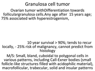
  • 1. Granulosa cell tumor Ovarian tumor withDifferentiation towards folliculargranulosa cells.Any age after 15 years age; 75% associated with hyperestrogenism, 10 year survival > 90%; tends to recur locally, - 25% risk of malignancy, cannot predict from histology M/S: Small, bland, cuboidal to polygonal cells in various patterns, including Call-Exner bodies (small follicle-like structures filled with acidophilic material), macrofollicular, trabecular, solid and insular patterns
  • 2. Malignant Theca tumor • Malignant thecomas are extremly rare. Thecomas having mitosis more than 4/10 hpf and one ormore of atypia, necrosis, rupture or adhesions is regarded as having malignant potential . In this case, there are sheets of round to spindle cells with ill-defined pale often vacuolated cytoplasm. The nuclei vary from round to spindle shape with moderate atypia and mitotic activity (arrow) is more than 4 per high power field. Hence, it has been classified as a malignant thecoma.
  • 3. Benign serous tumors • Includes cystadenoma, cystadenofibroma, adenofibroma, papillary cystadenoma, papillary cystadenofibroma, papillary adenofibroma • The term used depends on the relative amount of fibrous stroma, but distinctions are often arbitrary • Benign; 10-20% bilateral; most are partially or completely cystic; > 1 cm in size (< 1 cm signifies a cortical inclusion cyst or endosalpingiosis)
  • 4. Simple serous cyst • Usually small, uni- to multi-locular cysts lined by a single layer of tall, columnar, ciliated cells resembling normal tubal epithelium or cuboidal non-ciliated epithelium resembling ovarian surface epithelium • Stroma contains spindly fibroblasts
  • 5. Paillary serous cystadenoma • A case of papillary Serous cystadenoma Ovary showing papillary processes lined by a single layer of tubal type columnar epithelium with cilia and rounded nuclei. The stroma is edematous to fibrous. There is no atypia or mitotic activity.
  • 6. Serous cystadenofibroma • Serous Cystadenofibroma ovary showing a lining of a single layer of cuboidal cells with uniform basal nuclei. The stroma is myxoid to fibrous. There is no nuclear atypia or epithelial stratification. Few hypocellular edematous papillary projections can be seen in the lumen.
  • 7. adenofibroma • Adenofibroma: entirely solid appearing; if surface papillary projections are prominent, designate as surface papillary serous adenofibroma
  • 8. Borderline serous papillary tumor Serous tumors of low malignant potential, characterized by broad, branching papillae (hierarchical branching) focally covered by stratified epithelium with mild to moderate atypia with few mitoses Younger women, often pregnant.10% have microinvasion, but rarely has malignant behavior. Bilateral in 25%.patiets with borderline tumors may have peritoneal implants. Which may be noninvasive and have no influence on prognosis or implants may be invasive which are associated with tumor recurrence or transition to invasive carcinoma. Borderline serous papillary tumor ovary showing branching papillae of multiple lengths with edematous connective tissue cores. The papillae are lined by ciliated columnar epithelium with eosinophilic cytoplasm, round or oval nuclei showing mild to moderate atypia and cellular stratification. Mitotic figures are easily seen. There is no stromal invasion.
  • 9. Serous cystadenocarcinoma • Papillary serous carcinoma, Ovary- Low Grade malignant tumors showing irregularly branching papillae without much stroma lined by columnar cells with scant eosinophilic cytoplasm, moderate degree of pleomrphism with round to oval nuclei. Psammoma bodies (concentrically laminated calcified concretions) can be seen. There are two distinct types of ovarian serous carcinoma, low grade and high grade High grade serous carcinoma exhibits moderate to marked nuclear atypia and greater than 12 mitoses per 10 high power fields. Necrosis and multinucleate cells are often present.
  • 10. High grade serous adenocarcinoma,ov High grade: Branching papillary fronds, slit-like fenestrations, glandular complexity, moderate to marked nuclear atypia with marked pleomorphism, prominent nucleoli, stratification, frequent mitoses, stromal invasion (irregular or destructive infiltration by small glands or sheets of cells) • Variable psammoma bodies (calcium concretions with concentric laminations, may be intracellular due to autophagocytosis). Stroma may be fibrous, edematous, myxoid, or desmoplastic • A case of High Grade Serous Carcinoma showing marked necrosis. The tumor cells are arranged in microcystic pattern. There is marked nuclear atypia and mitosis (arrow) more than 12 mitoses per 10 high power fields can be seen.
  • 11. Benign mucinous tumor Includes cystadenoma, cystadenofibroma and adenofibroma (rare). Lesions are a spectrum of benign lesions with varying amounts of cysts, glands, and stroma - the distinction is arbitrary. 5% bilateral. Associations: carcinoid tumors in same ovary, dermoid cyst, Brenner tumor, endocervical adenocarcinoma; rarely with Zollinger-Ellison syndrome. Gross: Smooth surface with multiple cystic spaces, variable amounts of solid areas. Unilocular cysts are almost always benign. Filled with translucent viscous fluid. M/S: Tall, columnar, nonciliated cells, basal nuclei, abundant intracellular mucin. Usually endocervical type; also intestinal type (picket fence architecture with Paneth cells) or mixed. Stroma may be fibrous or mimic ovarian stroma. May have microscopic rupture of cysts with inflammatory reaction including histocytes and necrosis. Small bland papillae may be present, and mild, focal cytologic atypia may be present
  • 12. Borderline Mucinous tumor • Borderline mucinous tumor ovary is also known as atypical proliferative mucinous tumor of ovary or mucinous ovarian tumor of low malignant potential. 85% are intestinal type; 15% are endocervical type. 40-50% tumors are bilateral. • Classification: cllassified into 3 types:1. Borderline with atypia: grade 1 nuclei and papillae with cords. 2. Borderline with intraepithelial carcinoma: grade 2/3 nuclei, 4+ layers or cribriform or stroma-free papillary growth pattern. 3. Borderline with microinvasion: invasive glands usually accompanied by desmoplastic reaction In this case, The atypical epithelium is represented by tall columnar mucus secreting cells forming papillae and infoldings. Focally, Goblet cells (double arrow) are seen indicating intestinal type epithelium. The atypical epithelial lining cells are stratified in two or three layers (arrow) with moderate nuclear atypia. Focal cribriform architecture is seen (circle). Thick clear mucinous material fills the lumen
  • 13. Mucinous adenocarcinoma ovary 77% of ovarian mucinous carcinomas are metastases, 23% are ovarian primaries Of the ovarian primaries, most arise in a benign or borderline tumor; only 5-10% are pure Features favoring primary ovarian carcinoma vs. metastasis are: unilateral, "expansile" pattern of invasion, complex papillary pattern, size > 10 cm, smooth external surface, microscopic cystic glands, necrotic luminal debris, mural nodules and accompanying teratoma, adenofibroma, endometriosis or Brenner tumor .Stromal invasion > 10 mm2 distinguishes these tumors from borderline tumors. Two types of invasion - expansile or infiltrative:Expansile tum ors are usually stage I and behave "benign“. Infiltrative tumors may demonstrate malignant behavior and cause death even if stage I Present case--Ovary showing cysts lined with a mixture of goblet cells and tall, columnar intestinal- type epithelium. There is cellular stratification with atypia. The malignant glands (circle) can be seen lying within the fibrous stroma.
  • 14. Enddddometroid ca ovary • Endometroid Ovarian Carcinoma constitutes 10- 25% of primary ovarian carcinomas. 15-30% of patients have synchronous endometrial hyperplasia or carcinoma; these tumors are often well differentiated with squamous metaplasia.Stromal invasion is defined as confluent glandular growth, stromal disappearance or obvious stromal invasion. In this case, Crowded, back-to-back glands lined by tall columnar epithelium resembling endometrial epithelium with hyperchromatic nucleus are seen. There is squamous metaplasia (arrow).
  • 15. Clear cell adenocarcinoma ovary • Ovarian clear cell adenocarcinomas account for 5% of all ovarian malignancies. Here, neoplastic tumor cells are arranged in nests and glands. Clear cells are typically polyhedral with distinct cell membranes, central to eccentric rounded or slightly angular nuclei, and abundant clear cytoplasm. Hobnail cells can be seen. Stroma is fibrous. Numerous intracytoplasmic hyaline globules (arrow) can be seen. The cytoplasm of clear cells contains abundant glycogen which is Periodic acid-Schiff (PAS) positive, diastase digestion resistant. Microcalcifications are seen in 10-30% of cases
  • 16. Primary sq. cell ca ovary Squamous cell carcinoma of ovary can be pure arising denovo or may represent extreme expression of squamous metaplasia in endometriosis, endometrioid carcinomas, transformation of ovarian teratomas and HPV related transformation Pure SCC and those arising from endometriosis are included in surface epithelial tumor by WHO Classification SCC arising in dermoid cyst are included in germ cell tumor by WHO Poor prognosis. The stage of the tumor and its grade correlate best with overall survival Microscopy: Can be well to poorly differentiated Well-differentiated form- SCC will show squamous maturation, keratin formation and intracellular bridging Poorly differentiated form- few normal squamous features may be identifiable. Present case of SCC of ovary did not have any association with endometriotic cyst or mature teratoma.
  • 17. Brenner tumor • Benign Brenner Tumor Ovary:Derived from ovarian surface epithelium and metaplasia; similar to Walthard nests, 6% bilateral • Microscopy: Solid and cystic nests of urothelium-like cells surrounded by abundant dense, fibrous stroma • Epithelial cells have sharp outlines. Cells are uniform, polygonal with pale cytoplasm, small but distinct nucleoli, often grooved nuclei (similar to granulosa cell tumors) • Frequent microcysts within epithelial nests • Metaplastic - with prominent cystic formation, accompanied by florid mucinous changes similar to cystitis glandularis • Proliferating - with papillary pattern and low grade nuclear atypia resembling low grade urothelial carcinoma of bladder • Present case shows marked mucinous metaplasia
  • 18. Malignant Brenner tumor • Malignant Brenner tumour is a very rare malignancy and closely resembles transitional cell carcinoma of urinary bladder with squamous and undifferentiated variants. Transitional cell carcinoma, however, may • also occur as primary tumor of ovary and only presence or absence of areas with benign or borderline Brenner tumour differentiates the two. In this case, the malignant cells are arranged in nests and sheets, inflitrating and replacing the fibrous stroma. The tumour cells are displaying moderate degree of anaplasia, hyperchromatic nuclei with high mitotic activity. Areas of benign brenner tumor with characteristic coffee bean nuclei are marked with an arrow.
  • 19. Transitional cell ca ovary Transitional cell carcinoma (TCC) of ovary is a rare tumor, accounting for only 1% of surface epithelial carcinomas. It has been described as a primary ovarian carcinoma in which definite urothelial features are present but no benign, metaplastic and/or proliferating Brenner tumor can be identified. In this case, broad blunt papillae with fibrovascular cores lined by stratified and highly atypical transitional cells with round or oblong nucleus having prominent nucleoli can be seen. There are punched out microspaces and areas of necrosis. Mitotic activity is high. Some Authors are of view that Ovarian transitional cell carcinoma represents a poorly differentiated form of high-grade serous carcinomas ovary.
  • 20. Undifferentiated ca ovary Undifferentiated carcinomas are characterized by a patternless solid, sheet-like growth of tumor cells, with an aggressive clinical course There are no nests, papillae, glands, trabeculae or spindled patterns, no squamous or mucinous metaplasia, and no/minimal neuroendocrine differentiationIf areas of a differentiated component are found, the tumor is . called dedifferentiated carcinoma. Gross : Large tan- brown, fleshy adnexal masses with areas of necrosis, mostly with ovarian surface involvement m/s: Sheets of dyshesive round to ovoid cells, frequently resembling large cell lymphoma, separated by delicate fibrovascular septa No evidence of gland formation, trabecular or nested growth pattern May have foci of spindling / keratinization / necrosis. Vascular invasion in 20%. Frequent mitoses and apoptosis; mitotic rate of 5 to 50 per 10 high power fields
  • 21. Krukenberg tumor • Krukenberg tumor is a metastatic signet ring cell adenocarcinoma of the ovary, accounting for 1% to 2% of all ovarian tumors. Stomach is the primary site in most cases (70%). Carcinomas of colon, appendix and breast (mainly invasive lobular carcinoma) are the next most common primary sites. Microscopically, Krukenberg tumor has 2 components: epithelial and stromal.The epithelial component is composed of mucin-laden signet ring cells with eccentric hyperchromatic nuclei. The signet ring cells can be single, clustered, nested, or they can be arranged in tubules, acini, trabeculae, or cords. The intracytoplasmic mucins of the signet ring cells are neutral and acidic, they stain with mucicarmine, PAS and Alcian blue stains. The mesenchymal component of Krukenberg tumor is of ovarian stromal origin and is composed of plump and spindle-shaped cells. In this case, the above mentioned features are seen.
  • 22. dysgerminoma Less than 1% of ovarian malignancies. Counterpart of testicular seminoma. Usually young patients <30. 5% associated with gonadal dysgenesis androgen insensitivity or pseudohermaphroditism .Metastasize to opposite ovary, retroperitoneal nodes and peritoneal cavity. Rarely transforms to yolk sac tumor Survival: 95%. Mixture with choriocarcinoma, yolk sac or embryonal carcinoma worsens prognosis. 15% bilateral. Solid, nodular, small to huge and gray-pink (resembles cerebral cortex). Hemorrhage and necrosis common . M/S. Nests of tumor cells separated by fibrous stroma infiltated with T lymphocytes. Large vesicular cells with well defined cell borders, cleared cytoplasm containing glycogen and central nuclei. 1+ prominent nucleoli; occasional granulomas. May have syncytiotrophoblastic cells with raised beta HCG. May have abortive yolk sac elements with increased serum AFP Early carcinomatous differentiation: 30+ mitoses per 10 high power fields, may worsen prognosis
  • 23. Yolk sac tumor Yolk sac tumor (also known as endodermal sinus tumor) of the ovary is a malignant germ cell neoplasm. It often occurs in pure form but is also frequently found as a component of mixed malignant germ cell tumors. The most common ("typical") pattern consists of papillary projections, each of which contains a central blood vessel, surrounded by a thick layer of basement membrane material, and then covered by a layer of embryonic epithelial cells with clear or eosinophilic cytoplasm; this structure is referred to as a Schiller-Duval body. Another frequently encountered feature is hyaline globules, which are eosinophilic, PAS-positive, diastase-resistant globules. Histology reveals a wide range of patterns (microcystic, endodermal sinus, solid, alveolar-glandular, papillary, macrocystic, hepatoid, primitive endodermal). In this case, predominantly microcystic pattern is seen. In the lower half, ciliated pseudostratified columnar epithelial lining of dermoid cyst is seen.
  • 24. Mature teratoma Tumors arise from a single germ cell after first meiotic division. Mature if only contains adult tissues.If skin tissue predominant, called dermoid cyst. Usually teenage women (solid) or children (cystic). Excellent prognosis, even if peritoneal implants are present. May rupture into peritoneal cavity causing foreign body reaction that simulates metastatic carcinoma or miliary tuberculosis. Cystic tumors may contain squamous cell carcinoma, carcinoid tumor or adenocarcinoma. Gliomatosis peritonei: peritoneal implants exclusively composed of mature glial tissue; benign if all tissue is mature and other teratomatous elements are absent. Gross ; Solid or cystic. Cystic content may contain greasy material composed of keratin, hair and teeth. Micro Description: Ectodermal structures in 100%, mesodermal in 93% and endodermal in 71%. Skin and glial tissue common . Still considered mature if microscopic foci of immature tissue. A case of mature teratoma ovary showing mature, well-differentiated tissue elements from all three embryologic germ layers (ectoderm, mesoderm, endoderm).
  • 25. Struma ovarii • Rare monodermal teratoma composed predominantly of mature thyroid tissue. May show pathologic changes of thyroid gland including hyperfunctioning; malignancies are usually papillary thyroid carcinoma. Associated with mucinous cystadenoma, Brenner tumor, carcinoid tumor and dermoid cyst Gross ; Resembles red-brown thyroid tissue but usually multilocular cystic; usually unilateral Micro ; Thyroid follicles with colloid; other teratomatous elements may be present. Rarely has solid or pseudotubular patterns, microfollicles, abundant eosinophilic cytoplasm, abundant clear cytoplasm or minimal thyroid follicles • A case of Struma ovarii showing predominantly mature thyroid tissue consisting of acini of various sizes lined by a single layer of columnar or flattened epithelium. The acini contain eosinophilic, PAS positive colloid.Struma ovarii is a specialized or monodermal teratoma predominantly composed of mature thyroid tissue. Thyroid tissue must comprise more than 50 percent of the overall tissue to be classified as a struma ovarii.
  • 26. Sq cell ca in dermoid cyst • Mature cystic teratoma is a common benign adnexal tumour in females. Malignant transformation in a mature cystic teratoma of the ovary is rare, occurring in only 1-2% of cases. The most common malignancy is squamous cell carcinoma, which consists of about 75% of malignant transformations. In this case, squamous cell carcinoma arising from the lining of the cyst on left side of section can be seen. The tumor cells are showing moderate pleomorphism, abundant eosinophilic cytoplasm, roud to oval hyperchromatic nuclei, 0-1 prominent nucleoli. High mitotic activity (arrow), tumor giant cells (double arrow) and keratin pearls (circle) are seen.
  • 27. Melanoma arising from dermoid cyst • Malignant melanoma may originate from melanocytes in ovarian cystic teratomas (dermoid cysts). The extremely rare primary ovarian melanoma may be differentiated from the more common melanoma metastatic to the ovaries by its unilaterality, the presence of junctional change,associated with teratoid elements, and a detailed history and physical examination to exclude another primary site. Here, the tumor is seen arising from cyst wall The cells are arranged in nests and are pleomorphic oval to spindled cells with hyperchromatic nuclei, prominent nucleoli, and frequent mitosis. The cytoplasm contained abundant brown pigment, melanin.
  • 28. Immature teratoma Immature teratoma represents 3% of all teratomas and 1% of all ovarian cancers. An immature teratoma reveals mixtures of mature and immature tissues reminiscent of the developing stages of embryonic tissues. The most common immature element that is present and used for grading is primitive neuroepithelium. Norris grading system : 1: abundant mature tissue, loose mesenchymal tissue with occasional mitoses, immature cartilage and tooth anlage 2: less mature tissue than grade 1, rare foci of neuroepithelium with mitoses, less than 4 low power fields in any one slide 3: little / no mature tissue; numerous neuroepithelial elements merging with cellular stroma occupying more than low power fields micro images.
  • 29. Normal histology of F. Tube • Plica: delicate folds of mucosa on inner aspect of tube, most evident in ampulla, merge with fimbriae • Mucosa: consists predominately of single layer of cells including: 1. Ciliated columnar cells (~25%), most abundant in infundibulum and ampulla, estrogen increases production of cilia. 2. Secretory cells (~60%), non-ciliated, contain apical granules and produce tubular fluid, progesterone increases their number while estrogen increases their height and secretory activity 3. Intercalated cells (peg cells, < 10%), which may be inactive secretory cells Muscle Layer: 3 smooth muscle layers: Innermost - obliquely arranged Middle – circular Outermost - longitudinal, extends into fimbriae Serosal surface: mesothelium of visceral peritoneum. May contain Walthard cell rests
  • 30. Normal histology of F. Tube • Walthard cell rests (marked in ellipse); sometimes called Walthard cell nests, are a benign cluster of epithelial cells most commonly found in the connective tissue of the Fallopian tubes, but can also be seen in the mesovarium, mesosalpinx and ovarian hilus. They appear as white/yellow cysts or nodules of flat to cuboidal cells with minimal atypia that can reach a size of 2 millimeters. They typically have elliptical nuclei with a long groove (along the major axis) - so called - "coffee bean" nuclei. May be source of Brenner tumors or primary urethelial cell carcinomas (parafallopian tube transitional cell carcinoma).
  • 31. Endometriosis fallopian tube • Presence of endometrial tissue within fallopian tube; will cycle with normal endometrium • Typically seen during reproductive years, in 6-10% of women • Also occurs in 20-50% of tubes after ligation, • May be incidental finding, or associated with abdominal/pelvic pain, infertility • Gross; May have red nodules in wall or serosa, may be actual polyp, may have normal gross appearance • Micro; Endometrial glands, endometrial stroma, hemosiderin-laden macrophages: need 2 to make diagnosis
  • 32. Salpingitis isthmica nodosa • Salpingitis isthmica nodosa also called diverticulosis of fallopian tube is characterized by nodular thickening of the tunica muscularis of the isthmic portion of the tube enclosing cystically dilated glands leading to complete obliteration of tubal lumen. It is usually bilateral. May be analogous to uterine adenomyosis. Clinical ; Associated with infertility in 50% . May lead to ectopic pregnancy; Gross – Generally well delineated, yellow-white nodular swelling(s) up to 2 cm, usually isthmus, may be inconspicuous . Microscopy: Regularly spaced glands lined by normal appearing tubal epithelium within hypertrophied smooth muscle or surrounding fibrous tissue Glands may be cystically dilated and are true diverticula that communicate with tubal lumen.. Occasionally glands surrounded by endometrial-type stroma No significant atypia, stromal response minimal In this case, regularly spaced glands lined by normal appearing tubal epithelium within hypertrophied smooth muscle can be seen. Few of the glands are cystically dilated.
  • 33. hydrosalpinx • Hydrosalpinx is defined as dilatation of the ampullary segment of the fallopian tube with associated thinning of adherent fimbriae. Causes:- endometriosis, ovulation induction , pelvic inflammatory disease (e.g chlamydial or gonococcal infection): a hydrosalpinx is most commonly a sequela of adhesions from pelvic inflammatory disease, post hysterectomy (without salpingo-oophorectomy). this is from accumulation of tubal secretions caused by surgical blockage proximally and adhesion- related blockage distally, tubal ligation , tubal malignancy: primary or secondary tumours of the fallopian tubes • On microscopy, there is marked dilatation of the lumen, with flattening of mucosa.
  • 34. Ectopic tubal pregnancy Implantation of fertilized egg within fallopian tube , Ampulla (80%), isthmus (12%), fimbriae (5%), cornu (2%) Risk Factors: Tubal damage: due to Infections (PID) or salpingitis , Abdominal/pelvic surgery or tubal ligation, History of previous ectopic pregnancy Smoking as smoking may contribute to decreased tubal motility by damaging ciliated cells Altered tubal motility: due to smoking or hormonal contraception; progesterone-only contraception and progesterone intrauterine devices (IUDs) have been associated with increased risk of ectopic pregnancy; symptoms: abdominal or shoulder pain, cramps, vaginal bleeding, nausea, dizziness; Gross ; Distension of tube with thin or ruptured wall, dusky red serosa and hematosalpinx, possibly with fetal parts identified; Micro ; Intraluminal chorionic villi and extravillous trophoblast (may be degenerated); variable fetal parts; Decidual change in lamina propria in 1/3; mesothelial reactive proliferation Uterus: gestational hyperplasia with Arias-Stella reaction ; Present case-- showing thinned and dilated fallopian tube with Intraluminal chorionic villi (arrow), blood and extravillous trophoblast (double arrow)
  • 35. Epithelioid trophoblastic tumor,F. Tube• Microscopy: ETT is composed of nests of uniform mononucleate chorionic type intermediate trophoblastic cells, with eosinophilic or clear cytoplasm, round nuclei, and a well-defined cell membrane. Nests of trophoblastic cells are surrounded by extensive necrosis and a hyaline like matrix, resembling keratin, giving ETT its characteristic “geographic” appearance. Within the center of each tumor nest, there is often a small blood vessel, though overall the tumor lacks significant vascular invasion. Immunoperoxidase staining of the trophoblastic cells is positive for pancytokeratin AE1/AE3, p63, EMA, CD10, inhibin alpha, and Ki67.
  • 36. Acute salpingitis Acute inflammation of fallopian tube, most commonly due to chlamydia and gonorrhea Symptoms ; abdominal pain, abnormal smell and color of vaginal discharge, bloating, fever, lower back pain, nausea, vomiting , pain during ovulation, dysmanorrhea, dysprinaia, may develop PID oor may be asymptomatic PID has 3 principal complications: chronic pelvic pain (25%), infertility, ectopic pregnancy (15-50%) Impaired fertility is major concern; infection and inflammation can lead to scarring and adhesions within tubal lumens; May lead to tubule-ovarian abscess, pelvic peritonitis, perihepatitis Gross ; Enlarged, erythematous, edematous, Pyosalpinx , hematosalpinx ; May have fibrinous exudate ;Tubo-ovarian abscess common; Micro ; Marked neutrophilic infiltrate, congestion and edema , Mucosal ulceration, Reactive epithelial changes, Variable abscess formation, variable presence of microorganisms
  • 37. pyosalpinx Etiology same as for acute salpingitis .Inflammatiion is severe accompanied by suppuration and pus formationn Gross ;tube dilated and filled with pus A case of pyosalpinx showing dilated fallopian tube, thick wall, denudation of epithelium and pus in the lumen and wall.
  • 38. Chronic salpingitis • Chronic Salpingitis may follow prolonged acute suppurative salpingitis or result from repeated bouts of acute salpingitis. During acute salpingitis, fibrin exudation into the lumen results in the plicae adhering to each other. Subsequent healing and organization lead to permanent adhesions between the plicae producing follicular salpingitis. Plasma cells, lymphocytes or both are present in the mucosa. . When the inflammation subsides, the tube becomes distended with an acellular transudate producing a hydrosalpinx. The fimbriae may adhere to the ovary leading to direct involvement of the latter and a tubo- ovarian abscess may result. In the present case, numerous plasma cells (arrow), lymphocytes along with few eosinophils are present within plicae of the fallopian tube. Plasma cells must be present before the diagnosis of chronic salpingitis can be made.