Recomendados

Recomendados
Mais conteúdo relacionado
Mais procurados
Mais procurados (18)
Destaque
Destaque (16)
Semelhante a Hamster pinworm poster FINAL
Semelhante a Hamster pinworm poster FINAL (20)
Hamster pinworm poster FINAL
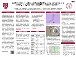
- 1. Identification of and surveillance for Syphacia pinworms in a colony of Syrian hamsters (Mesocricetus auratus) David K. Chu1*, Ramon A. Carreno2, Kristen M. Davis1, Sergio A. Koba1, Claude M. Nagamine1 Dept of Comparative Medicine, Stanford University1; Dept of Zoology, Ohio Wesleyan University2 Abstract Introduction Methods & Materials (continue) Results (continue) Discussion An investigator studying the biology and genetics of Sex-linked yellow (an X-linked gene that determines coat color) acquired a stock of hamsters from a private source. Pinworm ova of an unidentified species were discovered during health screening. These hamsters were quarantined to guard our laboratory mice and rats in case the helminthiasis was due to S. obvelata or S. muris, which were reported to infect Syrian hamsters. Primary goal of this study was to identify pinworm species that was infecting this hamster colony. Secondary goal was to determine if this pinworm may infect the mouse via dirty bedding transfer, a typical means to survey for murine disease agents. Answers to these questions may guide laboratory managers and veterinarians on how to assess the risks of infected hamsters to the laboratory mouse. This study was Stanford IACUC approved. Host Specificity and Surveillance ➢ Transfer one teaspoon of dirty bedding with feces from pinworm-infected hamsters to three different sentinel cages weekly. ➢ Sentinel composition: • Two Hsdhan:AURA female hamsters • Three AG129 (129.Ifnar1tm1Agt, Ifngr1tm1Agt) female mice • Five AG B6 (C57BL/6J.129 Ifnar1tm1Agt, Ifngr1tm1Agt) female mice ➢ Once per week for 5 weeks (beginning at 7 days post first dirty bedding transfer) we conducted perineal cellophane tape tests followed by perineal swabs from sentinels. ➢ Cellophane tapes were mounted on glass microscope slides for microscopy. ➢ Swab samples obtained using Charles River Labs (CRL) “pink sticky” swabs were sent to CRL for PCR. It may be necessary in a multi-species facility to co-house different rodent species free from adventitious infections. Goals of this study were to identify pinworms found in a closed golden hamster colony and to assess if a dirty bedding surveillance program can detect presence of this pinworm. For identification, we retrieved worms from the cecum of euthanized hamsters and fixed some either in 5% formalin for morphologic analysis or 90% ethanol for DNA sequencing. For surveillance, we transferred one teaspoon of dirty bedding with feces from pinworm- infected hamsters to three sentinel cages weekly. Sentinels were two female Hsdhan:AURA hamsters and three female 129.Ifnar1tm1Agt, Ifngr1tm1Agt and five C57BL/6.129 Ifnar1tm1Agt, Ifngr1tm1Agt mice, immunodeficient strains with a double knockout for Type I and II interferon receptors. We conducted weekly perineal cellophane tape tests plus we sent perineal swab samples to a commercial lab for PCR. Morphologically, the worms are consistent with Syphacia mesocriceti. However, worm dimensions are larger overall than in previously published descriptions. 1000 bases of the 28S rDNA locus were sequenced from four worms and all four were identical. A BLAST search revealed that the closest match to the sequences is S. agraria at 86% identity, followed by S. ohtaorum and S. obvelata. No evidence of pinworms was detected by tape tests or perineal swab PCR at 1 and 2 weeks post bedding transfer. Ova were observed on hamster cellophane tapes in weeks 3, 4, and 5 while no ova were seen in any of the mouse cellophane tapes. Hamster swab PCR was positive for generic pinworm in week 3 and mouse swab PCR was equivocal in week 5. Our data suggest this hamster colony is infected with S. mesocriceti and that this pinworm is non- transferable to the immunodeficient mouse strains tested by dirty bedding. *dchu98@stanford.edu ➢ GenBank BLAST search of 1000 bases of the 28S rDNA revealed that S. mesocricti 28S rDNA sequences were not in GenBank. The closest match to the sequences is S. agraria at 86% identity, followed by S. ohtaorum and S. obvelata. ➢ Scan this poster’s QR code for BLAST results and additional worm micrographs. Figure 2. Male worm with characteristic spicule (arrow) which facilitates sperm transfer. Syphacia demonstrates sexual dimorphism in which male pinworms are much smaller than females. This male measured 855 μm in length. Figure 1. Anterior of a cleared, mature female worm. More female worms were identified than males in our collection by a 15:2 ratio. References 1. Dick TA, Quentin JC, Freeman RS. 1973. Redescription Syphacia Mesocriceti (Nematoda: Oxyuroidea) parasite of the golden hamster. J Parasitol. 59(2):256-259. 2. Hasegawa H, Sato H, Iwakiri E, Ikeda Y, Une Y. 2008. Helminth collected from imported pet murids, with special reference to concomitant infection of the golden hamster with three pinworm species of the genus Syphacia (Nematoda: Oxyuridae). J Parasitol. 94(3):752-754. 3. Okamoto M, Urushima H, Hasegawa H. 2009. Phylogenetic relationships of rodent pinworms (genus Syphacia) in Japan inferred from 28S rDNA sequences. Parasitol Int. 58:330-333. 4. Pinto RM, Goncalves L, Gomes DC, Noronha D. 2001. Helminth fauna of golden hamster Mesocricetus auratus in Brazil. Contemp Top Lab Anim Sci. 40(2):21-26. P196 Naturally Infected Host Animals Closed colony of 5 to 8 month old mixed sex Syrian hamsters (private source, Fresno, CA) free from LCMV, PVM, Sendai virus, Reovirus, SV5, E. cuniculi, C. piliforme, external and other internal parasites housed in hardwood bedded (Teklad), filter top cages with feed (Teklad 2018) and RO filtered water. Parasite Characterization ➢ Retrieve worms from intestinal trace of CO2 euthanized hamsters. ➢ Fix worms in 5% formalin for morphologic analysis or 90% ethanol for DNA sequencing. ➢ Worms were cleared with lactic acid and viewed by light microscopy. ➢ Measure physical characteristics on 15 females and two males: Body length and max width, cephalic vesicle length and width, nerve ring width, excretory pore width, vulva distance from anterior, esophagus length, esophageal bulb length and width, uterine corpus length and width, tail length, ova length and width. ➢ Sequence 1000 bases of 28S rDNA locus. ➢ Survey GenBank for matching sequences using BLAST. Methods & Materials Results Table 1. Female worm morphometric measurements with reference values. All units are expressed as microns (μm). Table 2. Perineal cellophane tape tests and fecal PCR results. We consistently observed ova on hamster tapes since week 3. Hamster feces was PCR positive on Pinworm Screen PCR (but negative for Aspiculuris, S. obvelata, S. muris (CRL). Pooled mouse feces yielded equivocal PCR results on week 5. ➢ This was a mono-infection by S. mesocriceti. ➢ Although S. mesocriceti was reported to infect small intestine4, we found all of our worms in the large intestine. ➢ Female morphology is slightly larger than expected but overall is quite consistent with what has been previously reported (Table 1). ➢ We encountered a BLAST search zero match for S. mesocriceti because this pinworm species is not in GenBank. An effort will be made to deposit this species sequence into GenBank. ➢ The 86% 28S rDNA S. agaraia match is highly dissimilar for a relatively conserved locus and that most of the 28S should be identical or almost identical. Additionally, S. agaraia is known to infect Large Japanese field mouse3 and not likely to be pinworm species in our colony of Syrian hamster. However, polyinfection can occur2 and should not be ruled out. ➢ Despite the fact that sentinel mice do not express Type I and II interferon receptors (increases their susceptibility to parasite infection. Table 2 suggests that S. mesocriceti is host specific or the risk of fomite transmission to mice is low. ➢ Sentinel hamster tape tests and PCR don’t correlate and may be explained by 1) Tapes removed too much pinworm matter leaving insufficient DNA for PCR 2) Positive tapes in wks 4 and 5 were residuals from wk 3 or 3) Primers sensitivity was too low. ➢ Equivocal mouse fecal PCR result likely due to contamination during sample acquisition. Female Characteristics Mean Standard Deviation Reference 1 Reference 4 Body Length n=14 7261 742 5000 - 6900 3200 - 5200 Body Max Width n=15 262 28 149 - 165 150 - 180 Cephalic Vesicle Length n=14 173 30 - - Cephalic Vesicle Width n=14 143 16 - - Nerve Ring (from anterior) n=8 176 31 125 - 145 125 - 140 Excretory Pore (from anterior) n=13 575 108 530 - 650 490 - 560 Vulva Distance from Anterior n=11 926 170 810 - 890 700 - 830 Esophageal Length n=15 414 30 - 280 - 385 Uterine Corpus Length n=11 296 15 - - Uterine Corpus Width n=12 53 7 - - Esophogeal Bulb Length n=10 119 8 92 - 107 - Esophogeal Bulb Width n=15 98 9 77 - 88 - Tail Length n=13 973 70 - - Ova Length n= 40 128 4 115 - 118 130 - 140 Ova Width n= 40 33 3 31 - 46 40 - 50 Worms were visualized in cecum; some in colon; none in small intestine.