Microscope Biology I Lab
•Transferir como PPT, PDF•
5 gostaram•104,167 visualizações

Biology I Lab instructions: Microscope

Recomendados
Mais conteúdo relacionado
Mais procurados
Mais procurados (20)
Destaque
Destaque (20)
Semelhante a Microscope Biology I Lab
Semelhante a Microscope Biology I Lab (20)
Mais de Lumen Learning
Mais de Lumen Learning (20)
Último
Último (20)
Microscope Biology I Lab
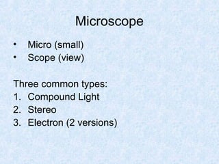
- 1. Microscope • Micro (small) • Scope (view) Three common types: 1. Compound Light 2. Stereo 3. Electron (2 versions)
- 2. 1. Compound Light • Most common • 2 lenses focusing at the same time 1. Ocular 2. Objective • 2D image • High magnification through light absorption
- 3. Parts of the microscope Arm Base
- 4. Term: Magnification • Ratio of image size to actual size • Ocular: 10X (“X” means times; for example, the ocular magnifies something 10 times) • 4 objectives – Scan: 4X – Low Power: 10X – High Power: 40X – Oil immersion: 100x • Multiply ocular x objective to get overall magnification
- 5. Field of View Amount of object you can see • Decreases with increasing magnification
- 6. Depth of Focus Thickness of specimen in focus at a given magnification • Decreases with increasing magnification • Lab says to make your own slide-we will use a prepared thread slide
- 7. Inversion Phenomenon • Objects appear upside down and backwards in the compound microscope • We will use prepared letter e slides
- 8. 2. Stereomicroscope • Used to get a better look at larger objects • 3D image • Specimen not mounted on a slide • Low magnification • Uses visible light through light scattering
- 9. 3. Electron Microscope • Uses electron streams focused by magnets to view specimens, not light • 2 types 1. Scanning 2. Transmission
- 10. Scanning Electron Microscope • 3D images • 1,000-10,000x magnification • Resolution of 5 nanometers pollen microscope
- 11. Transmission Electron Microscope • 2D • 10,000-100,000 magnification mitochondria
- 12. Wet mount • Way to create a simple slide • Need a clean slide and a coverslip • Procedure – Place specimen on slide – Add a drop of water – Place coverslip over specimen at an angle to avoid air bubbles – Wipe away excess water – View under microscope • Today: pond water wet mount
Notas do Editor
- Image of phase contrast microscope by GcG https://commons.wikimedia.org/wiki/File:Phase_contrast_microscope.jpg Public Domain
- Image of parts of the microscope by Peri Coleman https://commons.wikimedia.org/wiki/File:Labelledmicroscope.gif Public Domain
- Image of microscope lens by Halfblue https://commons.wikimedia.org/wiki/File:Eyepieces_random_selection.jpg CC-By SA Image of optic lenses on microscope by Rama https://commons.wikimedia.org/wiki/File:Loupe-binoculaire-p1030891.jpg CC-By SA
- Image of microscope lenses by PublicDomainPictures https://pixabay.com/en/microscope-slide-research-close-up-275984/ Public Domain
- Image of Optical stereo microscope nikon smz10 by GcG(jawp) https://commons.wikimedia.org/wiki/File:Optical_stereo_microscope_nikon_smz10.jpg Public Domain
- Image of Scanning Electron Microscope by Cjp24 https://commons.wikimedia.org/wiki/File:Scanning_electron_microscope2.jpg CC-By SA Image of pollen by Dartmouth College Electron Microscope Facility https://commons.wikimedia.org/wiki/File:Misc_pollen.jpg Public Domain
- Image of mitochondria, mammalian lung – TEM by Louisa Howard https://commons.wikimedia.org/wiki/File:Mitochondria,_mammalian_lung_-_TEM.jpg Public Domain Image of Transmission electron microscope (Morgangni 268D) 1pl by Pleple2000 https://commons.wikimedia.org/wiki/File:Transmission_electron_microscope_(Morgagni_268D)_1pl.jpg CC-By SA
- Image of Bdelloid Rotifer by Bob Blaylock https://commons.wikimedia.org/wiki/File:Bdelloid_Rotifer.jpg CC-By SA