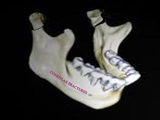
Condylar fractures /certified fixed orthodontic courses by Indian dental academy
•
32 gostaram•4,203 visualizações

The Indian Dental Academy is the Leader in continuing dental education , training dentists in all aspects of dentistry and offering a wide range of dental certified courses in different formats. Indian dental academy provides dental crown & Bridge,rotary endodontics,fixed orthodontics, Dental implants courses.for details pls visit www.indiandentalacademy.com ,or call 0091-9248678078

Recomendados
Recomendados
Mais conteúdo relacionado
Mais procurados
Mais procurados (20)
Destaque
Destaque (14)
Semelhante a Condylar fractures /certified fixed orthodontic courses by Indian dental academy
Semelhante a Condylar fractures /certified fixed orthodontic courses by Indian dental academy (20)
Mais de Indian dental academy
Mais de Indian dental academy (20)
Último
Último (20)
Condylar fractures /certified fixed orthodontic courses by Indian dental academy
- 2. INDIAN DENTAL ACADEMY Leader in continuing dental education www.indiandentalacademy.com Seminar On….. www.indiandentalacademy.com
- 3. Introduction: Condylar and Subcondylar fractures constitute 26-40% of all mandible fractures Given the unique geometry of the mandible and the temperomandibular joint, fractures of the condyle and subcondylar region can result in marked pain, dysfunction and deformity if not recognized and treated appropriately. These features may be associated with other injuries that have severe mobility( C- spine injuries,displacement of the condyle into the middle cranial fossa, injuries to the external auditory canal and occlusion of the internal www.indiandentalacademy.com carotid artery)
- 4. DEFINITION: Fracture is defined as a sudden violent solution in the continuity of bone which may be complete or incomplete resulting from direct or indirect causes. www.indiandentalacademy.com
- 5. Anatomy: Arises from the 1st Pharyngeal Arch-develops around the ventral cartilage of the 1st branchial arch. Continues to grow and develop throughout childhood and adolescence. Mandible is curved and articulates at both ends. TMJs are diarthroidal and allow both rotational and translatory movements. www.indiandentalacademy.com
- 9. Classification of Condylar Fractures: Rowe and Killey’s Classification (1968) a. Intracapsular Fractures or High Condylar i. Fractures involving the articular surface ii. Fractures above or through the anatomical neck, which do not involve the articular surfaces b. Extracapsular or Low Condylar Fractures c. Fractures associated with injury to the capsule, ligament and meniscus d. Fractures involving adjacent bone www.indiandentalacademy.com
- 10. Lindahl's Classification (1977) a. Fracture Level i. Condylar Head (Intracapsular) ii. Condylar Neck iii. Subcondylar 1. High 2. Low b. Relation of the condyle to the Mandible i. Non Displaced ii. Deviated or Angulated iii. Displaced 1. Medial overlap. 2. Lateral overlap www.indiandentalacademy.com
- 11. c. Relation of the condyle to the mandible i. Non Displaced ii. Displaced- still related to the fossa iii. Dislocation - completely out of fossa McLennan's Classification(1952) A. B. C. D. No displacement Deviation Displacement Dislocation www.indiandentalacademy.com
- 12. CLASSIFICATION OF FRACTURES OF MANDIBULAR CONDYLE ACCORDING TO SPIESSL AND SCHROLL Type I: fracture without displacement Type II: low fracture with displacement Type III: high fracture with displacement Type IV: low fracture with dislocation Type V: high fracture with dislocation Type VI: intracapsular fracture (diacapitular) www.indiandentalacademy.com
- 13. Etiology Adult Represents 20-30% of all mandibular fractures (Ellis et al, 1985) • Motor Vehicle Accidents • Assault • Sports related injuries • Falls Children Higher involvement ranging from 40-60% (Lehman and Saddawi, 1976) • Falls • Motor Vehicle Accidents • Sports related injuries www.indiandentalacademy.com • Assaults
- 14. Forces resulting in trauma to the TMJ A. Moving object striking a static individual B. Moving individual striking a static object C. Combination of forces www.indiandentalacademy.com
- 15. When a blow is given on the face resulting in fracture of the mandible condyle, the position of the fractured condyle in relation to the remainder of the ramal stump will depend on: 1. The direction and degree of force. 2. The precise point of application of force 3. Whether the teeth were in occlusion at the time of injury 4. Whether the patient is partially or fully edentulous. www.indiandentalacademy.com
- 16. Clinical Signs and Symptoms: Evidence of soft tissue injury - Chin Lacerations Facial Asymmetry with chin deviation Noticable palpable swelling over the affected TMJ Pain and tenderness over the affected TMJ Malocclusion Deviation of the mandible dental midline Muscle splinting due to pain with limited opening Bleeding from the external auditory canal Inability to palpate condylar movement. www.indiandentalacademy.com
- 18. Soft Tissue Injuries: The soft tissue injuries are characterized by: Localized pain at rest exacerbated by function Limited range of motion secondary to pain If effusion is present - palpable fluctuant swelling and decreased ability to occlude on ipsilateral posterior teeth with deviation away from the affected side www.indiandentalacademy.com
- 19. ENT and Neurological Signs In Displacement of Condvle into the Middle Cranial Fossa CSF Otorrhea Lacerations of the Extemai Auditory Canal Paralysis of the Facial Nerve Hearing Deficit Hemorrhage from the middle meningeal artery Dural Tears Subdural and epidural hematoma Altered level of consciousness Pupillary Dilatation Nausea In Condylar Fractures: Laceration of the External Auditory Canal www.indiandentalacademy.com
- 20. Imaging in the Diagnosis of Condylar Fractures: A. Conventional Radiography a. P A- View b. Lateral Oblique c. Panoramic view d. Reverse Towne's Projection B. CT C. MRI www.indiandentalacademy.com
- 21. Posterior anterior view Lateral view www.indiandentalacademy.com Panoramic view Reverse Towne’s view CT Scan
- 22. Treatment: Goals of Treatment: Relief from pain Stable occlusion Restoration of inter-inCIsal opening Full range of mandibular movements To minimize deviation Avoid growth disturbances Avoid Ankylosis Modes of Treatment: 1. Conservative treatment I Non-Surgical 2. Surgical treatment by Open Reduction www.indiandentalacademy.com
- 23. Conservative Treatment Unlike fractures of other bones, the exact anatomic reapproximation of the fractured segments may not be absolutely essential. There is no correlation between radiographic findings & either preoperative symptoms or post operative function. Complications are uncommon with conservative treatment. Normal occlusion with minimal discomfort: soft diet and maintain as near normal function as possible. Malocclusion, deviation with function, pain: period of immobilization (7-21 days) in the form of arch bars or ivy loops, followed by active mobilization and physical therapy. Period of immobilization depends on age of patient, level of fracture, & degree of displacement. www.indiandentalacademy.com
- 24. Surgical Treatment 1. 2. 3. 4. 5. Reduction Fixation Immobilization Control of Infection Rehabilitation www.indiandentalacademy.com
- 25. 0pen Reduction Indications for 0pen Reduction of Condylar Fractures (Zide & Kent -1983) Absolute Indications a. Displacement into the middle cranial fossa b. Impossiblity of obtaining adequate occlusion by closed reduction c. Lateral extracapsular displacement of the condyle d. Invasion of a foreign body (e.g.: gunshot wound) www.indiandentalacademy.com
- 26. Relative Indications a. Bilateral condylar fracture in edentulous patients when splinting is impossible b. Unilateral or bilateral condylar fractures when splinting is not recommended for medical reasons or adequate post operative physiotherapy is impossible c. Bilateral condylar fractures associated with comminuted mid-facial fractures. d. Bilateral condylar fractures associated with significant pre-injury malocclusion Perceived Benefits (Muller 1976) Early mobilization of the mandible ensures normal joint function and action. Restoration of normal mouth and jaw activity. www.indiandentalacademy.com
- 27. Possible Complications (Eckelt 1984) Potential visible scarring Damage to the facial nerve Intra-operative bleeding from the maxillary artery Loss of blood supply with avascular necrosis of the condyle www.indiandentalacademy.com
- 28. Posnick's Relative Indications for Open Reduction 1. Lateral displacement of the proximal fracture segment with cosmetic deformation or a decrease in range of motion. 2. Presence of foreign body in the joint capsule that will result in either infection or excessive scarring if left in place. ' 3. Fracture with dislocation into the middle cranial fossa / temporal fossa with expected clinical disability. 4. Inability to open/close the mouth because of mechanical blockage of the fractured segments. 5. Low condylar neck fracture with significant displacement/dislocation. www.indiandentalacademy.com
- 29. 6. In addition, if internal fixation is to be placed, it is assumed that: a. Fracture is extracapsular and low in the condylar neck b. The condylar neck is not split (medial & lateral pole fractures) c. Functional disability would be likely without ORIF d. Use of ORIF techniques will limit functional disability more than other options www.indiandentalacademy.com
- 30. Approaches: 1. 2. 3. 4. 5. 6. 7. Pre Auricular Submandibular Posterior Auricular Retromandibular Intra Oral Combination Face-Lift www.indiandentalacademy.com
- 31. Incisions Blair’s Inverted Hockey Stick Incision Thoma’s Angulated Incision Dingman’s Incision Endaural Incision Post ramal Submandibular Popowich & Crane Incision Posterior Auricular Incision www.indiandentalacademy.com
- 33. Selection of the Surgical Technique: The following factors influence the selection of the method of open reduction 1. Position of the condyle 2. Location of the fracture 3. Character of the patient 4. Amount of edema 5. Location of the incision 6. Type of fixation www.indiandentalacademy.com
- 34. Fixation Methods Miniplates Lag Screws Pin Fixation Interosseous Wire www.indiandentalacademy.com
- 35. Fixation Wire Osteosysnthesis Kirschner wire fixation Masser technique for wire osteosynthesis Plate & Screw www.indiandentalacademy.com fixation Axial anchor screw
- 36. Treatment for Patients upto 12 years of age Bony union & remodeling of condylar head in the glenoid fossa occurs spontaneously in children. This ability to regenerate & remodel declines after puberty. Conservative non immobilization (most cases) with active function. Brief immobilization (7-10 days) - for gross displacement with malocclusion, followed by active function & physical therapy to prevent ankylosis & growth disturbance. www.indiandentalacademy.com
- 39. COMPLICATIONS: -EARLY Complications that occur concurrent with or early after treatment of condylar fractures: 1. Fracture of the tympanic plate. 2. Fracture of the glenoid fossa with or without displacement of the condylar segment into the middle cranial fossa. 3. Damage to the Vth and VIIth cranial nerves. 4. Vascular injury. 5. Infection. 6. Drug Reaction. www.indiandentalacademy.com
- 40. LATE1. 2. 3. 4. 5. 6. 7. Malocclusion Growth disturbances TMJ dysfunction Ankylosis Delayed union Non-union Scars www.indiandentalacademy.com
- 41. References 1. Rowe and William’s Maxillofacillal Injuries Williams L. J., Rowe N. L. (Vol I) 2. Gray’s Anatomy - Williams P. L. (38th Ed.) 3. Textbook of Oral and Maxillofacial Surgery Neelima Malik 4. Oral and Maxillofacial Trauma - Raymond J. Fonseca (Vol I) 5. Oral Radiology – Principles and Interpretation – White S. C., Pharoah M. J. (4th Ed) www.indiandentalacademy.com
- 42. www.indiandentalacademy.com Leader in continuing dental education www.indiandentalacademy.com