SlideShare uma empresa Scribd logo
1 de 1
From: Post-Traumatic Epilepsy: Review of Risks, Pathophysiology, and Potential Biomarkers 
J Neuropsychiatry Clin Neurosci. 2014;26(2):iv-113. doi:10.1176/appi.neuropsych.260201 
Post-traumatic epilepsy (PTE) can develop following traumatic brain injuries (TBIs) of all levels of severity, as illustrated by these three cases. Case 1: A 58-year-old man who experienced a mild TBI as a result of a roll over collision as a teenager. He developed generalized convulsions within five years from injury. MRI obtained after onset of PTE demonstrated a single 3 mm nonspecific gliotic focus in the left frontal white matter (arrow) that is well visualized on both T2 weighted (T2W) and FLAIR sequences. Case 2: A 49-year-old man with mesial temporal sclerosis secondary to TBI as a result of a recent fall from a ladder. The temporal horns of the lateral ventricles are colored red. Note the dilation of the tip of the right temporal horn due to 
volume loss in the surrounding temporal lobe structures. The basic anatomy of the medial temporal lobe is illustrated on coronal and sagittal MRI of a healthy individual. The hippocampus (yellow) and temporal horn of the lateral ventricle (red) are color-coded and adjacent structures are labeled. Case 3: A 66-year-old man who experienced a severe TBI because of a gunshot wound to the left frontal lobe when he was 19 years of age. He developed seizures within 2 years following this injury. His seizures are described as generalized stiffening of upper and lower extremities followed by convulsive activity. CT demonstrates chronic postoperative changes with a large area of encephalomalacia involving the left frontal lobe. 
Date of download: 
9/2/2014 
Copyright © American Psychiatric Association. All rights 
reserved. 
Figure Legend:

Mais conteúdo relacionado

Mais procurados

Vertebral artery dissection
Vertebral artery dissectionVertebral artery dissection
Vertebral artery dissectionPS Deb
 
Central nervous system
Central nervous systemCentral nervous system
Central nervous systemghalan
 
Diagnostic Imaging of Cerebral Trauma
Diagnostic Imaging of Cerebral TraumaDiagnostic Imaging of Cerebral Trauma
Diagnostic Imaging of Cerebral TraumaMohamed M.A. Zaitoun
 
Late onset jugular foramen syndrome following head trauma
Late onset jugular foramen syndrome following head traumaLate onset jugular foramen syndrome following head trauma
Late onset jugular foramen syndrome following head traumaNeuro Surgeon
 
Clinical characteristics of symptomatic vertebral artery dissection
Clinical characteristics of symptomatic vertebral artery dissectionClinical characteristics of symptomatic vertebral artery dissection
Clinical characteristics of symptomatic vertebral artery dissectionMohammed El-Zalouey
 
Spinal Trauma (Spinal Cord Injury)
Spinal Trauma (Spinal Cord Injury)Spinal Trauma (Spinal Cord Injury)
Spinal Trauma (Spinal Cord Injury)Abdullah Mohammad
 
Intradural extramedullary mass - a case on MRI
Intradural extramedullary mass - a case on  MRIIntradural extramedullary mass - a case on  MRI
Intradural extramedullary mass - a case on MRIREKHAKHARE
 
Femoral Head Avascular Necrosis
Femoral Head Avascular NecrosisFemoral Head Avascular Necrosis
Femoral Head Avascular NecrosisAndrew Sill
 

Mais procurados (20)

Vol 11
Vol 11Vol 11
Vol 11
 
Vertebral artery dissection
Vertebral artery dissectionVertebral artery dissection
Vertebral artery dissection
 
Clinicopathologic features of jugular foramen tumors
Clinicopathologic features of jugular foramen tumorsClinicopathologic features of jugular foramen tumors
Clinicopathologic features of jugular foramen tumors
 
Vol 10 ppt
Vol 10 pptVol 10 ppt
Vol 10 ppt
 
Vertebral Artery Pathology
Vertebral Artery PathologyVertebral Artery Pathology
Vertebral Artery Pathology
 
Volume 16
Volume 16Volume 16
Volume 16
 
Central nervous system
Central nervous systemCentral nervous system
Central nervous system
 
Diagnostic Imaging of Cerebral Trauma
Diagnostic Imaging of Cerebral TraumaDiagnostic Imaging of Cerebral Trauma
Diagnostic Imaging of Cerebral Trauma
 
Late onset jugular foramen syndrome following head trauma
Late onset jugular foramen syndrome following head traumaLate onset jugular foramen syndrome following head trauma
Late onset jugular foramen syndrome following head trauma
 
TRAUMATIC BRAIN INJURY - F. Benoudiba, JL Sarrazin
TRAUMATIC BRAIN INJURY - F. Benoudiba, JL SarrazinTRAUMATIC BRAIN INJURY - F. Benoudiba, JL Sarrazin
TRAUMATIC BRAIN INJURY - F. Benoudiba, JL Sarrazin
 
Clinical characteristics of symptomatic vertebral artery dissection
Clinical characteristics of symptomatic vertebral artery dissectionClinical characteristics of symptomatic vertebral artery dissection
Clinical characteristics of symptomatic vertebral artery dissection
 
Vol 12 ppt
Vol 12 pptVol 12 ppt
Vol 12 ppt
 
Mri brain anatomy Dr Muhammad Bin Zulfiqar
Mri brain anatomy Dr Muhammad Bin ZulfiqarMri brain anatomy Dr Muhammad Bin Zulfiqar
Mri brain anatomy Dr Muhammad Bin Zulfiqar
 
Spinal Trauma (Spinal Cord Injury)
Spinal Trauma (Spinal Cord Injury)Spinal Trauma (Spinal Cord Injury)
Spinal Trauma (Spinal Cord Injury)
 
Vascular spinal rarity
Vascular spinal rarityVascular spinal rarity
Vascular spinal rarity
 
Intracranial lesions mimicking neoplasms
Intracranial lesions mimicking neoplasmsIntracranial lesions mimicking neoplasms
Intracranial lesions mimicking neoplasms
 
Volume 15
Volume 15Volume 15
Volume 15
 
Case record...Friedreich ataxia
Case record...Friedreich ataxiaCase record...Friedreich ataxia
Case record...Friedreich ataxia
 
Intradural extramedullary mass - a case on MRI
Intradural extramedullary mass - a case on  MRIIntradural extramedullary mass - a case on  MRI
Intradural extramedullary mass - a case on MRI
 
Femoral Head Avascular Necrosis
Femoral Head Avascular NecrosisFemoral Head Avascular Necrosis
Femoral Head Avascular Necrosis
 

Semelhante a Figure neuropsiquiatria__neuropsiquiatria__e__o.c.d.

Imaging of brachial plexus.Radiology pptx
Imaging of brachial plexus.Radiology pptxImaging of brachial plexus.Radiology pptx
Imaging of brachial plexus.Radiology pptxmohamededdhabazy
 
Akinetic mutism after subarachnoid haemorrhage
Akinetic mutism after subarachnoid haemorrhageAkinetic mutism after subarachnoid haemorrhage
Akinetic mutism after subarachnoid haemorrhageGuus Schoonman
 
1 hypoxic ischemic brain damage
1 hypoxic ischemic brain damage 1 hypoxic ischemic brain damage
1 hypoxic ischemic brain damage Anshul Kumar
 
Mr imaging findings in spinal ligamentous injury
Mr imaging findings in spinal ligamentous injuryMr imaging findings in spinal ligamentous injury
Mr imaging findings in spinal ligamentous injuryBattulga Munkhtsetseg
 
Radiological Investigations of spinal Trauma.pptx
Radiological Investigations of spinal Trauma.pptxRadiological Investigations of spinal Trauma.pptx
Radiological Investigations of spinal Trauma.pptxssusere6b07d
 
Paraplegia and spinal cord syndromes
Paraplegia and spinal cord syndromesParaplegia and spinal cord syndromes
Paraplegia and spinal cord syndromesramtinyoung
 
Imágenes cerebrales a través de Resonancia Magnética
Imágenes cerebrales a través de Resonancia MagnéticaImágenes cerebrales a través de Resonancia Magnética
Imágenes cerebrales a través de Resonancia Magnéticacristalina.segura
 
part 2- Neuroanatomy - Q And A clinical cases
part 2-  Neuroanatomy - Q And A clinical casespart 2-  Neuroanatomy - Q And A clinical cases
part 2- Neuroanatomy - Q And A clinical casesDr. Noura El Tahawy
 
Errores En Interpretacion De Tc Cráneo
Errores En Interpretacion De Tc CráneoErrores En Interpretacion De Tc Cráneo
Errores En Interpretacion De Tc Cráneorahterrazas
 
Umn lmn research article
Umn lmn research articleUmn lmn research article
Umn lmn research articleitsnotmee
 
Head & neck anatomy and neuroanatomy multiple choice questions
Head & neck anatomy and neuroanatomy multiple choice questionsHead & neck anatomy and neuroanatomy multiple choice questions
Head & neck anatomy and neuroanatomy multiple choice questionsShubhamm Baldha
 
Electrocution 1947-6-296
Electrocution 1947-6-296Electrocution 1947-6-296
Electrocution 1947-6-296DR RML DELHI
 
Myotonic muscle disorders
Myotonic muscle disordersMyotonic muscle disorders
Myotonic muscle disordersNeurologyKota
 

Semelhante a Figure neuropsiquiatria__neuropsiquiatria__e__o.c.d. (18)

Imaging of brachial plexus.Radiology pptx
Imaging of brachial plexus.Radiology pptxImaging of brachial plexus.Radiology pptx
Imaging of brachial plexus.Radiology pptx
 
Akinetic mutism after subarachnoid haemorrhage
Akinetic mutism after subarachnoid haemorrhageAkinetic mutism after subarachnoid haemorrhage
Akinetic mutism after subarachnoid haemorrhage
 
1 hypoxic ischemic brain damage
1 hypoxic ischemic brain damage 1 hypoxic ischemic brain damage
1 hypoxic ischemic brain damage
 
Mr imaging findings in spinal ligamentous injury
Mr imaging findings in spinal ligamentous injuryMr imaging findings in spinal ligamentous injury
Mr imaging findings in spinal ligamentous injury
 
Radiological Investigations of spinal Trauma.pptx
Radiological Investigations of spinal Trauma.pptxRadiological Investigations of spinal Trauma.pptx
Radiological Investigations of spinal Trauma.pptx
 
Paraplegia and spinal cord syndromes
Paraplegia and spinal cord syndromesParaplegia and spinal cord syndromes
Paraplegia and spinal cord syndromes
 
Imágenes cerebrales a través de Resonancia Magnética
Imágenes cerebrales a través de Resonancia MagnéticaImágenes cerebrales a través de Resonancia Magnética
Imágenes cerebrales a través de Resonancia Magnética
 
KUMMEL DISEASE.pptx
KUMMEL DISEASE.pptxKUMMEL DISEASE.pptx
KUMMEL DISEASE.pptx
 
Leiomyoma of Skull base
Leiomyoma of Skull baseLeiomyoma of Skull base
Leiomyoma of Skull base
 
part 2- Neuroanatomy - Q And A clinical cases
part 2-  Neuroanatomy - Q And A clinical casespart 2-  Neuroanatomy - Q And A clinical cases
part 2- Neuroanatomy - Q And A clinical cases
 
Errores En Interpretacion De Tc Cráneo
Errores En Interpretacion De Tc CráneoErrores En Interpretacion De Tc Cráneo
Errores En Interpretacion De Tc Cráneo
 
Ecr2017 c 0909
Ecr2017 c 0909Ecr2017 c 0909
Ecr2017 c 0909
 
Umn lmn research article
Umn lmn research articleUmn lmn research article
Umn lmn research article
 
Head & neck anatomy and neuroanatomy multiple choice questions
Head & neck anatomy and neuroanatomy multiple choice questionsHead & neck anatomy and neuroanatomy multiple choice questions
Head & neck anatomy and neuroanatomy multiple choice questions
 
Electrocution 1947-6-296
Electrocution 1947-6-296Electrocution 1947-6-296
Electrocution 1947-6-296
 
Myotonic muscle disorders
Myotonic muscle disordersMyotonic muscle disorders
Myotonic muscle disorders
 
23 intraventricular masses
23 intraventricular masses23 intraventricular masses
23 intraventricular masses
 
Anatomy - RW[1].pptx
Anatomy - RW[1].pptxAnatomy - RW[1].pptx
Anatomy - RW[1].pptx
 

Mais de m.o.a-associação mokiti okada, nippon--tokyo.

Mais de m.o.a-associação mokiti okada, nippon--tokyo. (11)

Ata 215a reuniao ordinaria
Ata 215a reuniao ordinariaAta 215a reuniao ordinaria
Ata 215a reuniao ordinaria
 
Lme formulario impressao_16_12_11
Lme formulario impressao_16_12_11Lme formulario impressao_16_12_11
Lme formulario impressao_16_12_11
 
Apresentacao2 cancerbucal-130424123419-phpapp02
Apresentacao2 cancerbucal-130424123419-phpapp02Apresentacao2 cancerbucal-130424123419-phpapp02
Apresentacao2 cancerbucal-130424123419-phpapp02
 
Seminariodegenetica 140611174419-phpapp02
Seminariodegenetica 140611174419-phpapp02Seminariodegenetica 140611174419-phpapp02
Seminariodegenetica 140611174419-phpapp02
 
Seminariodegenetica 140611174419-phpapp02
Seminariodegenetica 140611174419-phpapp02Seminariodegenetica 140611174419-phpapp02
Seminariodegenetica 140611174419-phpapp02
 
Fitoterapiasenac 130505162957-phpapp01
Fitoterapiasenac 130505162957-phpapp01Fitoterapiasenac 130505162957-phpapp01
Fitoterapiasenac 130505162957-phpapp01
 
Pssgina ic 2011 aluno externo
Pssgina ic 2011 aluno externoPssgina ic 2011 aluno externo
Pssgina ic 2011 aluno externo
 
Slidernovocamila 130702131228-phpapp01
Slidernovocamila 130702131228-phpapp01Slidernovocamila 130702131228-phpapp01
Slidernovocamila 130702131228-phpapp01
 
Freud reich freud e w. reichcompatibilidades e incompatibilidades.
Freud reich   freud  e w. reichcompatibilidades  e incompatibilidades.Freud reich   freud  e w. reichcompatibilidades  e incompatibilidades.
Freud reich freud e w. reichcompatibilidades e incompatibilidades.
 
Iprincipinutritivi 121212123144-phpapp01
Iprincipinutritivi 121212123144-phpapp01Iprincipinutritivi 121212123144-phpapp01
Iprincipinutritivi 121212123144-phpapp01
 
Freud reich freud e w. reichcompatibilidades e incompatibilidades.
Freud reich   freud  e w. reichcompatibilidades  e incompatibilidades.Freud reich   freud  e w. reichcompatibilidades  e incompatibilidades.
Freud reich freud e w. reichcompatibilidades e incompatibilidades.
 

Figure neuropsiquiatria__neuropsiquiatria__e__o.c.d.

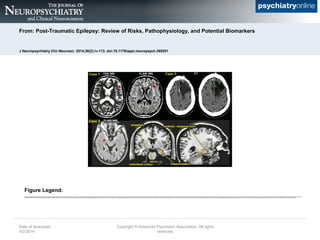
  • 1. From: Post-Traumatic Epilepsy: Review of Risks, Pathophysiology, and Potential Biomarkers J Neuropsychiatry Clin Neurosci. 2014;26(2):iv-113. doi:10.1176/appi.neuropsych.260201 Post-traumatic epilepsy (PTE) can develop following traumatic brain injuries (TBIs) of all levels of severity, as illustrated by these three cases. Case 1: A 58-year-old man who experienced a mild TBI as a result of a roll over collision as a teenager. He developed generalized convulsions within five years from injury. MRI obtained after onset of PTE demonstrated a single 3 mm nonspecific gliotic focus in the left frontal white matter (arrow) that is well visualized on both T2 weighted (T2W) and FLAIR sequences. Case 2: A 49-year-old man with mesial temporal sclerosis secondary to TBI as a result of a recent fall from a ladder. The temporal horns of the lateral ventricles are colored red. Note the dilation of the tip of the right temporal horn due to volume loss in the surrounding temporal lobe structures. The basic anatomy of the medial temporal lobe is illustrated on coronal and sagittal MRI of a healthy individual. The hippocampus (yellow) and temporal horn of the lateral ventricle (red) are color-coded and adjacent structures are labeled. Case 3: A 66-year-old man who experienced a severe TBI because of a gunshot wound to the left frontal lobe when he was 19 years of age. He developed seizures within 2 years following this injury. His seizures are described as generalized stiffening of upper and lower extremities followed by convulsive activity. CT demonstrates chronic postoperative changes with a large area of encephalomalacia involving the left frontal lobe. Date of download: 9/2/2014 Copyright © American Psychiatric Association. All rights reserved. Figure Legend: