SlideShare uma empresa Scribd logo
1 de 12
Baixar para ler offline
1 | P a g e
M- 6 Cell Cycle and Regulation
Cell Cycle
Cell cycle and cell division form the basis of life. Growth, regeneration, and reproduction depend
upon cell cycle and cell division. However, what happens when this cell cycle is disturbed or
when the cells fail to regulate the cell cycle? It can lead to uncontrolled cell growth and the
division which can finally lead to cancer
Scientists and microscopists have known cell cycle and cell division for over hundred years.
However, it was the pioneering and revolutionary work of Alma Howard and Stephen Pelc in the
1950s that revealed DNA replication took place at a particular phase of the cell cycle. They
demonstrated that cell cycle is distinct and separate from the mitosis process.
Definition
Cell cycle (cell division cycle) is an ordered sequence of events occurring in a cell. Cell cycle
results in cell growth and DNA replication thereby forming two daughter cells. It is an essential
process for the formation of a mature organism from single-celled fertilized eggs. The process of
cell cycle promotes renewal and regeneration of hair, blood cells, skin, and certain internal
organs. The formation of daughter cells by cell division indicates the start of a new interphase
cycle.
A cell splits after completing a sequence of events. For example, a cell grows, replicate its DNA
(genetic material), and then finally divides into two daughter cells. The cell cycle is termed a
2 | P a g e
cycle because the events repeat itself. After completing one complete round the newly formed
daughter cells begin the same process all over again. The two key phases of a cell cycle are
interphase and M phase or the mitotic phase.
Every phase will be successfully activated on proper progression and completion of the previous
phases. However, if a cell is temporarily stopped progressing or somehow stopped dividing then
the cell enter into another state termed as G0 phase, also called a “state of quiescence.” Let’s
discuss the steps or phases in details.
Interphase
In this phase, the cell grows and produces a copy of the genetic material (DNA). Interphase can
be further subdivided into three distinct phases: G1 phase, S phase (synthesis), G2 phase. The
cell cycle begins after the division of mother cell into two new daughter cells. If the newly formed
cell wants to move on then it must divide itself.
However, there are certain initial steps that occur before the actual division. In these phases, the
new daughter cell prepares itself for the division. The interphase usually seems like a resting
phase between the cell divisions but on contrary, it is a phase with a number of diverse activities.
The duration of interphase may vary from 12 to 24 hours in the mammalian tissues.
Sub-phases of Interphase
Gap 0: At times the cell will leave the cycle and temporarily stop dividing. This is called a resting
period. It can be for a short time or long more permanent period. For example neurons after
reaching the end stage of development stop dividing and enter into a more permanent resting
phase. G0 is a resting phase where the cell has left the cycle and has stopped dividing. The cell
cycle starts with this phase.
➢ The word "post-mitotic" is sometimes used to refer to both quiescent and senescent cells.
Non-proliferative (non-dividing) cells in multicellular eukaryotes generally enter the
quiescent G0 state from G1 and may remain quiescent for long periods of time, possibly
indefinitely (as is often the case for neurons). This is very common for cells that are fully
differentiated. Cellular senescence occurs in response to DNA damage and external stress
and usually constitutes an arrest in G1. Some cells enter the G0 phase semi-permanently
and are considered post-mitotic, e.g., some liver, kidney, and stomach cells. Many cells do
not enter G0 and continue to divide throughout an organism's life, e.g., epithelial cells.
Gap 1 (G1) Phase: It is also termed as the first gap phase. In this phase, the cell starts
growing and enlarges physically. It forms the copy of organelles, produces all the necessary
molecular building blocks such as RNA and also synthesizes proteins that are essential in
later stages. At this point, a control mechanism is activated to ensure proper DNA synthesis.
The control mechanism is termed as the G1 checkpoint. In this phase, the cell increases its
supply of proteins, increases the number of organelles (such as mitochondria, ribosomes),
and grows in size. In G1 phase, a cell has three options.
➢ To continue cell cycle and enter S phase
3 | P a g e
➢ Stop cell cycle and enter G0 phase for undergoing differentiation.
➢ Become arrested in G1 phase hence it may enter G0 phase or re-enter cell cycle. The
deciding point is called check point (Restriction point). This check point is called the
restriction point or START and is regulated by G1/S cyclins, which cause transition from
G1 to S phase. Passage through the G1 check point commits the cell to division.
S Phase: In this phase, a cell produces a complete copy of DNA in the nucleus to produce two
similar daughter cells. DNA replication begins in the S phase or the synthesis phase. The
microtubule-organizing structure (centrosome) is also copied in this phase. The centrosome
is the structure that helps in dividing the DNA during M phase.
➢ each chromosome consists of two sister chromatids. Thus, during this phase, the amount
of DNA in the cell has doubled, though the ploidy and number of chromosomes are
unchanged. Rates of RNA transcription and protein synthesis are very low during this
phase. An exception to this is histone production, most of which occurs during the S phase
Gap 2 (G2) phase: In G2 phase the cell grows further, produce proteins and organelles and
starts rearranging the constituents of the cell for mitosis phase. At the end of the G2 phase,
another checkpoint is activated called as G2 Checkpoint. G2 Checkpoint ensures everything is
ready for division and M phase. The end of the G2 phase ends when the mitosis process
begins.
➢ G2 phase occurs after DNA replication and is a period of protein synthesis and rapid cell
growth to prepare the cell for mitosis. During this phase microtubules begin to reorganize
to form a spindle (preprophase). Before proceeding to mitotic phase, cells must be
checked at the G2 checkpoint for any DNA damage within the chromosomes. The G2
checkpoint is mainly regulated by the tumor protein p53. If the DNA is damaged, p53 will
either repair the DNA or trigger the apoptosis of the cell. If p53 is dysfunctional or
mutated, cells with damaged DNA may continue through the cell cycle, leading to the
development of cancer.
M phase
n this phase, the cell splits its DNA into two copies. Additionally, the division of the cytoplasm
takes place thereby forming two daughter cells. M phase can be categorized into karyokinesis
(the division of cell chromosome) and cytokinesis (the division of cell cytoplasm to form new
daughter cells). M phase is categorized into two distinct phases: mitosis and cytokinesis. In this
phase, the cell divides the duplicated DNA and the cytoplasm into two new daughter cells.
• Mitosis: The cell’s “nuclear DNA” is condensed into chromosomes. These visible
chromosomes are pulled apart with the help of mitotic spindles (the special structures
formed from microtubules). Mitosis is further subdivided into 4 separate stages including
prophase, metaphase, anaphase, and telophase.
4 | P a g e
• Cytokinesis: Cytokinesis begins after mitosis is complete. In this phase, the cytoplasm of
the cell is divided into two daughter cells.
5 | P a g e
Cell Cycle Exit and Gap 0 Phase
By now we know the cell cycle results in the formation of two new daughter cells. Now the
question arises what happens to the newly formed daughter cells after one complete round of
cell cycle? This is entirely dependent on what cells are dividing. There are certain kinds of cells
that divide quickly and in these types of cells, the new daughter cells immediately enter into
another round of cell division cycle. Examples include embryo and tumour.
Alternatively, there are other types of cells that divide at a slow pace and sometimes completely
stop dividing and enter into another phase called as G0 phase or resting phase. The cell will
continue its usual function. Such as neuron after the end stage does not divide but continue to
conduct signals.
Duration of the Cell Cycle
The cell cycle duration will vary in different types of cells. The G1 phase will continue for
approximately 11 hours, S phase will continue for 8 hours, G2 phase for nearly 4 hours and the M
phase for nearly one hour in a rapidly dividing human cell with cell cycle duration of 24 hours.
Some cells may divide faster than human cells whereas some cells may take more time to
complete an entire cell cycle. For example “budding yeast” will complete the entire cell cycle (4
stages of the cell cycle) in about 90 minutes.
State Phase Abbreviation Description
Resting Gap 0 G0
A phase where the cell has left the cycle and has stopped
dividing.
Interphase
Gap 1 G1
Cells increase in size in Gap 1. The G1 checkpoint control
mechanism ensures that everything is ready for DNA
synthesis.
Synthesis S DNA replication occurs during this phase.
Gap 2 G2
During the gap between DNA synthesis and mitosis, the cell
will continue to grow. The G2 checkpoint control mechanism
ensures that everything is ready to enter the M (mitosis)
phase and divide.
Cell
division
Mitosis M
Cell growth stops at this stage and cellular energy is focused
on the orderly division into two daughter cells. A checkpoint in
the middle of mitosis (Metaphase Checkpoint) ensures that the
cell is ready to complete cell division.
6 | P a g e
Cell cycle regulation
The length of the cell cycle is highly variable, even within the cells of a single organism. In
humans, the frequency of cell turnover ranges from a few hours in early embryonic development,
to an average of two to five days for epithelial cells, and to an entire human lifetime spent in G0
by specialized cells, such as cortical neurons or cardiac muscle cells. There is also variation in the
time that a cell spends in each phase of the cell cycle. When fast-dividing mammalian cells are
grown in culture (outside the body under optimal growing conditions), the length of the cycle is
about 24 hours. In rapidly dividing human cells with a 24-hour cell cycle, the G1 phase lasts
approximately nine hours, the S phase lasts 10 hours, the G2 phase lasts about four and one-half
hours, and the M phase lasts approximately one-half hour. In early embryos of fruit flies, the cell
cycle is completed in about eight minutes. The timing of events in the cell cycle is controlled by
mechanisms that are both internal and external to the cell.
Regulation of the Cell Cycle by External Events
Both the initiation and inhibition of cell division are triggered by events external to the cell when it is
about to begin the replication process. An event may be as simple as the death of a nearby cell or as
sweeping as the release of growth-promoting hormones, such as human growth hormone (HGH). A
lack of HGH can inhibit cell division, resulting in dwarfism, whereas too much HGH can result in
gigantism. Crowding of cells can also inhibit cell division. Another factor that can initiate cell division
is the size of the cell; as a cell grows, it becomes inefficient due to its decreasing surface-to-volume
ratio. The solution to this problem is to divide.
Whatever the source of the message, the cell receives the signal, and a series of events within the cell
allows it to proceed into interphase. Moving forward from this initiation point, every parameter
required during each cell cycle phase must be met or the cycle cannot progress.
Regulation at Internal Checkpoints
It is essential that the daughter cells produced be exact duplicates of the parent cell. Mistakes in the
duplication or distribution of the chromosomes lead to mutations that may be passed forward to every
new cell produced from an abnormal cell. To prevent a compromised cell from continuing to divide,
there are internal control mechanisms that operate at three main cell cycle checkpoints. A checkpoint is
one of several points in the eukaryotic cell cycle at which the progression of a cell to the next stage in
the cycle can be halted until conditions are favorable. These checkpoints occur near the end of G1, at
the G2/M transition, and during metaphase
7 | P a g e
The G1 Checkpoint
The G1 checkpoint determines whether all conditions are favorable for cell division to proceed. The G1
checkpoint, also called the restriction point (in yeast), is a point at which the cell irreversibly commits
to the cell division process. External influences, such as growth factors, play a large role in carrying
the cell past the G1 checkpoint. In addition to adequate reserves and cell size, there is a check for
genomic DNA damage at the G1 checkpoint. A cell that does not meet all the requirements will not be
allowed to progress into the S phase. The cell can halt the cycle and attempt to remedy the problematic
condition, or the cell can advance into G0 and await further signals when conditions improve.
The G2 Checkpoint
The G2 checkpoint bars entry into the mitotic phase if certain conditions are not met. As at the G1
checkpoint, cell size and protein reserves are assessed. However, the most important role of the G2
checkpoint is to ensure that all of the chromosomes have been replicated and that the replicated DNA
is not damaged. If the checkpoint mechanisms detect problems with the DNA, the cell cycle is halted,
and the cell attempts to either complete DNA replication or repair the damaged DNA.
The M Checkpoint
The M checkpoint occurs near the end of the metaphase stage of karyokinesis. The M checkpoint is
also known as the spindle checkpoint, because it determines whether all the sister chromatids are
correctly attached to the spindle microtubules. Because the separation of the sister chromatids during
anaphase is an irreversible step, the cycle will not proceed until the kinetochores of each pair of sister
chromatids are firmly anchored to at least two spindle fibers arising from opposite poles of the cell.
Positive Regulation of the Cell Cycle
Two groups of proteins, called cyclins and cyclin-dependent kinases (Cdks), are responsible for the
progress of the cell through the various checkpoints. The levels of the four cyclin proteins fluctuate
throughout the cell cycle in a predictable pattern (Figure 2). Increases in the concentration of cyclin
proteins are triggered by both external and internal signals. After the cell moves to the next stage of the
cell cycle, the cyclins that were active in the previous stage are degraded.
The concentrations of cyclin proteins change throughout the cell cycle. There is a direct correlation between cyclin
accumulation and the three major cell cycle checkpoints. Also note the sharp decline of cyclin levels following each
checkpoint (the transition between phases of the cell cycle), as cyclin is degraded by cytoplasmic enzymes.
8 | P a g e
Cyclins
Cyclins are among the most important core cell cycle regulators. Cyclins are a group of related
proteins, and there are four basic types found in humans and most other eukaryotes: G1_11start
subscript, 1, end subscript cyclins, G1_11start subscript, 1, end subscript/S cyclins, S cyclins, and
M cyclins.
As the names suggest, each cyclin is associated with a particular phase, transition, or set of
phases in the cell cycle and helps drive the events of that phase or period. For instance, M cyclin
promotes the events of M phase, such as nuclear envelope breakdown and chromosome
condensation.
Cyclin-dependent kinases
In order to drive the cell cycle forward, a cyclin must activate or inactivate many target proteins
inside of the cell. Cyclins drive the events of the cell cycle by partnering with a family of enzymes
called the cyclin-dependent kinases (Cdks). A lone Cdk is inactive, but the binding of a cyclin
activates it, making it a functional enzyme and allowing it to modify target proteins.
How does this work?
Cdks are kinases, enzymes that phosphorylate (attach phosphate groups to) specific target
proteins. The attached phosphate group acts like a switch, making the target protein more or less
active. When a cyclin attaches to a Cdk, it has two important effects: it activates the Cdk as a
kinase, but it also directs the Cdk to a specific set of target proteins, ones appropriate to the cell
cycle period controlled by the cyclin. For instance, G1_11start subscript, 1, end subscript/S
cyclins send Cdks to S phase targets (e.g., promoting DNA replication), while M cyclins send Cdks
to M phase targets (e.g., making the nuclear membrane break down).
Cyclins and Cdks are very evolutionarily conserved, meaning that they are found in many different types
of species, from yeast to frogs to humans. The details of the system vary a little: for instance, yeast has just
9 | P a g e
one Cdk, while humans and other mammals have multiple Cdks that are used at different stages of the cell
cycle. (Yes, this kind of an exception to the "Cdks don't change in levels" rule!) But the basic principles are
quite similar, so that Cdks and the different types of cyclins can be found in each species.
Maturation-promoting factor (MPF)
A famous example of how cyclins and Cdks work together to control cell cycle transitions is that
of maturation-promoting factor (MPF). The name dates back to the 1970s, when researchers
found that cells in M phase contained an unknown factor that could force frog egg cells (stuck in
G2_to enter M phase. This mystery molecule, called MPF, was discovered in the 1980s to be a Cdk
bound to its M cyclin
MPF provides a good example of how cyclins and Cdks can work together to drive a cell cycle
transition. Like a typical cyclin, M cyclin stays at low levels for much of the cell cycle, but builds
up as the cell approaches the G2_M transition. As M cyclin accumulates, it binds to Cdks already
present in the cell, forming complexes that are poised to trigger M phase. Once these complexes
receive an additional signal (essentially, an all-clear confirming that the cell’s DNA is intact), they
become active and set the events of M phase in motion.
The MPF complexes add phosphate tags to several different proteins in the nuclear envelope,
resulting in its breakdown (a key event of early M phase), and also activate targets that promote
chromosome condensation and other M phase events. The role of MPF in nuclear envelope
breakdown is shown in simplified form in the diagram below.
The anaphase-promoting complex/cyclosome (APC/C)
In addition to driving the events of M phase, MPF also triggers its own destruction by activating
the anaphase-promoting complex/cyclosome (APC/C), a protein complex that causes M
10 | P a g e
cyclins to be destroyed starting in anaphase. The destruction of M cyclins pushes the cell out of
mitosis, allowing the new daughter cells to enter G1_The APC/C also causes destruction of the
proteins that hold the sister chromatids together, allowing them to separate in anaphase and
move to opposite poles of the cell.
How does the APC/C do its job? Like a Cdk, the APC/C is an enzyme, but it has different type of
function than a Cdk. Rather than attaching a phosphate group to its targets, it adds a small
protein tag called ubiquitin (Ub). When a target is tagged with ubiquitin, it is sent to the
proteasome, which can be thought of as the recycle bin of the cell, and destroyed. For example,
the APC/C attaches a ubiquitin tag to M cyclins, causing them to be chopped up by the
proteasome and allowing the newly forming daughter cells to enter G1.
The APC/C also uses ubiquitin tagging to trigger the separation of sister chromatids during
mitosis. If the APC/C gets the right signals at metaphase, it sets off a chain of events that destroys
cohesin, the protein glue that holds sister chromatids together
• The APC/C first adds a ubiquitin tag to a protein called securin, sending it for recycling.
Securin normally binds to, and inactivates, a protein called separase.
• When securin is sent for recycling, separase becomes active and can do its job. Separase chops
up the cohesin that holds sister chromatids together, allowing them to separate.
11 | P a g e
Checkpoints and regulators
Cdks, cyclins, and the APC/C are direct regulators of cell cycle transitions, but they aren’t always
in the driver’s seat. Instead, they respond to cues from inside and outside the cell. These cues
influence activity of the core regulators to determine whether the cell moves forward in the cell
cycle. Positive cues, like growth factors, typically increase activity of Cdks and cyclins, while
negative ones, like DNA damage, typically decrease or block activity.
As an example, let's examine how DNA damage halts the cell cycle in G1_. DNA damage can, and
will, happen in many cells of the body during a person’s lifetime (for example, due to UV rays
from the sun). Cells must be able to deal with this damage, fixing it if possible and preventing cell
division if not. Key to the DNA damage response is a protein called p53, a famous tumor
suppressor often described as “the guardian of the genome.”
p53 works on multiple levels to ensure that cells do not pass on their damaged DNA through cell
division First, it stops the cell cycle at the G1 checkpoint by triggering production of Cdk
inhibitor (CKI) proteins. The CKI proteins bind to Cdk-cyclin complexes and block their activity
(see diagram below), buying time for DNA repair. p53's second job is to activate DNA repair
enzymes. If DNA damage is not fixable, p53 will play its third and final role: triggering
programmed cell death so damaged DNA is not passed on.
Simplified diagram of how p53 halts the cell cycle at the G1/S checkpoint. p53 is activated by
DNA damage and causes production of a Cdk inhibitor, which binds to the Cdk-G1/S cyclin
complex and inactivates it. This halts the cell in G1 and prevents it from entering S phase,
allowing time for the DNA damage to be fixed.
By ensuring that cells don't divide when their DNA is damaged, p53 prevents mutations (changes
in DNA) from being passed on to daughter cells. When p53 is defective or missing, mutations can
accumulate quickly, potentially leading to cancer. Indeed, out of all the entire human genome,
p53 is the single gene most often mutated in cancers. p53 and cell cycle regulation are key topics
of study for researchers working on new treatments for cancer.
12 | P a g e
Negative Regulation of the Cell Cycle
The second group of cell cycle regulatory molecules are negative regulators. Negative regulators
halt the cell cycle. Remember that in positive regulation, active molecules cause the cycle to
progress.
The best understood negative regulatory molecules are retinoblastoma protein (Rb), p53, and
p21. Retinoblastoma proteins are a group of tumor-suppressor proteins common in many cells.
The 53 and 21 designations refer to the functional molecular masses of the proteins (p) in
kilodaltons. Much of what is known about cell cycle regulation comes from research conducted
with cells that have lost regulatory control. All three of these regulatory proteins were
discovered to be damaged or non-functional in cells that had begun to replicate uncontrollably
(became cancerous). In each case, the main cause of the unchecked progress through the cell
cycle was a faulty copy of the regulatory protein.
Rb, p53, and p21 act primarily at the G1 checkpoint. p53 is a multi-functional protein that has a
major impact on the commitment of a cell to division because it acts when there is damaged DNA
in cells that are undergoing the preparatory processes during G1. If damaged DNA is detected,
p53 halts the cell cycle and recruits enzymes to repair the DNA. If the DNA cannot be repaired,
p53 can trigger apoptosis, or cell suicide, to prevent the duplication of damaged chromosomes.
As p53 levels rise, the production of p21 is triggered. p21 enforces the halt in the cycle dictated
by p53 by binding to and inhibiting the activity of the Cdk/cyclin complexes. As a cell is exposed
to more stress, higher levels of p53 and p21 accumulate, making it less likely that the cell will
move into the S phase.
Rb exerts its regulatory influence on other positive regulator proteins. Chiefly, Rb monitors cell
size. In the active, dephosphorylated state, Rb binds to proteins called transcription factors, most
commonly, E2F (Figure 4). Transcription factors “turn on” specific genes, allowing the
production of proteins encoded by that gene. When Rb is bound to E2F, production of proteins
necessary for the G1/S transition is blocked. As the cell increases in size, Rb is slowly
phosphorylated until it becomes inactivated. Rb releases E2F, which can now turn on the gene
that produces the transition protein, and this particular block is removed. For the cell to move
past each of the checkpoints, all positive regulators must be “turned on,” and all negative
regulators must be “turned off.”

Mais conteúdo relacionado

Mais procurados

Cell cycle and control of cell division.
Cell cycle and control of cell division.Cell cycle and control of cell division.
Cell cycle and control of cell division.Irfana95
 
Cell cycle regulation
Cell cycle regulationCell cycle regulation
Cell cycle regulationRiddhi Datta
 
Cell cycle, check point,
Cell cycle, check point, Cell cycle, check point,
Cell cycle, check point, SoniaBajaj10
 
Cel cycle and its regulation
Cel cycle and its regulationCel cycle and its regulation
Cel cycle and its regulationarti yadav
 
Cell cycle and its checkpoints
Cell cycle and its checkpointsCell cycle and its checkpoints
Cell cycle and its checkpointsSrishti Aggrawal
 
cell cycle in detail (interphase )
cell cycle in detail (interphase ) cell cycle in detail (interphase )
cell cycle in detail (interphase ) Steffie Dmello
 
Cell cycle checkpoints, apoptosis and cancer
Cell cycle checkpoints, apoptosis and cancerCell cycle checkpoints, apoptosis and cancer
Cell cycle checkpoints, apoptosis and cancerSurender Rawat
 
Cell cycle seminar
Cell cycle seminarCell cycle seminar
Cell cycle seminartutun-nana
 
Cell division/Cell Cycle/ DNA duplication
Cell division/Cell Cycle/ DNA duplicationCell division/Cell Cycle/ DNA duplication
Cell division/Cell Cycle/ DNA duplicationPharmacy Universe
 
Cell Quiescense
Cell QuiescenseCell Quiescense
Cell QuiescenseZainabSaif
 
Cell cycle and it's checkpoints
Cell cycle and it's checkpointsCell cycle and it's checkpoints
Cell cycle and it's checkpointsRitisha Gupta
 

Mais procurados (20)

Cell cycle and control of cell division.
Cell cycle and control of cell division.Cell cycle and control of cell division.
Cell cycle and control of cell division.
 
Vacuoles
VacuolesVacuoles
Vacuoles
 
Cell cycle regulation
Cell cycle regulationCell cycle regulation
Cell cycle regulation
 
Cell cycle, check point,
Cell cycle, check point, Cell cycle, check point,
Cell cycle, check point,
 
Cel cycle and its regulation
Cel cycle and its regulationCel cycle and its regulation
Cel cycle and its regulation
 
Cell cycle and its checkpoints
Cell cycle and its checkpointsCell cycle and its checkpoints
Cell cycle and its checkpoints
 
Cell cycle
Cell cycle Cell cycle
Cell cycle
 
cell cycle in detail (interphase )
cell cycle in detail (interphase ) cell cycle in detail (interphase )
cell cycle in detail (interphase )
 
Cell cycle ppt.
Cell cycle ppt.Cell cycle ppt.
Cell cycle ppt.
 
Cell cycle checkpoints, apoptosis and cancer
Cell cycle checkpoints, apoptosis and cancerCell cycle checkpoints, apoptosis and cancer
Cell cycle checkpoints, apoptosis and cancer
 
Cell cycle seminar
Cell cycle seminarCell cycle seminar
Cell cycle seminar
 
Cell division/Cell Cycle/ DNA duplication
Cell division/Cell Cycle/ DNA duplicationCell division/Cell Cycle/ DNA duplication
Cell division/Cell Cycle/ DNA duplication
 
Cell cycle regulation ppt
Cell cycle regulation  pptCell cycle regulation  ppt
Cell cycle regulation ppt
 
Three stages of Interphase
Three stages of InterphaseThree stages of Interphase
Three stages of Interphase
 
Cell Quiescense
Cell QuiescenseCell Quiescense
Cell Quiescense
 
Cell cycle and it's checkpoints
Cell cycle and it's checkpointsCell cycle and it's checkpoints
Cell cycle and it's checkpoints
 
The Cell Cycle
The Cell CycleThe Cell Cycle
The Cell Cycle
 
Presentation on Cell Cycle
Presentation on Cell CyclePresentation on Cell Cycle
Presentation on Cell Cycle
 
Intron and types
Intron and typesIntron and types
Intron and types
 
Cell Cycle Regulation
Cell Cycle RegulationCell Cycle Regulation
Cell Cycle Regulation
 

Semelhante a Lecture 18 cell cycle and its regulation converted

Semelhante a Lecture 18 cell cycle and its regulation converted (20)

Cell cycle
Cell cycleCell cycle
Cell cycle
 
Eukaryotic cell division cycle
Eukaryotic cell division cycleEukaryotic cell division cycle
Eukaryotic cell division cycle
 
Cell cycle
Cell cycleCell cycle
Cell cycle
 
Concept Of Cell cycle phases.docx
Concept Of Cell cycle phases.docxConcept Of Cell cycle phases.docx
Concept Of Cell cycle phases.docx
 
cellcycleanditsregulation11.pptx
cellcycleanditsregulation11.pptxcellcycleanditsregulation11.pptx
cellcycleanditsregulation11.pptx
 
The Cell Cycle
The Cell CycleThe Cell Cycle
The Cell Cycle
 
Cell cycle
Cell cycleCell cycle
Cell cycle
 
18042020100230Cell cycle.pdf
18042020100230Cell cycle.pdf18042020100230Cell cycle.pdf
18042020100230Cell cycle.pdf
 
Cell Cycle and Cell Division Class 11 Important Notes for NEET Biology pdf
Cell Cycle and Cell Division Class 11 Important Notes for NEET Biology pdfCell Cycle and Cell Division Class 11 Important Notes for NEET Biology pdf
Cell Cycle and Cell Division Class 11 Important Notes for NEET Biology pdf
 
cell cycle.pdf
cell cycle.pdfcell cycle.pdf
cell cycle.pdf
 
Cell division
Cell divisionCell division
Cell division
 
Cell cycle and signaling
Cell cycle and signalingCell cycle and signaling
Cell cycle and signaling
 
cell cycel , and its stages
 cell cycel , and its stages  cell cycel , and its stages
cell cycel , and its stages
 
Cell cycle
Cell cycleCell cycle
Cell cycle
 
Cell cycle and cell division
Cell cycle and cell divisionCell cycle and cell division
Cell cycle and cell division
 
Cell cycle and signaling
Cell cycle and signalingCell cycle and signaling
Cell cycle and signaling
 
Cell cycle and mitosis presentation (3)
Cell cycle and mitosis presentation (3)Cell cycle and mitosis presentation (3)
Cell cycle and mitosis presentation (3)
 
Cell cycle ( B.Sc Biotech)
Cell cycle ( B.Sc Biotech)Cell cycle ( B.Sc Biotech)
Cell cycle ( B.Sc Biotech)
 
Cell Cycle
Cell CycleCell Cycle
Cell Cycle
 
Cell cycle and regulation
Cell cycle and regulationCell cycle and regulation
Cell cycle and regulation
 

Último

Call Girls in Dwarka Mor Delhi Contact Us 9654467111
Call Girls in Dwarka Mor Delhi Contact Us 9654467111Call Girls in Dwarka Mor Delhi Contact Us 9654467111
Call Girls in Dwarka Mor Delhi Contact Us 9654467111Sapana Sha
 
mini mental status format.docx
mini    mental       status     format.docxmini    mental       status     format.docx
mini mental status format.docxPoojaSen20
 
microwave assisted reaction. General introduction
microwave assisted reaction. General introductionmicrowave assisted reaction. General introduction
microwave assisted reaction. General introductionMaksud Ahmed
 
Sanyam Choudhary Chemistry practical.pdf
Sanyam Choudhary Chemistry practical.pdfSanyam Choudhary Chemistry practical.pdf
Sanyam Choudhary Chemistry practical.pdfsanyamsingh5019
 
Employee wellbeing at the workplace.pptx
Employee wellbeing at the workplace.pptxEmployee wellbeing at the workplace.pptx
Employee wellbeing at the workplace.pptxNirmalaLoungPoorunde1
 
A Critique of the Proposed National Education Policy Reform
A Critique of the Proposed National Education Policy ReformA Critique of the Proposed National Education Policy Reform
A Critique of the Proposed National Education Policy ReformChameera Dedduwage
 
Interactive Powerpoint_How to Master effective communication
Interactive Powerpoint_How to Master effective communicationInteractive Powerpoint_How to Master effective communication
Interactive Powerpoint_How to Master effective communicationnomboosow
 
Paris 2024 Olympic Geographies - an activity
Paris 2024 Olympic Geographies - an activityParis 2024 Olympic Geographies - an activity
Paris 2024 Olympic Geographies - an activityGeoBlogs
 
Arihant handbook biology for class 11 .pdf
Arihant handbook biology for class 11 .pdfArihant handbook biology for class 11 .pdf
Arihant handbook biology for class 11 .pdfchloefrazer622
 
Kisan Call Centre - To harness potential of ICT in Agriculture by answer farm...
Kisan Call Centre - To harness potential of ICT in Agriculture by answer farm...Kisan Call Centre - To harness potential of ICT in Agriculture by answer farm...
Kisan Call Centre - To harness potential of ICT in Agriculture by answer farm...Krashi Coaching
 
BASLIQ CURRENT LOOKBOOK LOOKBOOK(1) (1).pdf
BASLIQ CURRENT LOOKBOOK  LOOKBOOK(1) (1).pdfBASLIQ CURRENT LOOKBOOK  LOOKBOOK(1) (1).pdf
BASLIQ CURRENT LOOKBOOK LOOKBOOK(1) (1).pdfSoniaTolstoy
 
Privatization and Disinvestment - Meaning, Objectives, Advantages and Disadva...
Privatization and Disinvestment - Meaning, Objectives, Advantages and Disadva...Privatization and Disinvestment - Meaning, Objectives, Advantages and Disadva...
Privatization and Disinvestment - Meaning, Objectives, Advantages and Disadva...RKavithamani
 
Activity 01 - Artificial Culture (1).pdf
Activity 01 - Artificial Culture (1).pdfActivity 01 - Artificial Culture (1).pdf
Activity 01 - Artificial Culture (1).pdfciinovamais
 
Mastering the Unannounced Regulatory Inspection
Mastering the Unannounced Regulatory InspectionMastering the Unannounced Regulatory Inspection
Mastering the Unannounced Regulatory InspectionSafetyChain Software
 
18-04-UA_REPORT_MEDIALITERAСY_INDEX-DM_23-1-final-eng.pdf
18-04-UA_REPORT_MEDIALITERAСY_INDEX-DM_23-1-final-eng.pdf18-04-UA_REPORT_MEDIALITERAСY_INDEX-DM_23-1-final-eng.pdf
18-04-UA_REPORT_MEDIALITERAСY_INDEX-DM_23-1-final-eng.pdfssuser54595a
 
URLs and Routing in the Odoo 17 Website App
URLs and Routing in the Odoo 17 Website AppURLs and Routing in the Odoo 17 Website App
URLs and Routing in the Odoo 17 Website AppCeline George
 
Web & Social Media Analytics Previous Year Question Paper.pdf
Web & Social Media Analytics Previous Year Question Paper.pdfWeb & Social Media Analytics Previous Year Question Paper.pdf
Web & Social Media Analytics Previous Year Question Paper.pdfJayanti Pande
 
Separation of Lanthanides/ Lanthanides and Actinides
Separation of Lanthanides/ Lanthanides and ActinidesSeparation of Lanthanides/ Lanthanides and Actinides
Separation of Lanthanides/ Lanthanides and ActinidesFatimaKhan178732
 

Último (20)

Call Girls in Dwarka Mor Delhi Contact Us 9654467111
Call Girls in Dwarka Mor Delhi Contact Us 9654467111Call Girls in Dwarka Mor Delhi Contact Us 9654467111
Call Girls in Dwarka Mor Delhi Contact Us 9654467111
 
mini mental status format.docx
mini    mental       status     format.docxmini    mental       status     format.docx
mini mental status format.docx
 
microwave assisted reaction. General introduction
microwave assisted reaction. General introductionmicrowave assisted reaction. General introduction
microwave assisted reaction. General introduction
 
Sanyam Choudhary Chemistry practical.pdf
Sanyam Choudhary Chemistry practical.pdfSanyam Choudhary Chemistry practical.pdf
Sanyam Choudhary Chemistry practical.pdf
 
TataKelola dan KamSiber Kecerdasan Buatan v022.pdf
TataKelola dan KamSiber Kecerdasan Buatan v022.pdfTataKelola dan KamSiber Kecerdasan Buatan v022.pdf
TataKelola dan KamSiber Kecerdasan Buatan v022.pdf
 
Employee wellbeing at the workplace.pptx
Employee wellbeing at the workplace.pptxEmployee wellbeing at the workplace.pptx
Employee wellbeing at the workplace.pptx
 
A Critique of the Proposed National Education Policy Reform
A Critique of the Proposed National Education Policy ReformA Critique of the Proposed National Education Policy Reform
A Critique of the Proposed National Education Policy Reform
 
Interactive Powerpoint_How to Master effective communication
Interactive Powerpoint_How to Master effective communicationInteractive Powerpoint_How to Master effective communication
Interactive Powerpoint_How to Master effective communication
 
Paris 2024 Olympic Geographies - an activity
Paris 2024 Olympic Geographies - an activityParis 2024 Olympic Geographies - an activity
Paris 2024 Olympic Geographies - an activity
 
Arihant handbook biology for class 11 .pdf
Arihant handbook biology for class 11 .pdfArihant handbook biology for class 11 .pdf
Arihant handbook biology for class 11 .pdf
 
Kisan Call Centre - To harness potential of ICT in Agriculture by answer farm...
Kisan Call Centre - To harness potential of ICT in Agriculture by answer farm...Kisan Call Centre - To harness potential of ICT in Agriculture by answer farm...
Kisan Call Centre - To harness potential of ICT in Agriculture by answer farm...
 
BASLIQ CURRENT LOOKBOOK LOOKBOOK(1) (1).pdf
BASLIQ CURRENT LOOKBOOK  LOOKBOOK(1) (1).pdfBASLIQ CURRENT LOOKBOOK  LOOKBOOK(1) (1).pdf
BASLIQ CURRENT LOOKBOOK LOOKBOOK(1) (1).pdf
 
Privatization and Disinvestment - Meaning, Objectives, Advantages and Disadva...
Privatization and Disinvestment - Meaning, Objectives, Advantages and Disadva...Privatization and Disinvestment - Meaning, Objectives, Advantages and Disadva...
Privatization and Disinvestment - Meaning, Objectives, Advantages and Disadva...
 
Activity 01 - Artificial Culture (1).pdf
Activity 01 - Artificial Culture (1).pdfActivity 01 - Artificial Culture (1).pdf
Activity 01 - Artificial Culture (1).pdf
 
Mastering the Unannounced Regulatory Inspection
Mastering the Unannounced Regulatory InspectionMastering the Unannounced Regulatory Inspection
Mastering the Unannounced Regulatory Inspection
 
18-04-UA_REPORT_MEDIALITERAСY_INDEX-DM_23-1-final-eng.pdf
18-04-UA_REPORT_MEDIALITERAСY_INDEX-DM_23-1-final-eng.pdf18-04-UA_REPORT_MEDIALITERAСY_INDEX-DM_23-1-final-eng.pdf
18-04-UA_REPORT_MEDIALITERAСY_INDEX-DM_23-1-final-eng.pdf
 
URLs and Routing in the Odoo 17 Website App
URLs and Routing in the Odoo 17 Website AppURLs and Routing in the Odoo 17 Website App
URLs and Routing in the Odoo 17 Website App
 
Web & Social Media Analytics Previous Year Question Paper.pdf
Web & Social Media Analytics Previous Year Question Paper.pdfWeb & Social Media Analytics Previous Year Question Paper.pdf
Web & Social Media Analytics Previous Year Question Paper.pdf
 
Separation of Lanthanides/ Lanthanides and Actinides
Separation of Lanthanides/ Lanthanides and ActinidesSeparation of Lanthanides/ Lanthanides and Actinides
Separation of Lanthanides/ Lanthanides and Actinides
 
Mattingly "AI & Prompt Design: The Basics of Prompt Design"
Mattingly "AI & Prompt Design: The Basics of Prompt Design"Mattingly "AI & Prompt Design: The Basics of Prompt Design"
Mattingly "AI & Prompt Design: The Basics of Prompt Design"
 

Lecture 18 cell cycle and its regulation converted

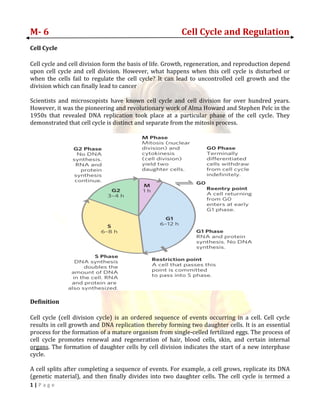
  • 1. 1 | P a g e M- 6 Cell Cycle and Regulation Cell Cycle Cell cycle and cell division form the basis of life. Growth, regeneration, and reproduction depend upon cell cycle and cell division. However, what happens when this cell cycle is disturbed or when the cells fail to regulate the cell cycle? It can lead to uncontrolled cell growth and the division which can finally lead to cancer Scientists and microscopists have known cell cycle and cell division for over hundred years. However, it was the pioneering and revolutionary work of Alma Howard and Stephen Pelc in the 1950s that revealed DNA replication took place at a particular phase of the cell cycle. They demonstrated that cell cycle is distinct and separate from the mitosis process. Definition Cell cycle (cell division cycle) is an ordered sequence of events occurring in a cell. Cell cycle results in cell growth and DNA replication thereby forming two daughter cells. It is an essential process for the formation of a mature organism from single-celled fertilized eggs. The process of cell cycle promotes renewal and regeneration of hair, blood cells, skin, and certain internal organs. The formation of daughter cells by cell division indicates the start of a new interphase cycle. A cell splits after completing a sequence of events. For example, a cell grows, replicate its DNA (genetic material), and then finally divides into two daughter cells. The cell cycle is termed a
  • 2. 2 | P a g e cycle because the events repeat itself. After completing one complete round the newly formed daughter cells begin the same process all over again. The two key phases of a cell cycle are interphase and M phase or the mitotic phase. Every phase will be successfully activated on proper progression and completion of the previous phases. However, if a cell is temporarily stopped progressing or somehow stopped dividing then the cell enter into another state termed as G0 phase, also called a “state of quiescence.” Let’s discuss the steps or phases in details. Interphase In this phase, the cell grows and produces a copy of the genetic material (DNA). Interphase can be further subdivided into three distinct phases: G1 phase, S phase (synthesis), G2 phase. The cell cycle begins after the division of mother cell into two new daughter cells. If the newly formed cell wants to move on then it must divide itself. However, there are certain initial steps that occur before the actual division. In these phases, the new daughter cell prepares itself for the division. The interphase usually seems like a resting phase between the cell divisions but on contrary, it is a phase with a number of diverse activities. The duration of interphase may vary from 12 to 24 hours in the mammalian tissues. Sub-phases of Interphase Gap 0: At times the cell will leave the cycle and temporarily stop dividing. This is called a resting period. It can be for a short time or long more permanent period. For example neurons after reaching the end stage of development stop dividing and enter into a more permanent resting phase. G0 is a resting phase where the cell has left the cycle and has stopped dividing. The cell cycle starts with this phase. ➢ The word "post-mitotic" is sometimes used to refer to both quiescent and senescent cells. Non-proliferative (non-dividing) cells in multicellular eukaryotes generally enter the quiescent G0 state from G1 and may remain quiescent for long periods of time, possibly indefinitely (as is often the case for neurons). This is very common for cells that are fully differentiated. Cellular senescence occurs in response to DNA damage and external stress and usually constitutes an arrest in G1. Some cells enter the G0 phase semi-permanently and are considered post-mitotic, e.g., some liver, kidney, and stomach cells. Many cells do not enter G0 and continue to divide throughout an organism's life, e.g., epithelial cells. Gap 1 (G1) Phase: It is also termed as the first gap phase. In this phase, the cell starts growing and enlarges physically. It forms the copy of organelles, produces all the necessary molecular building blocks such as RNA and also synthesizes proteins that are essential in later stages. At this point, a control mechanism is activated to ensure proper DNA synthesis. The control mechanism is termed as the G1 checkpoint. In this phase, the cell increases its supply of proteins, increases the number of organelles (such as mitochondria, ribosomes), and grows in size. In G1 phase, a cell has three options. ➢ To continue cell cycle and enter S phase
  • 3. 3 | P a g e ➢ Stop cell cycle and enter G0 phase for undergoing differentiation. ➢ Become arrested in G1 phase hence it may enter G0 phase or re-enter cell cycle. The deciding point is called check point (Restriction point). This check point is called the restriction point or START and is regulated by G1/S cyclins, which cause transition from G1 to S phase. Passage through the G1 check point commits the cell to division. S Phase: In this phase, a cell produces a complete copy of DNA in the nucleus to produce two similar daughter cells. DNA replication begins in the S phase or the synthesis phase. The microtubule-organizing structure (centrosome) is also copied in this phase. The centrosome is the structure that helps in dividing the DNA during M phase. ➢ each chromosome consists of two sister chromatids. Thus, during this phase, the amount of DNA in the cell has doubled, though the ploidy and number of chromosomes are unchanged. Rates of RNA transcription and protein synthesis are very low during this phase. An exception to this is histone production, most of which occurs during the S phase Gap 2 (G2) phase: In G2 phase the cell grows further, produce proteins and organelles and starts rearranging the constituents of the cell for mitosis phase. At the end of the G2 phase, another checkpoint is activated called as G2 Checkpoint. G2 Checkpoint ensures everything is ready for division and M phase. The end of the G2 phase ends when the mitosis process begins. ➢ G2 phase occurs after DNA replication and is a period of protein synthesis and rapid cell growth to prepare the cell for mitosis. During this phase microtubules begin to reorganize to form a spindle (preprophase). Before proceeding to mitotic phase, cells must be checked at the G2 checkpoint for any DNA damage within the chromosomes. The G2 checkpoint is mainly regulated by the tumor protein p53. If the DNA is damaged, p53 will either repair the DNA or trigger the apoptosis of the cell. If p53 is dysfunctional or mutated, cells with damaged DNA may continue through the cell cycle, leading to the development of cancer. M phase n this phase, the cell splits its DNA into two copies. Additionally, the division of the cytoplasm takes place thereby forming two daughter cells. M phase can be categorized into karyokinesis (the division of cell chromosome) and cytokinesis (the division of cell cytoplasm to form new daughter cells). M phase is categorized into two distinct phases: mitosis and cytokinesis. In this phase, the cell divides the duplicated DNA and the cytoplasm into two new daughter cells. • Mitosis: The cell’s “nuclear DNA” is condensed into chromosomes. These visible chromosomes are pulled apart with the help of mitotic spindles (the special structures formed from microtubules). Mitosis is further subdivided into 4 separate stages including prophase, metaphase, anaphase, and telophase.
  • 4. 4 | P a g e • Cytokinesis: Cytokinesis begins after mitosis is complete. In this phase, the cytoplasm of the cell is divided into two daughter cells.
  • 5. 5 | P a g e Cell Cycle Exit and Gap 0 Phase By now we know the cell cycle results in the formation of two new daughter cells. Now the question arises what happens to the newly formed daughter cells after one complete round of cell cycle? This is entirely dependent on what cells are dividing. There are certain kinds of cells that divide quickly and in these types of cells, the new daughter cells immediately enter into another round of cell division cycle. Examples include embryo and tumour. Alternatively, there are other types of cells that divide at a slow pace and sometimes completely stop dividing and enter into another phase called as G0 phase or resting phase. The cell will continue its usual function. Such as neuron after the end stage does not divide but continue to conduct signals. Duration of the Cell Cycle The cell cycle duration will vary in different types of cells. The G1 phase will continue for approximately 11 hours, S phase will continue for 8 hours, G2 phase for nearly 4 hours and the M phase for nearly one hour in a rapidly dividing human cell with cell cycle duration of 24 hours. Some cells may divide faster than human cells whereas some cells may take more time to complete an entire cell cycle. For example “budding yeast” will complete the entire cell cycle (4 stages of the cell cycle) in about 90 minutes. State Phase Abbreviation Description Resting Gap 0 G0 A phase where the cell has left the cycle and has stopped dividing. Interphase Gap 1 G1 Cells increase in size in Gap 1. The G1 checkpoint control mechanism ensures that everything is ready for DNA synthesis. Synthesis S DNA replication occurs during this phase. Gap 2 G2 During the gap between DNA synthesis and mitosis, the cell will continue to grow. The G2 checkpoint control mechanism ensures that everything is ready to enter the M (mitosis) phase and divide. Cell division Mitosis M Cell growth stops at this stage and cellular energy is focused on the orderly division into two daughter cells. A checkpoint in the middle of mitosis (Metaphase Checkpoint) ensures that the cell is ready to complete cell division.
  • 6. 6 | P a g e Cell cycle regulation The length of the cell cycle is highly variable, even within the cells of a single organism. In humans, the frequency of cell turnover ranges from a few hours in early embryonic development, to an average of two to five days for epithelial cells, and to an entire human lifetime spent in G0 by specialized cells, such as cortical neurons or cardiac muscle cells. There is also variation in the time that a cell spends in each phase of the cell cycle. When fast-dividing mammalian cells are grown in culture (outside the body under optimal growing conditions), the length of the cycle is about 24 hours. In rapidly dividing human cells with a 24-hour cell cycle, the G1 phase lasts approximately nine hours, the S phase lasts 10 hours, the G2 phase lasts about four and one-half hours, and the M phase lasts approximately one-half hour. In early embryos of fruit flies, the cell cycle is completed in about eight minutes. The timing of events in the cell cycle is controlled by mechanisms that are both internal and external to the cell. Regulation of the Cell Cycle by External Events Both the initiation and inhibition of cell division are triggered by events external to the cell when it is about to begin the replication process. An event may be as simple as the death of a nearby cell or as sweeping as the release of growth-promoting hormones, such as human growth hormone (HGH). A lack of HGH can inhibit cell division, resulting in dwarfism, whereas too much HGH can result in gigantism. Crowding of cells can also inhibit cell division. Another factor that can initiate cell division is the size of the cell; as a cell grows, it becomes inefficient due to its decreasing surface-to-volume ratio. The solution to this problem is to divide. Whatever the source of the message, the cell receives the signal, and a series of events within the cell allows it to proceed into interphase. Moving forward from this initiation point, every parameter required during each cell cycle phase must be met or the cycle cannot progress. Regulation at Internal Checkpoints It is essential that the daughter cells produced be exact duplicates of the parent cell. Mistakes in the duplication or distribution of the chromosomes lead to mutations that may be passed forward to every new cell produced from an abnormal cell. To prevent a compromised cell from continuing to divide, there are internal control mechanisms that operate at three main cell cycle checkpoints. A checkpoint is one of several points in the eukaryotic cell cycle at which the progression of a cell to the next stage in the cycle can be halted until conditions are favorable. These checkpoints occur near the end of G1, at the G2/M transition, and during metaphase
  • 7. 7 | P a g e The G1 Checkpoint The G1 checkpoint determines whether all conditions are favorable for cell division to proceed. The G1 checkpoint, also called the restriction point (in yeast), is a point at which the cell irreversibly commits to the cell division process. External influences, such as growth factors, play a large role in carrying the cell past the G1 checkpoint. In addition to adequate reserves and cell size, there is a check for genomic DNA damage at the G1 checkpoint. A cell that does not meet all the requirements will not be allowed to progress into the S phase. The cell can halt the cycle and attempt to remedy the problematic condition, or the cell can advance into G0 and await further signals when conditions improve. The G2 Checkpoint The G2 checkpoint bars entry into the mitotic phase if certain conditions are not met. As at the G1 checkpoint, cell size and protein reserves are assessed. However, the most important role of the G2 checkpoint is to ensure that all of the chromosomes have been replicated and that the replicated DNA is not damaged. If the checkpoint mechanisms detect problems with the DNA, the cell cycle is halted, and the cell attempts to either complete DNA replication or repair the damaged DNA. The M Checkpoint The M checkpoint occurs near the end of the metaphase stage of karyokinesis. The M checkpoint is also known as the spindle checkpoint, because it determines whether all the sister chromatids are correctly attached to the spindle microtubules. Because the separation of the sister chromatids during anaphase is an irreversible step, the cycle will not proceed until the kinetochores of each pair of sister chromatids are firmly anchored to at least two spindle fibers arising from opposite poles of the cell. Positive Regulation of the Cell Cycle Two groups of proteins, called cyclins and cyclin-dependent kinases (Cdks), are responsible for the progress of the cell through the various checkpoints. The levels of the four cyclin proteins fluctuate throughout the cell cycle in a predictable pattern (Figure 2). Increases in the concentration of cyclin proteins are triggered by both external and internal signals. After the cell moves to the next stage of the cell cycle, the cyclins that were active in the previous stage are degraded. The concentrations of cyclin proteins change throughout the cell cycle. There is a direct correlation between cyclin accumulation and the three major cell cycle checkpoints. Also note the sharp decline of cyclin levels following each checkpoint (the transition between phases of the cell cycle), as cyclin is degraded by cytoplasmic enzymes.
  • 8. 8 | P a g e Cyclins Cyclins are among the most important core cell cycle regulators. Cyclins are a group of related proteins, and there are four basic types found in humans and most other eukaryotes: G1_11start subscript, 1, end subscript cyclins, G1_11start subscript, 1, end subscript/S cyclins, S cyclins, and M cyclins. As the names suggest, each cyclin is associated with a particular phase, transition, or set of phases in the cell cycle and helps drive the events of that phase or period. For instance, M cyclin promotes the events of M phase, such as nuclear envelope breakdown and chromosome condensation. Cyclin-dependent kinases In order to drive the cell cycle forward, a cyclin must activate or inactivate many target proteins inside of the cell. Cyclins drive the events of the cell cycle by partnering with a family of enzymes called the cyclin-dependent kinases (Cdks). A lone Cdk is inactive, but the binding of a cyclin activates it, making it a functional enzyme and allowing it to modify target proteins. How does this work? Cdks are kinases, enzymes that phosphorylate (attach phosphate groups to) specific target proteins. The attached phosphate group acts like a switch, making the target protein more or less active. When a cyclin attaches to a Cdk, it has two important effects: it activates the Cdk as a kinase, but it also directs the Cdk to a specific set of target proteins, ones appropriate to the cell cycle period controlled by the cyclin. For instance, G1_11start subscript, 1, end subscript/S cyclins send Cdks to S phase targets (e.g., promoting DNA replication), while M cyclins send Cdks to M phase targets (e.g., making the nuclear membrane break down). Cyclins and Cdks are very evolutionarily conserved, meaning that they are found in many different types of species, from yeast to frogs to humans. The details of the system vary a little: for instance, yeast has just
  • 9. 9 | P a g e one Cdk, while humans and other mammals have multiple Cdks that are used at different stages of the cell cycle. (Yes, this kind of an exception to the "Cdks don't change in levels" rule!) But the basic principles are quite similar, so that Cdks and the different types of cyclins can be found in each species. Maturation-promoting factor (MPF) A famous example of how cyclins and Cdks work together to control cell cycle transitions is that of maturation-promoting factor (MPF). The name dates back to the 1970s, when researchers found that cells in M phase contained an unknown factor that could force frog egg cells (stuck in G2_to enter M phase. This mystery molecule, called MPF, was discovered in the 1980s to be a Cdk bound to its M cyclin MPF provides a good example of how cyclins and Cdks can work together to drive a cell cycle transition. Like a typical cyclin, M cyclin stays at low levels for much of the cell cycle, but builds up as the cell approaches the G2_M transition. As M cyclin accumulates, it binds to Cdks already present in the cell, forming complexes that are poised to trigger M phase. Once these complexes receive an additional signal (essentially, an all-clear confirming that the cell’s DNA is intact), they become active and set the events of M phase in motion. The MPF complexes add phosphate tags to several different proteins in the nuclear envelope, resulting in its breakdown (a key event of early M phase), and also activate targets that promote chromosome condensation and other M phase events. The role of MPF in nuclear envelope breakdown is shown in simplified form in the diagram below. The anaphase-promoting complex/cyclosome (APC/C) In addition to driving the events of M phase, MPF also triggers its own destruction by activating the anaphase-promoting complex/cyclosome (APC/C), a protein complex that causes M
  • 10. 10 | P a g e cyclins to be destroyed starting in anaphase. The destruction of M cyclins pushes the cell out of mitosis, allowing the new daughter cells to enter G1_The APC/C also causes destruction of the proteins that hold the sister chromatids together, allowing them to separate in anaphase and move to opposite poles of the cell. How does the APC/C do its job? Like a Cdk, the APC/C is an enzyme, but it has different type of function than a Cdk. Rather than attaching a phosphate group to its targets, it adds a small protein tag called ubiquitin (Ub). When a target is tagged with ubiquitin, it is sent to the proteasome, which can be thought of as the recycle bin of the cell, and destroyed. For example, the APC/C attaches a ubiquitin tag to M cyclins, causing them to be chopped up by the proteasome and allowing the newly forming daughter cells to enter G1. The APC/C also uses ubiquitin tagging to trigger the separation of sister chromatids during mitosis. If the APC/C gets the right signals at metaphase, it sets off a chain of events that destroys cohesin, the protein glue that holds sister chromatids together • The APC/C first adds a ubiquitin tag to a protein called securin, sending it for recycling. Securin normally binds to, and inactivates, a protein called separase. • When securin is sent for recycling, separase becomes active and can do its job. Separase chops up the cohesin that holds sister chromatids together, allowing them to separate.
  • 11. 11 | P a g e Checkpoints and regulators Cdks, cyclins, and the APC/C are direct regulators of cell cycle transitions, but they aren’t always in the driver’s seat. Instead, they respond to cues from inside and outside the cell. These cues influence activity of the core regulators to determine whether the cell moves forward in the cell cycle. Positive cues, like growth factors, typically increase activity of Cdks and cyclins, while negative ones, like DNA damage, typically decrease or block activity. As an example, let's examine how DNA damage halts the cell cycle in G1_. DNA damage can, and will, happen in many cells of the body during a person’s lifetime (for example, due to UV rays from the sun). Cells must be able to deal with this damage, fixing it if possible and preventing cell division if not. Key to the DNA damage response is a protein called p53, a famous tumor suppressor often described as “the guardian of the genome.” p53 works on multiple levels to ensure that cells do not pass on their damaged DNA through cell division First, it stops the cell cycle at the G1 checkpoint by triggering production of Cdk inhibitor (CKI) proteins. The CKI proteins bind to Cdk-cyclin complexes and block their activity (see diagram below), buying time for DNA repair. p53's second job is to activate DNA repair enzymes. If DNA damage is not fixable, p53 will play its third and final role: triggering programmed cell death so damaged DNA is not passed on. Simplified diagram of how p53 halts the cell cycle at the G1/S checkpoint. p53 is activated by DNA damage and causes production of a Cdk inhibitor, which binds to the Cdk-G1/S cyclin complex and inactivates it. This halts the cell in G1 and prevents it from entering S phase, allowing time for the DNA damage to be fixed. By ensuring that cells don't divide when their DNA is damaged, p53 prevents mutations (changes in DNA) from being passed on to daughter cells. When p53 is defective or missing, mutations can accumulate quickly, potentially leading to cancer. Indeed, out of all the entire human genome, p53 is the single gene most often mutated in cancers. p53 and cell cycle regulation are key topics of study for researchers working on new treatments for cancer.
  • 12. 12 | P a g e Negative Regulation of the Cell Cycle The second group of cell cycle regulatory molecules are negative regulators. Negative regulators halt the cell cycle. Remember that in positive regulation, active molecules cause the cycle to progress. The best understood negative regulatory molecules are retinoblastoma protein (Rb), p53, and p21. Retinoblastoma proteins are a group of tumor-suppressor proteins common in many cells. The 53 and 21 designations refer to the functional molecular masses of the proteins (p) in kilodaltons. Much of what is known about cell cycle regulation comes from research conducted with cells that have lost regulatory control. All three of these regulatory proteins were discovered to be damaged or non-functional in cells that had begun to replicate uncontrollably (became cancerous). In each case, the main cause of the unchecked progress through the cell cycle was a faulty copy of the regulatory protein. Rb, p53, and p21 act primarily at the G1 checkpoint. p53 is a multi-functional protein that has a major impact on the commitment of a cell to division because it acts when there is damaged DNA in cells that are undergoing the preparatory processes during G1. If damaged DNA is detected, p53 halts the cell cycle and recruits enzymes to repair the DNA. If the DNA cannot be repaired, p53 can trigger apoptosis, or cell suicide, to prevent the duplication of damaged chromosomes. As p53 levels rise, the production of p21 is triggered. p21 enforces the halt in the cycle dictated by p53 by binding to and inhibiting the activity of the Cdk/cyclin complexes. As a cell is exposed to more stress, higher levels of p53 and p21 accumulate, making it less likely that the cell will move into the S phase. Rb exerts its regulatory influence on other positive regulator proteins. Chiefly, Rb monitors cell size. In the active, dephosphorylated state, Rb binds to proteins called transcription factors, most commonly, E2F (Figure 4). Transcription factors “turn on” specific genes, allowing the production of proteins encoded by that gene. When Rb is bound to E2F, production of proteins necessary for the G1/S transition is blocked. As the cell increases in size, Rb is slowly phosphorylated until it becomes inactivated. Rb releases E2F, which can now turn on the gene that produces the transition protein, and this particular block is removed. For the cell to move past each of the checkpoints, all positive regulators must be “turned on,” and all negative regulators must be “turned off.”