SlideShare uma empresa Scribd logo
1 de 4
Baixar para ler offline
Short case publication... version 2.6 | Edited by professor Yasser Metwally | November 2008


                                                                                                      Short case

                                                                                                     Edited by
                                                                                             Professor Yasser Metwally
                                                                                               Professor of neurology
                                                                                           Ain Shams university school of medicine
                                                                                                      Cairo, Egypt

                                                                                                 Visit my web site at:
                                                                                              http://yassermetwally.com


A 30 years old female patient presented clinically with oro-facial dyskinesia, parkinsonian manifestations (with rigidity
and hypokinesia). Slit lamb examination of the cornea revealed Kayser-Fleischer rings. Serum ceruloplasmin was reduced
and urinary excretion of copper was increased. Liver biopsy revealed increased concentration of copper.

DIAGNOSIS: WILSON DISEASE
Figure 1. Wilson's disease has a relatively characteristic appearance, particularly on high field strength MR imaging
studies. Signal hyperintensity on T2-weighted and FLAIR images exists in the putamen symmetrically and
bilaterally, presumably representing chronic gliotic and edematous changes. Within the putaminal high signal
intensity, irregular areas of low signal intensity are also frequently observed on the T2-weighted images. These areas
of low signal intensity are somewhat characteristic of Wilson's disease and most likely represent increased iron
accumulation, occurring secondary to the increased copper distribution in the putamen. Central atrophy is also
noted. Notice the signal hypointensity that is observed in the corticospinal tract bilaterally, The signal hypointensity
is anatomically mapping the corticospinal tract bilaterally.


Figure 2. Wilson's disease has a relatively characteristic appearance, particularly on high field strength MR
imaging studies. Signal hyperintensity on T2-weighted and FLAIR images exists in the putamen symmetrically
and bilaterally, presumably representing chronic gliotic and edematous changes. Within the putaminal high
signal intensity, irregular areas of low signal intensity are also frequently observed on the T2-weighted images.
These areas of low signal intensity are somewhat characteristic of Wilson's disease and most likely represent
increased iron accumulation, occurring secondary to the increased copper distribution in the putamen. Central
atrophy is also noted. Notice the signal hypointensity that is observed in the corticospinal tract bilaterally, The
signal hypointensity is anatomically mapping the corticospinal tract bilaterally.
Figure 3. MRI FLAIR image showing signal hyperintensity in the basal ganglia,
                                    bithalamic and hypothalamic areas. Notice the lateral ventricular dilatation.


T 2 hyperintense zones are primarily due to cavitations, astrogliosis and spongy degeneration

T2 hypointense zones, are primarily due to excessive iron deposition in the same areas of copper deposition. This occurs
due to low to low-normal levels of plasma iron-binding globulin. Ceruloplasmin directly affects the transfer of iron from
tissue cells to plasma transferrin.

Signal abnormalities in the basal ganglia (in this patient) on the T2 images were in the form of a mixture of hypointensities
and hyperintensities. while T2 signal abnormalities in the corticospinal tract were in the form of T2 hypointensity only. It
must be noted also that the clinical picture of the patient were dominated with extrapyramidal features with no clinical
evidence of pyramidal tract involvement. Pyramidal tract involvement by MRI was a subclinical one.

In the reported patient, ganglionic involvement with astrogliosis and iron deposition (a mixture of hypointensities and
hyperintensities on the T2 images) was different from extra-ganglionic involvement (T2 hypointensity only) which probably
reflects iron deposition only. Involvement of the basal ganglia was associated with extrapyramidal symptoms and signs
while involvement of the pyramidal tract was a subclinical one. Astrogliosis (T2 hyperintensity) is probably a reaction of
copper deposition and occurs mainly in the basal ganglia in Wilson disease. T2 hyperintensities are also found in
dentatothalamic tract, pontocerebellar tract and the cortex of the frontal lobe. Astrogliosis and spongy degeneration are
always associated with copper deposition and it is probably induced by it, thus explaining the T2 signal abnormalities (T2
hyperintesity) associated with copper deposition. Copper itself does not induce any MRI signal changes at least at the tissue
concentration noticed in Wilson changes.

In Wilson disease, an abnormal striatum depicted on MR images correlated with pseudoparkinsonian signs, an abnormal
dentatothalamic tract correlated with cerebellar signs, and an abnormal pontocerebellar tract correlated with
pseudoparkinsonian signs.

The T2 hypointensity observed in some anatomical areas in the brain in Wilson disease probably reflects increased iron
concentration in these areas. Iron deposition is a secondary phenomenon in Wilson disease and probably does not have any
clinical associates, however it does induce observable MRI T2 signal changes. Iron deposition probably does not induce any
structural changes as copper deposition does, thus explaining its asymptomatic nature. The pathophysiology and the
anatomical distribution of both copper and iron deposition in Wilson disease are different. Although T2 hypointensity and
hyperintensity might coincide in the same anatomical areas (the basal ganglia in this patient), however the T2 hypointensity
might occur in isolation in some other areas (the pyramidal tract in this patient), and this simply means that the
pathophysiology of copper deposition is different from that of iron deposition. Iron deposition is not just a phenomenon
secondary to copper deposition.

In brain, the basal ganglia show the most striking alterations. They have a brick-red pigmentation; spongy degeneration of
the putamen frequently leads to the formation of small cavities. Microscopic studies reveal a loss of neurons, axonal
degeneration, and large numbers of protoplasmic astrocytes, including giant forms known as Alzheimer cells. The cortex
of the frontal lobe may also show spongy degeneration and astrocytosis. Copper is deposited in the pericapillary area and
within astrocytes, where it is located in the subcellular soluble fraction and bound not only to cerebrocuprein but also to
other cerebral proteins. Copper is uniformly absent from neurons and ground substance.


Addendum

    A new version of short case is uploaded in my web site every week (every Saturday and remains available till Friday.)
    To download the current version follow the link quot;http://pdf.yassermetwally.com/short.pdfquot;.
    You can download the long case version of this short case during the same week from: http://pdf.yassermetwally.com/case.pdf or visit
    web site: http://pdf.yassermetwally.com
    To download the software version of the publication (crow.exe) follow the link: http://neurology.yassermetwally.com/crow.zip
    At the end of each year, all the publications are compiled on a single CD-ROM, please contact the author to know more
    details.
    Screen resolution is better set at 1024*768 pixel screen area for optimum display
    For an archive of the previously reported cases go to www.yassermetwally.net, then under pages in the right panel,
    scroll down and click on the text entry quot;downloadable short cases in PDF formatquot;
    Also to view a list of the previously published case records follow the following link (http://wordpress.com/tag/case-
    record/) or click on it if it appears as a link in your PDF reader


References

1. Metwally, MYM: Textbook of neurimaging, A CD-ROM publication, (Metwally, MYM editor) WEB-CD agency for
electronic publishing, version 9.4a October 2008

Mais conteúdo relacionado

Destaque

Clinical pathology case presentation
Clinical pathology case presentationClinical pathology case presentation
Clinical pathology case presentationAnnie Thomson
 
Clinical case study presentation
Clinical case study presentationClinical case study presentation
Clinical case study presentationSunny Schemery
 
Clinical Case study
Clinical Case study Clinical Case study
Clinical Case study kmonique801
 
Hematology case studies: Platelets
Hematology case studies: PlateletsHematology case studies: Platelets
Hematology case studies: PlateletsEmmanuel Koma
 
Wilson disease
Wilson diseaseWilson disease
Wilson diseaseAmr Hassan
 
Case scenarios in wilson disease by Dr Aabha Nagral
Case scenarios in wilson disease by Dr Aabha NagralCase scenarios in wilson disease by Dr Aabha Nagral
Case scenarios in wilson disease by Dr Aabha NagralSanjeev Kumar
 
Case control study
Case control studyCase control study
Case control studyTimiresh Das
 
clinical case presentation on anterior uveitis
clinical case presentation on anterior uveitisclinical case presentation on anterior uveitis
clinical case presentation on anterior uveitisSamten Dorji
 
Wilson’s disease
Wilson’s disease Wilson’s disease
Wilson’s disease PS Deb
 
Hemophagocytic lymphohistiocytosis
Hemophagocytic lymphohistiocytosisHemophagocytic lymphohistiocytosis
Hemophagocytic lymphohistiocytosisJagjit Khosla
 
Kawasaki with hlh,an unusual case of fever
Kawasaki with hlh,an unusual case of feverKawasaki with hlh,an unusual case of fever
Kawasaki with hlh,an unusual case of feverGnandas Barman
 

Destaque (15)

Clinical pathology case presentation
Clinical pathology case presentationClinical pathology case presentation
Clinical pathology case presentation
 
Clinical Case Study PPT
Clinical Case Study PPTClinical Case Study PPT
Clinical Case Study PPT
 
Case record...Wilson disease
Case record...Wilson diseaseCase record...Wilson disease
Case record...Wilson disease
 
Clinical case study presentation
Clinical case study presentationClinical case study presentation
Clinical case study presentation
 
Wilsons disease
Wilsons diseaseWilsons disease
Wilsons disease
 
Clinical Case study
Clinical Case study Clinical Case study
Clinical Case study
 
Hematology case studies: Platelets
Hematology case studies: PlateletsHematology case studies: Platelets
Hematology case studies: Platelets
 
Wilson disease
Wilson diseaseWilson disease
Wilson disease
 
Wilson disease
Wilson diseaseWilson disease
Wilson disease
 
Case scenarios in wilson disease by Dr Aabha Nagral
Case scenarios in wilson disease by Dr Aabha NagralCase scenarios in wilson disease by Dr Aabha Nagral
Case scenarios in wilson disease by Dr Aabha Nagral
 
Case control study
Case control studyCase control study
Case control study
 
clinical case presentation on anterior uveitis
clinical case presentation on anterior uveitisclinical case presentation on anterior uveitis
clinical case presentation on anterior uveitis
 
Wilson’s disease
Wilson’s disease Wilson’s disease
Wilson’s disease
 
Hemophagocytic lymphohistiocytosis
Hemophagocytic lymphohistiocytosisHemophagocytic lymphohistiocytosis
Hemophagocytic lymphohistiocytosis
 
Kawasaki with hlh,an unusual case of fever
Kawasaki with hlh,an unusual case of feverKawasaki with hlh,an unusual case of fever
Kawasaki with hlh,an unusual case of fever
 

Semelhante a Short case...Wilson disease

Short case...Cerebellopontine angle meningioma
Short case...Cerebellopontine angle meningiomaShort case...Cerebellopontine angle meningioma
Short case...Cerebellopontine angle meningiomaProfessor Yasser Metwally
 
Short case...Cervical vascular spondylotic myelopathy
Short case...Cervical vascular spondylotic myelopathyShort case...Cervical vascular spondylotic myelopathy
Short case...Cervical vascular spondylotic myelopathyProfessor Yasser Metwally
 
Short case...Multiple dural arteriovenous fistulas
Short case...Multiple dural arteriovenous fistulasShort case...Multiple dural arteriovenous fistulas
Short case...Multiple dural arteriovenous fistulasProfessor Yasser Metwally
 
Short case...Spinal dural arteriovenous fistula
Short case...Spinal dural arteriovenous fistulaShort case...Spinal dural arteriovenous fistula
Short case...Spinal dural arteriovenous fistulaProfessor Yasser Metwally
 
Short case...Spinal dural arteriovenous fistula withn congestive myelopathy
Short case...Spinal dural arteriovenous fistula withn congestive myelopathyShort case...Spinal dural arteriovenous fistula withn congestive myelopathy
Short case...Spinal dural arteriovenous fistula withn congestive myelopathyProfessor Yasser Metwally
 
Short case......Primary subependymal CNS lymphoma (MRI approach)
Short case......Primary subependymal CNS lymphoma (MRI approach)Short case......Primary subependymal CNS lymphoma (MRI approach)
Short case......Primary subependymal CNS lymphoma (MRI approach)Professor Yasser Metwally
 
Short case...Multiple craniocervical anomalies
Short case...Multiple craniocervical anomaliesShort case...Multiple craniocervical anomalies
Short case...Multiple craniocervical anomaliesProfessor Yasser Metwally
 

Semelhante a Short case...Wilson disease (20)

Case record...Wilson disease
Case record...Wilson diseaseCase record...Wilson disease
Case record...Wilson disease
 
Short case...Multiple meningiomas
Short case...Multiple meningiomasShort case...Multiple meningiomas
Short case...Multiple meningiomas
 
Short case...Cerebellopontine angle meningioma
Short case...Cerebellopontine angle meningiomaShort case...Cerebellopontine angle meningioma
Short case...Cerebellopontine angle meningioma
 
Short case...Aicardi syndrome
Short case...Aicardi syndromeShort case...Aicardi syndrome
Short case...Aicardi syndrome
 
Short case...Cervical vascular spondylotic myelopathy
Short case...Cervical vascular spondylotic myelopathyShort case...Cervical vascular spondylotic myelopathy
Short case...Cervical vascular spondylotic myelopathy
 
Short case...Multiple dural arteriovenous fistulas
Short case...Multiple dural arteriovenous fistulasShort case...Multiple dural arteriovenous fistulas
Short case...Multiple dural arteriovenous fistulas
 
Short case...Spinal dural arteriovenous fistula
Short case...Spinal dural arteriovenous fistulaShort case...Spinal dural arteriovenous fistula
Short case...Spinal dural arteriovenous fistula
 
Short case...Spinal multiple sclerosis
Short case...Spinal multiple sclerosisShort case...Spinal multiple sclerosis
Short case...Spinal multiple sclerosis
 
Short case...Hypoparathyroidism
Short case...HypoparathyroidismShort case...Hypoparathyroidism
Short case...Hypoparathyroidism
 
Short case...Cortical dysplasia
Short case...Cortical dysplasiaShort case...Cortical dysplasia
Short case...Cortical dysplasia
 
Short case...Wilson disease
Short case...Wilson diseaseShort case...Wilson disease
Short case...Wilson disease
 
Short case...Occipital lobe cavernoma
Short case...Occipital lobe cavernomaShort case...Occipital lobe cavernoma
Short case...Occipital lobe cavernoma
 
Short case...Spinal meningioma
Short case...Spinal meningiomaShort case...Spinal meningioma
Short case...Spinal meningioma
 
Short case...Multiple sclerosis
Short case...Multiple sclerosisShort case...Multiple sclerosis
Short case...Multiple sclerosis
 
Short case...Spinal dural arteriovenous fistula withn congestive myelopathy
Short case...Spinal dural arteriovenous fistula withn congestive myelopathyShort case...Spinal dural arteriovenous fistula withn congestive myelopathy
Short case...Spinal dural arteriovenous fistula withn congestive myelopathy
 
Short case...Neuro-Behcet
Short case...Neuro-BehcetShort case...Neuro-Behcet
Short case...Neuro-Behcet
 
Short case...Lymphomatous leptomeningitis
Short case...Lymphomatous leptomeningitisShort case...Lymphomatous leptomeningitis
Short case...Lymphomatous leptomeningitis
 
Short case......Primary subependymal CNS lymphoma (MRI approach)
Short case......Primary subependymal CNS lymphoma (MRI approach)Short case......Primary subependymal CNS lymphoma (MRI approach)
Short case......Primary subependymal CNS lymphoma (MRI approach)
 
Short case...Multiple craniocervical anomalies
Short case...Multiple craniocervical anomaliesShort case...Multiple craniocervical anomalies
Short case...Multiple craniocervical anomalies
 
Short case...Friedreich ataxia
Short case...Friedreich ataxiaShort case...Friedreich ataxia
Short case...Friedreich ataxia
 

Mais de Professor Yasser Metwally

The Snake, the Scorpion, the turtle in Egypt
The Snake, the Scorpion, the turtle in EgyptThe Snake, the Scorpion, the turtle in Egypt
The Snake, the Scorpion, the turtle in EgyptProfessor Yasser Metwally
 
Radiological pathology of epileptic disorders
Radiological pathology of epileptic disordersRadiological pathology of epileptic disorders
Radiological pathology of epileptic disordersProfessor Yasser Metwally
 
Radiological pathology of cerebrovascular disorders
Radiological pathology of cerebrovascular disordersRadiological pathology of cerebrovascular disorders
Radiological pathology of cerebrovascular disordersProfessor Yasser Metwally
 
Radiological pathology of spontaneous cerebral hemorrhage
Radiological pathology of spontaneous cerebral hemorrhageRadiological pathology of spontaneous cerebral hemorrhage
Radiological pathology of spontaneous cerebral hemorrhageProfessor Yasser Metwally
 
Radiological pathology of cerebral amyloid angiography
Radiological pathology of cerebral amyloid angiographyRadiological pathology of cerebral amyloid angiography
Radiological pathology of cerebral amyloid angiographyProfessor Yasser Metwally
 
Radiological pathology of cerebral microbleeds
Radiological pathology of cerebral microbleedsRadiological pathology of cerebral microbleeds
Radiological pathology of cerebral microbleedsProfessor Yasser Metwally
 
Issues in radiological pathology: Radiological pathology of watershed infarct...
Issues in radiological pathology: Radiological pathology of watershed infarct...Issues in radiological pathology: Radiological pathology of watershed infarct...
Issues in radiological pathology: Radiological pathology of watershed infarct...Professor Yasser Metwally
 
Radiological pathology of cortical laminar necrosis
Radiological pathology of cortical laminar necrosisRadiological pathology of cortical laminar necrosis
Radiological pathology of cortical laminar necrosisProfessor Yasser Metwally
 

Mais de Professor Yasser Metwally (20)

The Egyptian Zoo in Cairo 2015
The Egyptian Zoo in Cairo 2015The Egyptian Zoo in Cairo 2015
The Egyptian Zoo in Cairo 2015
 
End of the great nile river in Ras Elbar
End of the great nile river in Ras ElbarEnd of the great nile river in Ras Elbar
End of the great nile river in Ras Elbar
 
The Lion and The tiger in Egypt
The Lion and The tiger in EgyptThe Lion and The tiger in Egypt
The Lion and The tiger in Egypt
 
The monkeys in Egypt
The monkeys in EgyptThe monkeys in Egypt
The monkeys in Egypt
 
The Snake, the Scorpion, the turtle in Egypt
The Snake, the Scorpion, the turtle in EgyptThe Snake, the Scorpion, the turtle in Egypt
The Snake, the Scorpion, the turtle in Egypt
 
The Egyptian Parrot
The Egyptian ParrotThe Egyptian Parrot
The Egyptian Parrot
 
The Egyptian Deer
The Egyptian DeerThe Egyptian Deer
The Egyptian Deer
 
The Egyptian Pelican
The Egyptian PelicanThe Egyptian Pelican
The Egyptian Pelican
 
The Flamingo bird in Egypt
The Flamingo bird in EgyptThe Flamingo bird in Egypt
The Flamingo bird in Egypt
 
Egyptian Cats
Egyptian CatsEgyptian Cats
Egyptian Cats
 
Radiological pathology of epileptic disorders
Radiological pathology of epileptic disordersRadiological pathology of epileptic disorders
Radiological pathology of epileptic disorders
 
Radiological pathology of cerebrovascular disorders
Radiological pathology of cerebrovascular disordersRadiological pathology of cerebrovascular disorders
Radiological pathology of cerebrovascular disorders
 
Radiological pathology of spontaneous cerebral hemorrhage
Radiological pathology of spontaneous cerebral hemorrhageRadiological pathology of spontaneous cerebral hemorrhage
Radiological pathology of spontaneous cerebral hemorrhage
 
Radiological pathology of cerebral amyloid angiography
Radiological pathology of cerebral amyloid angiographyRadiological pathology of cerebral amyloid angiography
Radiological pathology of cerebral amyloid angiography
 
Radiological pathology of cerebral microbleeds
Radiological pathology of cerebral microbleedsRadiological pathology of cerebral microbleeds
Radiological pathology of cerebral microbleeds
 
The Egyptian Zoo in Cairo
The Egyptian Zoo in CairoThe Egyptian Zoo in Cairo
The Egyptian Zoo in Cairo
 
Progressive multifocal leukoencephalopathy
Progressive multifocal leukoencephalopathyProgressive multifocal leukoencephalopathy
Progressive multifocal leukoencephalopathy
 
Progressive multifocal leukoencephalopathy
Progressive multifocal leukoencephalopathyProgressive multifocal leukoencephalopathy
Progressive multifocal leukoencephalopathy
 
Issues in radiological pathology: Radiological pathology of watershed infarct...
Issues in radiological pathology: Radiological pathology of watershed infarct...Issues in radiological pathology: Radiological pathology of watershed infarct...
Issues in radiological pathology: Radiological pathology of watershed infarct...
 
Radiological pathology of cortical laminar necrosis
Radiological pathology of cortical laminar necrosisRadiological pathology of cortical laminar necrosis
Radiological pathology of cortical laminar necrosis
 

Último

HMCS Max Bernays Pre-Deployment Brief (May 2024).pptx
HMCS Max Bernays Pre-Deployment Brief (May 2024).pptxHMCS Max Bernays Pre-Deployment Brief (May 2024).pptx
HMCS Max Bernays Pre-Deployment Brief (May 2024).pptxEsquimalt MFRC
 
Explore beautiful and ugly buildings. Mathematics helps us create beautiful d...
Explore beautiful and ugly buildings. Mathematics helps us create beautiful d...Explore beautiful and ugly buildings. Mathematics helps us create beautiful d...
Explore beautiful and ugly buildings. Mathematics helps us create beautiful d...christianmathematics
 
ComPTIA Overview | Comptia Security+ Book SY0-701
ComPTIA Overview | Comptia Security+ Book SY0-701ComPTIA Overview | Comptia Security+ Book SY0-701
ComPTIA Overview | Comptia Security+ Book SY0-701bronxfugly43
 
Understanding Accommodations and Modifications
Understanding  Accommodations and ModificationsUnderstanding  Accommodations and Modifications
Understanding Accommodations and ModificationsMJDuyan
 
Unit-V; Pricing (Pharma Marketing Management).pptx
Unit-V; Pricing (Pharma Marketing Management).pptxUnit-V; Pricing (Pharma Marketing Management).pptx
Unit-V; Pricing (Pharma Marketing Management).pptxVishalSingh1417
 
Sociology 101 Demonstration of Learning Exhibit
Sociology 101 Demonstration of Learning ExhibitSociology 101 Demonstration of Learning Exhibit
Sociology 101 Demonstration of Learning Exhibitjbellavia9
 
How to Manage Global Discount in Odoo 17 POS
How to Manage Global Discount in Odoo 17 POSHow to Manage Global Discount in Odoo 17 POS
How to Manage Global Discount in Odoo 17 POSCeline George
 
The basics of sentences session 3pptx.pptx
The basics of sentences session 3pptx.pptxThe basics of sentences session 3pptx.pptx
The basics of sentences session 3pptx.pptxheathfieldcps1
 
Dyslexia AI Workshop for Slideshare.pptx
Dyslexia AI Workshop for Slideshare.pptxDyslexia AI Workshop for Slideshare.pptx
Dyslexia AI Workshop for Slideshare.pptxcallscotland1987
 
Graduate Outcomes Presentation Slides - English
Graduate Outcomes Presentation Slides - EnglishGraduate Outcomes Presentation Slides - English
Graduate Outcomes Presentation Slides - Englishneillewis46
 
Google Gemini An AI Revolution in Education.pptx
Google Gemini An AI Revolution in Education.pptxGoogle Gemini An AI Revolution in Education.pptx
Google Gemini An AI Revolution in Education.pptxDr. Sarita Anand
 
ICT Role in 21st Century Education & its Challenges.pptx
ICT Role in 21st Century Education & its Challenges.pptxICT Role in 21st Century Education & its Challenges.pptx
ICT Role in 21st Century Education & its Challenges.pptxAreebaZafar22
 
Kodo Millet PPT made by Ghanshyam bairwa college of Agriculture kumher bhara...
Kodo Millet  PPT made by Ghanshyam bairwa college of Agriculture kumher bhara...Kodo Millet  PPT made by Ghanshyam bairwa college of Agriculture kumher bhara...
Kodo Millet PPT made by Ghanshyam bairwa college of Agriculture kumher bhara...pradhanghanshyam7136
 
ICT role in 21st century education and it's challenges.
ICT role in 21st century education and it's challenges.ICT role in 21st century education and it's challenges.
ICT role in 21st century education and it's challenges.MaryamAhmad92
 
Key note speaker Neum_Admir Softic_ENG.pdf
Key note speaker Neum_Admir Softic_ENG.pdfKey note speaker Neum_Admir Softic_ENG.pdf
Key note speaker Neum_Admir Softic_ENG.pdfAdmir Softic
 
Salient Features of India constitution especially power and functions
Salient Features of India constitution especially power and functionsSalient Features of India constitution especially power and functions
Salient Features of India constitution especially power and functionsKarakKing
 
SKILL OF INTRODUCING THE LESSON MICRO SKILLS.pptx
SKILL OF INTRODUCING THE LESSON MICRO SKILLS.pptxSKILL OF INTRODUCING THE LESSON MICRO SKILLS.pptx
SKILL OF INTRODUCING THE LESSON MICRO SKILLS.pptxAmanpreet Kaur
 
On National Teacher Day, meet the 2024-25 Kenan Fellows
On National Teacher Day, meet the 2024-25 Kenan FellowsOn National Teacher Day, meet the 2024-25 Kenan Fellows
On National Teacher Day, meet the 2024-25 Kenan FellowsMebane Rash
 
UGC NET Paper 1 Mathematical Reasoning & Aptitude.pdf
UGC NET Paper 1 Mathematical Reasoning & Aptitude.pdfUGC NET Paper 1 Mathematical Reasoning & Aptitude.pdf
UGC NET Paper 1 Mathematical Reasoning & Aptitude.pdfNirmal Dwivedi
 
Mixin Classes in Odoo 17 How to Extend Models Using Mixin Classes
Mixin Classes in Odoo 17  How to Extend Models Using Mixin ClassesMixin Classes in Odoo 17  How to Extend Models Using Mixin Classes
Mixin Classes in Odoo 17 How to Extend Models Using Mixin ClassesCeline George
 

Último (20)

HMCS Max Bernays Pre-Deployment Brief (May 2024).pptx
HMCS Max Bernays Pre-Deployment Brief (May 2024).pptxHMCS Max Bernays Pre-Deployment Brief (May 2024).pptx
HMCS Max Bernays Pre-Deployment Brief (May 2024).pptx
 
Explore beautiful and ugly buildings. Mathematics helps us create beautiful d...
Explore beautiful and ugly buildings. Mathematics helps us create beautiful d...Explore beautiful and ugly buildings. Mathematics helps us create beautiful d...
Explore beautiful and ugly buildings. Mathematics helps us create beautiful d...
 
ComPTIA Overview | Comptia Security+ Book SY0-701
ComPTIA Overview | Comptia Security+ Book SY0-701ComPTIA Overview | Comptia Security+ Book SY0-701
ComPTIA Overview | Comptia Security+ Book SY0-701
 
Understanding Accommodations and Modifications
Understanding  Accommodations and ModificationsUnderstanding  Accommodations and Modifications
Understanding Accommodations and Modifications
 
Unit-V; Pricing (Pharma Marketing Management).pptx
Unit-V; Pricing (Pharma Marketing Management).pptxUnit-V; Pricing (Pharma Marketing Management).pptx
Unit-V; Pricing (Pharma Marketing Management).pptx
 
Sociology 101 Demonstration of Learning Exhibit
Sociology 101 Demonstration of Learning ExhibitSociology 101 Demonstration of Learning Exhibit
Sociology 101 Demonstration of Learning Exhibit
 
How to Manage Global Discount in Odoo 17 POS
How to Manage Global Discount in Odoo 17 POSHow to Manage Global Discount in Odoo 17 POS
How to Manage Global Discount in Odoo 17 POS
 
The basics of sentences session 3pptx.pptx
The basics of sentences session 3pptx.pptxThe basics of sentences session 3pptx.pptx
The basics of sentences session 3pptx.pptx
 
Dyslexia AI Workshop for Slideshare.pptx
Dyslexia AI Workshop for Slideshare.pptxDyslexia AI Workshop for Slideshare.pptx
Dyslexia AI Workshop for Slideshare.pptx
 
Graduate Outcomes Presentation Slides - English
Graduate Outcomes Presentation Slides - EnglishGraduate Outcomes Presentation Slides - English
Graduate Outcomes Presentation Slides - English
 
Google Gemini An AI Revolution in Education.pptx
Google Gemini An AI Revolution in Education.pptxGoogle Gemini An AI Revolution in Education.pptx
Google Gemini An AI Revolution in Education.pptx
 
ICT Role in 21st Century Education & its Challenges.pptx
ICT Role in 21st Century Education & its Challenges.pptxICT Role in 21st Century Education & its Challenges.pptx
ICT Role in 21st Century Education & its Challenges.pptx
 
Kodo Millet PPT made by Ghanshyam bairwa college of Agriculture kumher bhara...
Kodo Millet  PPT made by Ghanshyam bairwa college of Agriculture kumher bhara...Kodo Millet  PPT made by Ghanshyam bairwa college of Agriculture kumher bhara...
Kodo Millet PPT made by Ghanshyam bairwa college of Agriculture kumher bhara...
 
ICT role in 21st century education and it's challenges.
ICT role in 21st century education and it's challenges.ICT role in 21st century education and it's challenges.
ICT role in 21st century education and it's challenges.
 
Key note speaker Neum_Admir Softic_ENG.pdf
Key note speaker Neum_Admir Softic_ENG.pdfKey note speaker Neum_Admir Softic_ENG.pdf
Key note speaker Neum_Admir Softic_ENG.pdf
 
Salient Features of India constitution especially power and functions
Salient Features of India constitution especially power and functionsSalient Features of India constitution especially power and functions
Salient Features of India constitution especially power and functions
 
SKILL OF INTRODUCING THE LESSON MICRO SKILLS.pptx
SKILL OF INTRODUCING THE LESSON MICRO SKILLS.pptxSKILL OF INTRODUCING THE LESSON MICRO SKILLS.pptx
SKILL OF INTRODUCING THE LESSON MICRO SKILLS.pptx
 
On National Teacher Day, meet the 2024-25 Kenan Fellows
On National Teacher Day, meet the 2024-25 Kenan FellowsOn National Teacher Day, meet the 2024-25 Kenan Fellows
On National Teacher Day, meet the 2024-25 Kenan Fellows
 
UGC NET Paper 1 Mathematical Reasoning & Aptitude.pdf
UGC NET Paper 1 Mathematical Reasoning & Aptitude.pdfUGC NET Paper 1 Mathematical Reasoning & Aptitude.pdf
UGC NET Paper 1 Mathematical Reasoning & Aptitude.pdf
 
Mixin Classes in Odoo 17 How to Extend Models Using Mixin Classes
Mixin Classes in Odoo 17  How to Extend Models Using Mixin ClassesMixin Classes in Odoo 17  How to Extend Models Using Mixin Classes
Mixin Classes in Odoo 17 How to Extend Models Using Mixin Classes
 

Short case...Wilson disease

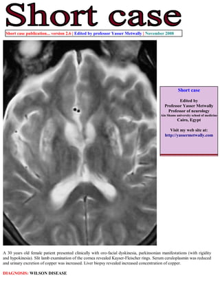
  • 1. Short case publication... version 2.6 | Edited by professor Yasser Metwally | November 2008 Short case Edited by Professor Yasser Metwally Professor of neurology Ain Shams university school of medicine Cairo, Egypt Visit my web site at: http://yassermetwally.com A 30 years old female patient presented clinically with oro-facial dyskinesia, parkinsonian manifestations (with rigidity and hypokinesia). Slit lamb examination of the cornea revealed Kayser-Fleischer rings. Serum ceruloplasmin was reduced and urinary excretion of copper was increased. Liver biopsy revealed increased concentration of copper. DIAGNOSIS: WILSON DISEASE
  • 2. Figure 1. Wilson's disease has a relatively characteristic appearance, particularly on high field strength MR imaging studies. Signal hyperintensity on T2-weighted and FLAIR images exists in the putamen symmetrically and bilaterally, presumably representing chronic gliotic and edematous changes. Within the putaminal high signal intensity, irregular areas of low signal intensity are also frequently observed on the T2-weighted images. These areas of low signal intensity are somewhat characteristic of Wilson's disease and most likely represent increased iron accumulation, occurring secondary to the increased copper distribution in the putamen. Central atrophy is also noted. Notice the signal hypointensity that is observed in the corticospinal tract bilaterally, The signal hypointensity is anatomically mapping the corticospinal tract bilaterally. Figure 2. Wilson's disease has a relatively characteristic appearance, particularly on high field strength MR imaging studies. Signal hyperintensity on T2-weighted and FLAIR images exists in the putamen symmetrically and bilaterally, presumably representing chronic gliotic and edematous changes. Within the putaminal high signal intensity, irregular areas of low signal intensity are also frequently observed on the T2-weighted images. These areas of low signal intensity are somewhat characteristic of Wilson's disease and most likely represent increased iron accumulation, occurring secondary to the increased copper distribution in the putamen. Central atrophy is also noted. Notice the signal hypointensity that is observed in the corticospinal tract bilaterally, The signal hypointensity is anatomically mapping the corticospinal tract bilaterally.
  • 3. Figure 3. MRI FLAIR image showing signal hyperintensity in the basal ganglia, bithalamic and hypothalamic areas. Notice the lateral ventricular dilatation. T 2 hyperintense zones are primarily due to cavitations, astrogliosis and spongy degeneration T2 hypointense zones, are primarily due to excessive iron deposition in the same areas of copper deposition. This occurs due to low to low-normal levels of plasma iron-binding globulin. Ceruloplasmin directly affects the transfer of iron from tissue cells to plasma transferrin. Signal abnormalities in the basal ganglia (in this patient) on the T2 images were in the form of a mixture of hypointensities and hyperintensities. while T2 signal abnormalities in the corticospinal tract were in the form of T2 hypointensity only. It must be noted also that the clinical picture of the patient were dominated with extrapyramidal features with no clinical evidence of pyramidal tract involvement. Pyramidal tract involvement by MRI was a subclinical one. In the reported patient, ganglionic involvement with astrogliosis and iron deposition (a mixture of hypointensities and hyperintensities on the T2 images) was different from extra-ganglionic involvement (T2 hypointensity only) which probably reflects iron deposition only. Involvement of the basal ganglia was associated with extrapyramidal symptoms and signs while involvement of the pyramidal tract was a subclinical one. Astrogliosis (T2 hyperintensity) is probably a reaction of copper deposition and occurs mainly in the basal ganglia in Wilson disease. T2 hyperintensities are also found in dentatothalamic tract, pontocerebellar tract and the cortex of the frontal lobe. Astrogliosis and spongy degeneration are always associated with copper deposition and it is probably induced by it, thus explaining the T2 signal abnormalities (T2 hyperintesity) associated with copper deposition. Copper itself does not induce any MRI signal changes at least at the tissue concentration noticed in Wilson changes. In Wilson disease, an abnormal striatum depicted on MR images correlated with pseudoparkinsonian signs, an abnormal dentatothalamic tract correlated with cerebellar signs, and an abnormal pontocerebellar tract correlated with pseudoparkinsonian signs. The T2 hypointensity observed in some anatomical areas in the brain in Wilson disease probably reflects increased iron concentration in these areas. Iron deposition is a secondary phenomenon in Wilson disease and probably does not have any clinical associates, however it does induce observable MRI T2 signal changes. Iron deposition probably does not induce any structural changes as copper deposition does, thus explaining its asymptomatic nature. The pathophysiology and the anatomical distribution of both copper and iron deposition in Wilson disease are different. Although T2 hypointensity and hyperintensity might coincide in the same anatomical areas (the basal ganglia in this patient), however the T2 hypointensity might occur in isolation in some other areas (the pyramidal tract in this patient), and this simply means that the pathophysiology of copper deposition is different from that of iron deposition. Iron deposition is not just a phenomenon secondary to copper deposition. In brain, the basal ganglia show the most striking alterations. They have a brick-red pigmentation; spongy degeneration of
  • 4. the putamen frequently leads to the formation of small cavities. Microscopic studies reveal a loss of neurons, axonal degeneration, and large numbers of protoplasmic astrocytes, including giant forms known as Alzheimer cells. The cortex of the frontal lobe may also show spongy degeneration and astrocytosis. Copper is deposited in the pericapillary area and within astrocytes, where it is located in the subcellular soluble fraction and bound not only to cerebrocuprein but also to other cerebral proteins. Copper is uniformly absent from neurons and ground substance. Addendum A new version of short case is uploaded in my web site every week (every Saturday and remains available till Friday.) To download the current version follow the link quot;http://pdf.yassermetwally.com/short.pdfquot;. You can download the long case version of this short case during the same week from: http://pdf.yassermetwally.com/case.pdf or visit web site: http://pdf.yassermetwally.com To download the software version of the publication (crow.exe) follow the link: http://neurology.yassermetwally.com/crow.zip At the end of each year, all the publications are compiled on a single CD-ROM, please contact the author to know more details. Screen resolution is better set at 1024*768 pixel screen area for optimum display For an archive of the previously reported cases go to www.yassermetwally.net, then under pages in the right panel, scroll down and click on the text entry quot;downloadable short cases in PDF formatquot; Also to view a list of the previously published case records follow the following link (http://wordpress.com/tag/case- record/) or click on it if it appears as a link in your PDF reader References 1. Metwally, MYM: Textbook of neurimaging, A CD-ROM publication, (Metwally, MYM editor) WEB-CD agency for electronic publishing, version 9.4a October 2008