SlideShare uma empresa Scribd logo
1 de 5
Baixar para ler offline
Short case publication... version 1.12 | Edited by professor Yasser Metwally | February 2008


                                                                                                     Short case

                                                                                                    Edited by
                                                                                            Professor Yasser Metwally
                                                                                              Professor of neurology
                                                                                          Ain Shams university school of medicine
                                                                                                     Cairo, Egypt

                                                                                               Visit my web site at:
                                                                                            http://yassermetwally.com


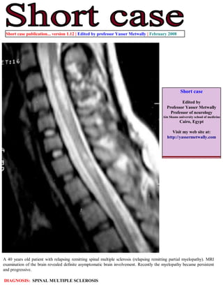
A 40 years old patient with relapsing remitting spinal multiple sclerosis (relapsing remitting partial myelopathy). MRI
examination of the brain revealed definite asymptomatic brain involvement. Recently the myelopathy became persistent
and progressive.

DIAGNOSIS: SPINAL MULTIPLE SCLEROSIS
Figure 1. A case of spinal multiple sclerosis. Precontrast MRI T1 images showing peripherally located, pencil
shaped multiple sclerosis plaques that occupy 2-3 spinal segments. The plaques are well demarcated and
hypointense. No evidence of spinal cord atrophy and postcontrast studies did not show evidence of contrast
enhancement.


Figure 2. A case of spinal multiple sclerosis. MRI T2 images showing peripherally located, pencil shaped multiple
sclerosis plaques that occupy 2-3 spinal segments The plaques are orientated along the longitudinal axis of the
spinal cord on sagittal sections. The plaques are short (less than two spinal segments), pencil shaped, multiple and
well demarcated. No evidence of spinal cord atrophy. The spinal cord parenchyma is asymmetrically involved.
Diffuse abnormalities seen as poorly demarcated areas of increased signal intensity on MRI T2 images are also
seen in this study. Diffuse abnormalities are more common in primary progressive MS and secondary progressive
MS.
Figure 3. A case of spinal multiple sclerosis. MRI T2 and precontrast T1 cross section images showing diffuse
abnormalities seen as poorly demarcated areas of increased signal intensity on MRI T2 images and low signal
on the MRI T1 images. Diffuse abnormalities are more common in primary progressive MS and secondary
progressive MS.


                                                   Figure 4. Atrophy affecting the upper cervical spinal
                                                   segments.


Over the past decade, researchers and clinicians have gained new insights into the core of demyelinating diseases of
the spinal cord, and much progress has been made in the management of these diseases. Although we are starting
to uncover some of the structural and physiologic substrates of demyelination of the CNS, we are far from
understanding what causes many of these demyelinating disorders and how to prevent their progression. With
further development of new techniques, such as DTI and more potent MR units, spinal cord diseases may be
distinguished from each other, and effective therapeutic strategies may be initiated before any cord damage occurs.

In particular MRI is very helpful in differentiation between Spinal multiple sclerosis and transverse myelitis In the
series reported by Choi et al, [2] the centrally located MRI T2 high signal intensity occupied more than two thirds
of the cross-sectional area of the cord in transverse myelitis. In multiple sclerosis, plaques are usually located
peripherally and occupy less than half the cross-sectional area of the cord. The central isointensity, or dot
(commonly seen in transverse myelitis), represents central gray matter squeezed by the uniform, evenly distributed
oedematous changes of the cord. Choi and colleagues [2] have demonstrated the role of contrast media in
differentiating transverse myelitis from multiple sclerosis. In transverse myelitis, enhancement is in the periphery of
a centrally located area of high T2 weighted images. In multiple sclerosis, the lesions show enhancement in the
central zone of peripherally located high signal intensity on T2 weighted images.

In conclusion, certain MRI characteristics help in differentiating acute transverse myelitis from spinal form of
multiple sclerosis. These include: 1) centrally located high intensity signal extending over 3 to 4 segments and
occupying more than two thirds of the cord cross-sectional area and 2) peripheral contrast enhancement of high
intensity signal.


                                                                                    Figure 5.       Differential diagnoses of
                                                                                    intramedullary lesions based on their
                                                                                    location at the cross-sectional area of the
                                                                                    cord. (A) MS: Dorsally located wedge-shaped
                                                                                    lesion involving less then two thirds of the
                                                                                    cross-sectional area of the spinal cord seen on
                                                                                    axial T2-Wi MR image. (B) Poliomyelitis:
                                                                                    Bilateral enhancing anterior nerve roots
                                                                                    demonstrated on postcontrast T1-Wi MR
                                                                                    image. (C) Vacuolar myelopathy: Bilateral,
                                                                                    symmetrical,              high-signal-intensity
         MS               Poliomyelitis            Sbacute combined degeneration
                                                                                    abnormality located dorsally in the spinal
                                                                                    cord in an HIV-positive patient. DD:
                                                                                    Subacute combined degeneration. (D) ATM:
                                                                                    On axial T2-Wi, a high-signal-intensity lesion
                                                                                    involving more than two thirds of cross-
                                                                                    sectional area of the spinal cord is observed.
                                                                                    (E)       Herpes-simplex-virus         myelitis:
                                                                                    Postcontrast T1-Wi axial MR image showing
                                                                                    nodular enhancing lesion located in the
                                                                                    lateral part of the cervical spinal cord. DD:
                                                                                    active MS plaque. (F) Spinal cord infarction:
                                                                                    Swelling of the anterior parts of the spinal
                                                                                    cord is shown on axial T2-Wi MR images,
                                                                                    indicating vulnerability of the anterior
                                                                                    portions of the spinal cord to ischemia.
 Transverse myelitis       HIV myelitis                     Infarction

Addendum

    A new version of short case is uploaded in my web site every week (every Saturday and remains available till Friday.)
    To download the current version follow the link quot;http://pdf.yassermetwally.com/short.pdfquot;.
    You can download the long case version of this short case during the same week from: http://pdf.yassermetwally.com/case.pdf or visit web
    site: http://pdf.yassermetwally.com
    To download the software version of the publication (crow.exe) follow the link: http://neurology.yassermetwally.com/crow.zip
    At the end of each year, all the publications are compiled on a single CD-ROM, please contact the author to know more
    details.
    Screen resolution is better set at 1024*768 pixel screen area for optimum display
    For an archive of the previously reported cases go to www.yassermetwally.net, then under pages in the right panel,
    scroll down and click on the text entry quot;downloadable short cases in PDF formatquot;
References

1. Metwally, MYM: Textbook of neurimaging, A CD-ROM publication, (Metwally, MYM editor) WEB-CD agency for
electronic publishing, version 9.1a January 2008

2. Choi KH, Lee KS, Chung SO, et al.. Idiopathic transverse myelitis: MR characteristics. AJNR Am J Neuroradiol.
1996;17:1151–1160.

Mais conteúdo relacionado

Semelhante a Short case...Spinal multiple sclerosis

Case record...Idiopathic postinfectious transverse myelitis'
Case record...Idiopathic postinfectious transverse myelitis'Case record...Idiopathic postinfectious transverse myelitis'
Case record...Idiopathic postinfectious transverse myelitis'Professor Yasser Metwally
 
Atlas of peripheral nerve ultrasound
Atlas of peripheral nerve ultrasoundAtlas of peripheral nerve ultrasound
Atlas of peripheral nerve ultrasoundSpringer
 
Congenital Anomalies Of Spine And Spinal Cord
Congenital Anomalies Of Spine And Spinal CordCongenital Anomalies Of Spine And Spinal Cord
Congenital Anomalies Of Spine And Spinal CordRoshan Valentine
 
Short case...Acute postinfectious transverse myelitis
Short case...Acute postinfectious transverse myelitisShort case...Acute postinfectious transverse myelitis
Short case...Acute postinfectious transverse myelitisProfessor Yasser Metwally
 
Short case...Multiple craniocervical anomalies
Short case...Multiple craniocervical anomaliesShort case...Multiple craniocervical anomalies
Short case...Multiple craniocervical anomaliesProfessor Yasser Metwally
 
Imaging in spinal dysraphism
Imaging in spinal dysraphismImaging in spinal dysraphism
Imaging in spinal dysraphismPooja Saji
 
Short case...Cerebellopontine angle meningioma
Short case...Cerebellopontine angle meningiomaShort case...Cerebellopontine angle meningioma
Short case...Cerebellopontine angle meningiomaProfessor Yasser Metwally
 
Short case...Acute disseminated encephalomyelitis
Short case...Acute disseminated encephalomyelitisShort case...Acute disseminated encephalomyelitis
Short case...Acute disseminated encephalomyelitisProfessor Yasser Metwally
 

Semelhante a Short case...Spinal multiple sclerosis (20)

Case record...Spinal multiple sclerosis
Case record...Spinal multiple sclerosisCase record...Spinal multiple sclerosis
Case record...Spinal multiple sclerosis
 
Short case...Intramedullary cavernoma
Short case...Intramedullary cavernomaShort case...Intramedullary cavernoma
Short case...Intramedullary cavernoma
 
Case record...Idiopathic postinfectious transverse myelitis'
Case record...Idiopathic postinfectious transverse myelitis'Case record...Idiopathic postinfectious transverse myelitis'
Case record...Idiopathic postinfectious transverse myelitis'
 
Atlas of peripheral nerve ultrasound
Atlas of peripheral nerve ultrasoundAtlas of peripheral nerve ultrasound
Atlas of peripheral nerve ultrasound
 
Short case...Focal midbrain glioma
Short case...Focal midbrain gliomaShort case...Focal midbrain glioma
Short case...Focal midbrain glioma
 
Short case...Congenital syringomyelia
Short case...Congenital syringomyeliaShort case...Congenital syringomyelia
Short case...Congenital syringomyelia
 
Benign bone tumors
Benign bone tumorsBenign bone tumors
Benign bone tumors
 
Case record...Spinal meningioma
Case record...Spinal meningiomaCase record...Spinal meningioma
Case record...Spinal meningioma
 
Congenital Anomalies Of Spine And Spinal Cord
Congenital Anomalies Of Spine And Spinal CordCongenital Anomalies Of Spine And Spinal Cord
Congenital Anomalies Of Spine And Spinal Cord
 
Short case...Multiple meningiomas
Short case...Multiple meningiomasShort case...Multiple meningiomas
Short case...Multiple meningiomas
 
Short case...Acute postinfectious transverse myelitis
Short case...Acute postinfectious transverse myelitisShort case...Acute postinfectious transverse myelitis
Short case...Acute postinfectious transverse myelitis
 
Foramen Magnum Meningioma
Foramen Magnum MeningiomaForamen Magnum Meningioma
Foramen Magnum Meningioma
 
Case record...Neuromyelitis optica
Case record...Neuromyelitis opticaCase record...Neuromyelitis optica
Case record...Neuromyelitis optica
 
Short case...Multiple craniocervical anomalies
Short case...Multiple craniocervical anomaliesShort case...Multiple craniocervical anomalies
Short case...Multiple craniocervical anomalies
 
Short case...tectal plate glioma
Short case...tectal plate gliomaShort case...tectal plate glioma
Short case...tectal plate glioma
 
Case record...Spinal metastasis
Case record...Spinal metastasisCase record...Spinal metastasis
Case record...Spinal metastasis
 
Imaging in spinal dysraphism
Imaging in spinal dysraphismImaging in spinal dysraphism
Imaging in spinal dysraphism
 
Short case...Cerebellopontine angle meningioma
Short case...Cerebellopontine angle meningiomaShort case...Cerebellopontine angle meningioma
Short case...Cerebellopontine angle meningioma
 
Short case...Spinal meningioma
Short case...Spinal meningiomaShort case...Spinal meningioma
Short case...Spinal meningioma
 
Short case...Acute disseminated encephalomyelitis
Short case...Acute disseminated encephalomyelitisShort case...Acute disseminated encephalomyelitis
Short case...Acute disseminated encephalomyelitis
 

Mais de Professor Yasser Metwally

The Snake, the Scorpion, the turtle in Egypt
The Snake, the Scorpion, the turtle in EgyptThe Snake, the Scorpion, the turtle in Egypt
The Snake, the Scorpion, the turtle in EgyptProfessor Yasser Metwally
 
Radiological pathology of epileptic disorders
Radiological pathology of epileptic disordersRadiological pathology of epileptic disorders
Radiological pathology of epileptic disordersProfessor Yasser Metwally
 
Radiological pathology of cerebrovascular disorders
Radiological pathology of cerebrovascular disordersRadiological pathology of cerebrovascular disorders
Radiological pathology of cerebrovascular disordersProfessor Yasser Metwally
 
Radiological pathology of spontaneous cerebral hemorrhage
Radiological pathology of spontaneous cerebral hemorrhageRadiological pathology of spontaneous cerebral hemorrhage
Radiological pathology of spontaneous cerebral hemorrhageProfessor Yasser Metwally
 
Radiological pathology of cerebral amyloid angiography
Radiological pathology of cerebral amyloid angiographyRadiological pathology of cerebral amyloid angiography
Radiological pathology of cerebral amyloid angiographyProfessor Yasser Metwally
 
Radiological pathology of cerebral microbleeds
Radiological pathology of cerebral microbleedsRadiological pathology of cerebral microbleeds
Radiological pathology of cerebral microbleedsProfessor Yasser Metwally
 
Issues in radiological pathology: Radiological pathology of watershed infarct...
Issues in radiological pathology: Radiological pathology of watershed infarct...Issues in radiological pathology: Radiological pathology of watershed infarct...
Issues in radiological pathology: Radiological pathology of watershed infarct...Professor Yasser Metwally
 
Radiological pathology of cortical laminar necrosis
Radiological pathology of cortical laminar necrosisRadiological pathology of cortical laminar necrosis
Radiological pathology of cortical laminar necrosisProfessor Yasser Metwally
 

Mais de Professor Yasser Metwally (20)

The Egyptian Zoo in Cairo 2015
The Egyptian Zoo in Cairo 2015The Egyptian Zoo in Cairo 2015
The Egyptian Zoo in Cairo 2015
 
End of the great nile river in Ras Elbar
End of the great nile river in Ras ElbarEnd of the great nile river in Ras Elbar
End of the great nile river in Ras Elbar
 
The Lion and The tiger in Egypt
The Lion and The tiger in EgyptThe Lion and The tiger in Egypt
The Lion and The tiger in Egypt
 
The monkeys in Egypt
The monkeys in EgyptThe monkeys in Egypt
The monkeys in Egypt
 
The Snake, the Scorpion, the turtle in Egypt
The Snake, the Scorpion, the turtle in EgyptThe Snake, the Scorpion, the turtle in Egypt
The Snake, the Scorpion, the turtle in Egypt
 
The Egyptian Parrot
The Egyptian ParrotThe Egyptian Parrot
The Egyptian Parrot
 
The Egyptian Deer
The Egyptian DeerThe Egyptian Deer
The Egyptian Deer
 
The Egyptian Pelican
The Egyptian PelicanThe Egyptian Pelican
The Egyptian Pelican
 
The Flamingo bird in Egypt
The Flamingo bird in EgyptThe Flamingo bird in Egypt
The Flamingo bird in Egypt
 
Egyptian Cats
Egyptian CatsEgyptian Cats
Egyptian Cats
 
Radiological pathology of epileptic disorders
Radiological pathology of epileptic disordersRadiological pathology of epileptic disorders
Radiological pathology of epileptic disorders
 
Radiological pathology of cerebrovascular disorders
Radiological pathology of cerebrovascular disordersRadiological pathology of cerebrovascular disorders
Radiological pathology of cerebrovascular disorders
 
Radiological pathology of spontaneous cerebral hemorrhage
Radiological pathology of spontaneous cerebral hemorrhageRadiological pathology of spontaneous cerebral hemorrhage
Radiological pathology of spontaneous cerebral hemorrhage
 
Radiological pathology of cerebral amyloid angiography
Radiological pathology of cerebral amyloid angiographyRadiological pathology of cerebral amyloid angiography
Radiological pathology of cerebral amyloid angiography
 
Radiological pathology of cerebral microbleeds
Radiological pathology of cerebral microbleedsRadiological pathology of cerebral microbleeds
Radiological pathology of cerebral microbleeds
 
The Egyptian Zoo in Cairo
The Egyptian Zoo in CairoThe Egyptian Zoo in Cairo
The Egyptian Zoo in Cairo
 
Progressive multifocal leukoencephalopathy
Progressive multifocal leukoencephalopathyProgressive multifocal leukoencephalopathy
Progressive multifocal leukoencephalopathy
 
Progressive multifocal leukoencephalopathy
Progressive multifocal leukoencephalopathyProgressive multifocal leukoencephalopathy
Progressive multifocal leukoencephalopathy
 
Issues in radiological pathology: Radiological pathology of watershed infarct...
Issues in radiological pathology: Radiological pathology of watershed infarct...Issues in radiological pathology: Radiological pathology of watershed infarct...
Issues in radiological pathology: Radiological pathology of watershed infarct...
 
Radiological pathology of cortical laminar necrosis
Radiological pathology of cortical laminar necrosisRadiological pathology of cortical laminar necrosis
Radiological pathology of cortical laminar necrosis
 

Último

Proudly South Africa powerpoint Thorisha.pptx
Proudly South Africa powerpoint Thorisha.pptxProudly South Africa powerpoint Thorisha.pptx
Proudly South Africa powerpoint Thorisha.pptxthorishapillay1
 
ECONOMIC CONTEXT - LONG FORM TV DRAMA - PPT
ECONOMIC CONTEXT - LONG FORM TV DRAMA - PPTECONOMIC CONTEXT - LONG FORM TV DRAMA - PPT
ECONOMIC CONTEXT - LONG FORM TV DRAMA - PPTiammrhaywood
 
Influencing policy (training slides from Fast Track Impact)
Influencing policy (training slides from Fast Track Impact)Influencing policy (training slides from Fast Track Impact)
Influencing policy (training slides from Fast Track Impact)Mark Reed
 
Computed Fields and api Depends in the Odoo 17
Computed Fields and api Depends in the Odoo 17Computed Fields and api Depends in the Odoo 17
Computed Fields and api Depends in the Odoo 17Celine George
 
Barangay Council for the Protection of Children (BCPC) Orientation.pptx
Barangay Council for the Protection of Children (BCPC) Orientation.pptxBarangay Council for the Protection of Children (BCPC) Orientation.pptx
Barangay Council for the Protection of Children (BCPC) Orientation.pptxCarlos105
 
USPS® Forced Meter Migration - How to Know if Your Postage Meter Will Soon be...
USPS® Forced Meter Migration - How to Know if Your Postage Meter Will Soon be...USPS® Forced Meter Migration - How to Know if Your Postage Meter Will Soon be...
USPS® Forced Meter Migration - How to Know if Your Postage Meter Will Soon be...Postal Advocate Inc.
 
Science 7 Quarter 4 Module 2: Natural Resources.pptx
Science 7 Quarter 4 Module 2: Natural Resources.pptxScience 7 Quarter 4 Module 2: Natural Resources.pptx
Science 7 Quarter 4 Module 2: Natural Resources.pptxMaryGraceBautista27
 
THEORIES OF ORGANIZATION-PUBLIC ADMINISTRATION
THEORIES OF ORGANIZATION-PUBLIC ADMINISTRATIONTHEORIES OF ORGANIZATION-PUBLIC ADMINISTRATION
THEORIES OF ORGANIZATION-PUBLIC ADMINISTRATIONHumphrey A Beña
 
ANG SEKTOR NG agrikultura.pptx QUARTER 4
ANG SEKTOR NG agrikultura.pptx QUARTER 4ANG SEKTOR NG agrikultura.pptx QUARTER 4
ANG SEKTOR NG agrikultura.pptx QUARTER 4MiaBumagat1
 
Field Attribute Index Feature in Odoo 17
Field Attribute Index Feature in Odoo 17Field Attribute Index Feature in Odoo 17
Field Attribute Index Feature in Odoo 17Celine George
 
Grade 9 Q4-MELC1-Active and Passive Voice.pptx
Grade 9 Q4-MELC1-Active and Passive Voice.pptxGrade 9 Q4-MELC1-Active and Passive Voice.pptx
Grade 9 Q4-MELC1-Active and Passive Voice.pptxChelloAnnAsuncion2
 
Incoming and Outgoing Shipments in 3 STEPS Using Odoo 17
Incoming and Outgoing Shipments in 3 STEPS Using Odoo 17Incoming and Outgoing Shipments in 3 STEPS Using Odoo 17
Incoming and Outgoing Shipments in 3 STEPS Using Odoo 17Celine George
 
ENGLISH 7_Q4_LESSON 2_ Employing a Variety of Strategies for Effective Interp...
ENGLISH 7_Q4_LESSON 2_ Employing a Variety of Strategies for Effective Interp...ENGLISH 7_Q4_LESSON 2_ Employing a Variety of Strategies for Effective Interp...
ENGLISH 7_Q4_LESSON 2_ Employing a Variety of Strategies for Effective Interp...JhezDiaz1
 
ACC 2024 Chronicles. Cardiology. Exam.pdf
ACC 2024 Chronicles. Cardiology. Exam.pdfACC 2024 Chronicles. Cardiology. Exam.pdf
ACC 2024 Chronicles. Cardiology. Exam.pdfSpandanaRallapalli
 
Like-prefer-love -hate+verb+ing & silent letters & citizenship text.pdf
Like-prefer-love -hate+verb+ing & silent letters & citizenship text.pdfLike-prefer-love -hate+verb+ing & silent letters & citizenship text.pdf
Like-prefer-love -hate+verb+ing & silent letters & citizenship text.pdfMr Bounab Samir
 

Último (20)

Proudly South Africa powerpoint Thorisha.pptx
Proudly South Africa powerpoint Thorisha.pptxProudly South Africa powerpoint Thorisha.pptx
Proudly South Africa powerpoint Thorisha.pptx
 
ECONOMIC CONTEXT - LONG FORM TV DRAMA - PPT
ECONOMIC CONTEXT - LONG FORM TV DRAMA - PPTECONOMIC CONTEXT - LONG FORM TV DRAMA - PPT
ECONOMIC CONTEXT - LONG FORM TV DRAMA - PPT
 
YOUVE_GOT_EMAIL_PRELIMS_EL_DORADO_2024.pptx
YOUVE_GOT_EMAIL_PRELIMS_EL_DORADO_2024.pptxYOUVE_GOT_EMAIL_PRELIMS_EL_DORADO_2024.pptx
YOUVE_GOT_EMAIL_PRELIMS_EL_DORADO_2024.pptx
 
Influencing policy (training slides from Fast Track Impact)
Influencing policy (training slides from Fast Track Impact)Influencing policy (training slides from Fast Track Impact)
Influencing policy (training slides from Fast Track Impact)
 
Computed Fields and api Depends in the Odoo 17
Computed Fields and api Depends in the Odoo 17Computed Fields and api Depends in the Odoo 17
Computed Fields and api Depends in the Odoo 17
 
Barangay Council for the Protection of Children (BCPC) Orientation.pptx
Barangay Council for the Protection of Children (BCPC) Orientation.pptxBarangay Council for the Protection of Children (BCPC) Orientation.pptx
Barangay Council for the Protection of Children (BCPC) Orientation.pptx
 
USPS® Forced Meter Migration - How to Know if Your Postage Meter Will Soon be...
USPS® Forced Meter Migration - How to Know if Your Postage Meter Will Soon be...USPS® Forced Meter Migration - How to Know if Your Postage Meter Will Soon be...
USPS® Forced Meter Migration - How to Know if Your Postage Meter Will Soon be...
 
Science 7 Quarter 4 Module 2: Natural Resources.pptx
Science 7 Quarter 4 Module 2: Natural Resources.pptxScience 7 Quarter 4 Module 2: Natural Resources.pptx
Science 7 Quarter 4 Module 2: Natural Resources.pptx
 
THEORIES OF ORGANIZATION-PUBLIC ADMINISTRATION
THEORIES OF ORGANIZATION-PUBLIC ADMINISTRATIONTHEORIES OF ORGANIZATION-PUBLIC ADMINISTRATION
THEORIES OF ORGANIZATION-PUBLIC ADMINISTRATION
 
ANG SEKTOR NG agrikultura.pptx QUARTER 4
ANG SEKTOR NG agrikultura.pptx QUARTER 4ANG SEKTOR NG agrikultura.pptx QUARTER 4
ANG SEKTOR NG agrikultura.pptx QUARTER 4
 
Field Attribute Index Feature in Odoo 17
Field Attribute Index Feature in Odoo 17Field Attribute Index Feature in Odoo 17
Field Attribute Index Feature in Odoo 17
 
Grade 9 Q4-MELC1-Active and Passive Voice.pptx
Grade 9 Q4-MELC1-Active and Passive Voice.pptxGrade 9 Q4-MELC1-Active and Passive Voice.pptx
Grade 9 Q4-MELC1-Active and Passive Voice.pptx
 
Model Call Girl in Tilak Nagar Delhi reach out to us at 🔝9953056974🔝
Model Call Girl in Tilak Nagar Delhi reach out to us at 🔝9953056974🔝Model Call Girl in Tilak Nagar Delhi reach out to us at 🔝9953056974🔝
Model Call Girl in Tilak Nagar Delhi reach out to us at 🔝9953056974🔝
 
Raw materials used in Herbal Cosmetics.pptx
Raw materials used in Herbal Cosmetics.pptxRaw materials used in Herbal Cosmetics.pptx
Raw materials used in Herbal Cosmetics.pptx
 
Incoming and Outgoing Shipments in 3 STEPS Using Odoo 17
Incoming and Outgoing Shipments in 3 STEPS Using Odoo 17Incoming and Outgoing Shipments in 3 STEPS Using Odoo 17
Incoming and Outgoing Shipments in 3 STEPS Using Odoo 17
 
ENGLISH 7_Q4_LESSON 2_ Employing a Variety of Strategies for Effective Interp...
ENGLISH 7_Q4_LESSON 2_ Employing a Variety of Strategies for Effective Interp...ENGLISH 7_Q4_LESSON 2_ Employing a Variety of Strategies for Effective Interp...
ENGLISH 7_Q4_LESSON 2_ Employing a Variety of Strategies for Effective Interp...
 
ACC 2024 Chronicles. Cardiology. Exam.pdf
ACC 2024 Chronicles. Cardiology. Exam.pdfACC 2024 Chronicles. Cardiology. Exam.pdf
ACC 2024 Chronicles. Cardiology. Exam.pdf
 
LEFT_ON_C'N_ PRELIMS_EL_DORADO_2024.pptx
LEFT_ON_C'N_ PRELIMS_EL_DORADO_2024.pptxLEFT_ON_C'N_ PRELIMS_EL_DORADO_2024.pptx
LEFT_ON_C'N_ PRELIMS_EL_DORADO_2024.pptx
 
Like-prefer-love -hate+verb+ing & silent letters & citizenship text.pdf
Like-prefer-love -hate+verb+ing & silent letters & citizenship text.pdfLike-prefer-love -hate+verb+ing & silent letters & citizenship text.pdf
Like-prefer-love -hate+verb+ing & silent letters & citizenship text.pdf
 
TataKelola dan KamSiber Kecerdasan Buatan v022.pdf
TataKelola dan KamSiber Kecerdasan Buatan v022.pdfTataKelola dan KamSiber Kecerdasan Buatan v022.pdf
TataKelola dan KamSiber Kecerdasan Buatan v022.pdf
 

Short case...Spinal multiple sclerosis

  • 1. Short case publication... version 1.12 | Edited by professor Yasser Metwally | February 2008 Short case Edited by Professor Yasser Metwally Professor of neurology Ain Shams university school of medicine Cairo, Egypt Visit my web site at: http://yassermetwally.com A 40 years old patient with relapsing remitting spinal multiple sclerosis (relapsing remitting partial myelopathy). MRI examination of the brain revealed definite asymptomatic brain involvement. Recently the myelopathy became persistent and progressive. DIAGNOSIS: SPINAL MULTIPLE SCLEROSIS
  • 2. Figure 1. A case of spinal multiple sclerosis. Precontrast MRI T1 images showing peripherally located, pencil shaped multiple sclerosis plaques that occupy 2-3 spinal segments. The plaques are well demarcated and hypointense. No evidence of spinal cord atrophy and postcontrast studies did not show evidence of contrast enhancement. Figure 2. A case of spinal multiple sclerosis. MRI T2 images showing peripherally located, pencil shaped multiple sclerosis plaques that occupy 2-3 spinal segments The plaques are orientated along the longitudinal axis of the spinal cord on sagittal sections. The plaques are short (less than two spinal segments), pencil shaped, multiple and well demarcated. No evidence of spinal cord atrophy. The spinal cord parenchyma is asymmetrically involved. Diffuse abnormalities seen as poorly demarcated areas of increased signal intensity on MRI T2 images are also seen in this study. Diffuse abnormalities are more common in primary progressive MS and secondary progressive MS.
  • 3. Figure 3. A case of spinal multiple sclerosis. MRI T2 and precontrast T1 cross section images showing diffuse abnormalities seen as poorly demarcated areas of increased signal intensity on MRI T2 images and low signal on the MRI T1 images. Diffuse abnormalities are more common in primary progressive MS and secondary progressive MS. Figure 4. Atrophy affecting the upper cervical spinal segments. Over the past decade, researchers and clinicians have gained new insights into the core of demyelinating diseases of the spinal cord, and much progress has been made in the management of these diseases. Although we are starting to uncover some of the structural and physiologic substrates of demyelination of the CNS, we are far from understanding what causes many of these demyelinating disorders and how to prevent their progression. With further development of new techniques, such as DTI and more potent MR units, spinal cord diseases may be distinguished from each other, and effective therapeutic strategies may be initiated before any cord damage occurs. In particular MRI is very helpful in differentiation between Spinal multiple sclerosis and transverse myelitis In the series reported by Choi et al, [2] the centrally located MRI T2 high signal intensity occupied more than two thirds
  • 4. of the cross-sectional area of the cord in transverse myelitis. In multiple sclerosis, plaques are usually located peripherally and occupy less than half the cross-sectional area of the cord. The central isointensity, or dot (commonly seen in transverse myelitis), represents central gray matter squeezed by the uniform, evenly distributed oedematous changes of the cord. Choi and colleagues [2] have demonstrated the role of contrast media in differentiating transverse myelitis from multiple sclerosis. In transverse myelitis, enhancement is in the periphery of a centrally located area of high T2 weighted images. In multiple sclerosis, the lesions show enhancement in the central zone of peripherally located high signal intensity on T2 weighted images. In conclusion, certain MRI characteristics help in differentiating acute transverse myelitis from spinal form of multiple sclerosis. These include: 1) centrally located high intensity signal extending over 3 to 4 segments and occupying more than two thirds of the cord cross-sectional area and 2) peripheral contrast enhancement of high intensity signal. Figure 5. Differential diagnoses of intramedullary lesions based on their location at the cross-sectional area of the cord. (A) MS: Dorsally located wedge-shaped lesion involving less then two thirds of the cross-sectional area of the spinal cord seen on axial T2-Wi MR image. (B) Poliomyelitis: Bilateral enhancing anterior nerve roots demonstrated on postcontrast T1-Wi MR image. (C) Vacuolar myelopathy: Bilateral, symmetrical, high-signal-intensity MS Poliomyelitis Sbacute combined degeneration abnormality located dorsally in the spinal cord in an HIV-positive patient. DD: Subacute combined degeneration. (D) ATM: On axial T2-Wi, a high-signal-intensity lesion involving more than two thirds of cross- sectional area of the spinal cord is observed. (E) Herpes-simplex-virus myelitis: Postcontrast T1-Wi axial MR image showing nodular enhancing lesion located in the lateral part of the cervical spinal cord. DD: active MS plaque. (F) Spinal cord infarction: Swelling of the anterior parts of the spinal cord is shown on axial T2-Wi MR images, indicating vulnerability of the anterior portions of the spinal cord to ischemia. Transverse myelitis HIV myelitis Infarction Addendum A new version of short case is uploaded in my web site every week (every Saturday and remains available till Friday.) To download the current version follow the link quot;http://pdf.yassermetwally.com/short.pdfquot;. You can download the long case version of this short case during the same week from: http://pdf.yassermetwally.com/case.pdf or visit web site: http://pdf.yassermetwally.com To download the software version of the publication (crow.exe) follow the link: http://neurology.yassermetwally.com/crow.zip At the end of each year, all the publications are compiled on a single CD-ROM, please contact the author to know more details. Screen resolution is better set at 1024*768 pixel screen area for optimum display For an archive of the previously reported cases go to www.yassermetwally.net, then under pages in the right panel, scroll down and click on the text entry quot;downloadable short cases in PDF formatquot;
  • 5. References 1. Metwally, MYM: Textbook of neurimaging, A CD-ROM publication, (Metwally, MYM editor) WEB-CD agency for electronic publishing, version 9.1a January 2008 2. Choi KH, Lee KS, Chung SO, et al.. Idiopathic transverse myelitis: MR characteristics. AJNR Am J Neuroradiol. 1996;17:1151–1160.