SlideShare uma empresa Scribd logo
1 de 4
Baixar para ler offline
Short case publication... version 1.2 | Edited by professor Yasser Metwally | December 2007


                                                                                                   Short case

                                                                                                  Edited by
                                                                                          Professor Yasser Metwally
                                                                                            Professor of neurology
                                                                                        Ain Shams university school of medicine
                                                                                                   Cairo, Egypt

                                                                                              Visit my web site at:
                                                                                           http://yassermetwally.com


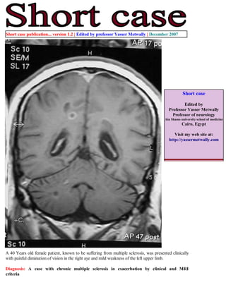
A 40 Years old female patient, known to be suffering from multiple sclerosis, was presented clinically
with painful diminution of vision in the right aye and mild weakness of the left upper limb.

Diagnosis: A case with chronic multiple sclerosis in exacerbation by clinical and MRI
criteria
Figure 1. A case of chronic multiple sclerosis. MRI T1 precontrast images showing multiple periventricular oval
or rounded plaques. The plaques are well-defined, hypointense to markedly hypointense (black holes) with mild
central atrophy. Some plaques are perpendicular to the ventricular surface (Dawson fingers). Tl hypointensities
serve as a surrogate marker of disability in multiple sclerosis because they are specific for matrix destruction
and axonal loss. Development of Tl hypointensities implies not only focal damage to axons within lesions, but
also more widespread tissue loss, leading to generalized atrophy of the brain.


Figure 2. Postcontrast MRI T1 images showing contrast enhancement of multiple sclerosis plaques. Two patterns
are seen. Diffuse contrast enhancement (A) and ring enhancement (either complete ring enhancement or
incomplete ring enhancement, both patterns are seen in this patient) (B,C). Enhanced plaques are active plaques
and is a manifestation of disease exacerbation even when not associated with clinical manifestations. Ring
enhancement demonstrates how MRI can show dissemination in time as well as place. Plaques with ring
enhancement are plaques with different ages. The central unenhanced parts are older than the peripherally
enhanced parts. Reactivation of old plaques takes place at the periphery of plaques and that explains ring
enhancement. Plaques with ring enhancement represent dissemination in time. Non enhanced plaques are plaques
in remission while enhanced plaques are plaques in exacerbation. Plaques with ring enhancement are plaques
which contain central parts in remission and peripheral parts in exacerbation. The open, incomplete, ring
enhancement that is seen in the cerebellum in this patient (B) is more indicative of an MS lesion than metastatic
disease or infection which almost always has a complete, fully enclosed ring of enhancement.
Figure 3. A case of chronic multiple sclerosis. MRI T2 (A,B) and MRI FLAIR (C) images showing
multiple periventricular oval or rounded plaques. The plaques are well-defined and hyperintense. Some
plaques are perpendicular to the ventricular surface (Dawson fingers).

USING MRI CRITERIA, THE DIAGNOSIS OF THE CASE IS CHRONIC MULTIPLE SCLEROSIS BECAUSE
OF:

    The existence of dissemination in place: MRI plaques that are widespread within the brain, in the
    periventricular regions, Some plaques are perpendicular to the ventricular surface (Dawson fingers). In
    particular Dawson fingers are rarely seen in other white matter diseases.

    The existence of dissemination in time: As manifested by some plaques showing contrast enhancement (active
    plaques) and other are not enhanced (chronic inactive plaques). Ring enhancement in a single plaque is also a
    sign of dissemination in time.


Multiple sclerosis is characterized by dissemination in time and place. Dissemination in time and place
(either clinically or by MRI) is not seen in other white matter diseases.

DISCUSSION

MR imaging plays a key and significant role in the assessment of patients with MS. Because of its high sensitivity,
MR imaging of the brain is the single most useful investigation during the diagnostic work-up of the patient with
clinically isolated syndrome. Knowledge of the characteristics of MS lesions, anatomic distribution of lesions,
differential diagnosis, and most important, the clinical context, markedly improve the specificity of the MR
imaging examination. It is clear that findings on MR imaging can be used to predict future conversion to clinically
definite MS before a second clinical exacerbation occurs. The data suggest a valuable role for counseling
individual patients at presentation, possibly selecting patients for clinical trials aimed at delaying disease
progression. Potential MR imaging markers of inflammation, demyelination, and axonal loss are helping to further
our understanding of the disease process and the resulting clinical disability. The use of serial MR imaging studies
to monitor therapy in individual patients will depend on the ability in the future to better understand the
relationship between disease activity detected in the short term by MR imaging and long-term disability or
response to therapy.
While commenting on a neuroimaging study for a multiple sclerosis patient, four parameters must be commented
upon. The anatomical distribution of the lesions, The presence or absence of brain atrophy, T2 and FLAIR
appearance of the lesions and the appearance of the lesions on the pre and postcontrast T1 images.


Table 1.

Parameter        Comment
Anatomy          Usually, MS lesions extend along medullary veins that drain into the subependymal veins lining the
                 lateral ventricle at the callososeptal interface. MS lesions are characteristically oriented along the fibers
                 of the corpus callosum (although the veins themselves are not visible on routine MR imaging)
                 paralleling these veins. This appearance corresponds to the so-called Dawson's finger identified at
                 pathology, and is well demonstrated on MR imaging.
Atrophy          With an extensive lesion burden, marked atrophy may be present. The corpus callosum may be severely
                 thinned throughout. To what extent the atrophy represents primary white matter disease or secondary
                 loss due to Wallerian degeneration is not clear.
T2, FLAIR        Increased water content of tissue leads to an increase in the tissue T2 relaxation time. This results in
                 hyperintense signal on FLAIR, PD- and T2-weighted imaging. Because most pathologic processes lead
                 to increased tissue water, initially through edema and inflammation, and later through gliosis and frank
                 replacement of tissue with fluid, T2 imaging is a sensitive but not particularly specific indicator of
                 pathology.
T1 precontrast   Initially, MS lesions are isointense to mildly hypointense on T1-weighted imaging. With time, the mild
                 hypointensity may progress to develop into a so-called T1 hole or black hole in which the lesion has
                 completely been replaced by CSF. Alternatively, the lesion may remain stable in intensity. In many
                 cases, there is a slightly hyperintense rim surrounding a lesion. This hyperintensity is typically ascribed
                 to the presence of free radicals in the surrounding inflammatory tissue, but paramagnetic effects from
                 petechial hemorrhage and fat signal from myelin breakdown also may contribute to this phenomenon.
                 Tl hypointensities serve as a surrogate marker of disability in multiple sclerosis because they are
                 specific for matrix destruction and axonal loss. Development of Tl hypointensities implies not only
                 focal damage to axons within lesions, but also more widespread tissue loss, leading to generalized
                 atrophy of the brain.
T1 postcontrast Contrast enhancement represents a disruption in the blood-brain barrier that allows for the leakage of
                 gadolinium-chelates. The presence of enhancement can play an important role in the evaluation of MS,
                 depending on the clinical context. Enhancement results from a local breakdown of the blood-brain
                 barrier, and indicates the presence of an active inflammatory process; therefore, enhancement identifies
                 quot;active lesions,quot; which often correlate with the acute symptomatology. These active lesions also may
                 be clinically silent and are frequently seen on serial imaging performed as part of research protocols.
                 Because nonenhancing lesions identified on T2-weighted images are thought to be chronic lesions, the
                 simultaneous presence of enhancing and nonenhancing lesions is strong evidence to indicate that these
                 are quot;multiple lesions separated in time,quot; supporting a putative diagnosis of MS at the time of initial
                 presentation. The presence of ring-enhancement suggests the reactivation of an old lesion-the central
                 nonenhancing portion of the lesion representing the quot;burnt outquot; portion of the lesion. An incomplete or
                 open ring of enhancement is more indicative of an MS lesion than metastatic disease or infection, which
                 almost always has a complete, fully enclosed ring of enhancement.

  References

  1. Metwally, MYM: Textbook of neurimaging, A CD-ROM publication, (Metwally, MYM editor) WEB-CD agency
  for electronic publishing, version 9.1a January 2008

Mais conteúdo relacionado

Mais procurados

Spinal neoplasms
Spinal neoplasmsSpinal neoplasms
Spinal neoplasmsfahad shafi
 
PVNS,SYNOVIAL CHONDROMATOSIS & LOOSE BODIES
PVNS,SYNOVIAL CHONDROMATOSIS & LOOSE BODIESPVNS,SYNOVIAL CHONDROMATOSIS & LOOSE BODIES
PVNS,SYNOVIAL CHONDROMATOSIS & LOOSE BODIESdarshanck89
 
Presentation2, radiological imaging of intra cranial meningioma.
Presentation2, radiological imaging of intra cranial meningioma.Presentation2, radiological imaging of intra cranial meningioma.
Presentation2, radiological imaging of intra cranial meningioma.Abdellah Nazeer
 
Inflammatory processes &spondyloarthropathies eva pharma
Inflammatory  processes &spondyloarthropathies eva pharmaInflammatory  processes &spondyloarthropathies eva pharma
Inflammatory processes &spondyloarthropathies eva pharmaSelf-employed
 
Hemangiopericytoma
HemangiopericytomaHemangiopericytoma
Hemangiopericytomasuhas k r
 
Meningeal Based Intracranial Masses Beyond Meningioma
Meningeal Based Intracranial Masses Beyond MeningiomaMeningeal Based Intracranial Masses Beyond Meningioma
Meningeal Based Intracranial Masses Beyond MeningiomaDr Varun Bansal
 
Primary vertebral body...........
Primary vertebral body...........Primary vertebral body...........
Primary vertebral body...........Yashveer Singh
 
Ewings sarcoma_UTSAV
Ewings sarcoma_UTSAVEwings sarcoma_UTSAV
Ewings sarcoma_UTSAVUtsav Agrawal
 
Imaging of Intracranial Meningioma
Imaging of Intracranial MeningiomaImaging of Intracranial Meningioma
Imaging of Intracranial Meningiomahazem youssef
 
Pvns dr.sameer
Pvns dr.sameerPvns dr.sameer
Pvns dr.sameerDr Sameer
 
Topic of the month: Radiological pathology of meningiomas
Topic of the month: Radiological pathology of meningiomasTopic of the month: Radiological pathology of meningiomas
Topic of the month: Radiological pathology of meningiomasProfessor Yasser Metwally
 
extra axial Meningioma
extra axial Meningiomaextra axial Meningioma
extra axial MeningiomaHassan Alqarni
 
Spinal tumors drsant91
Spinal tumors drsant91Spinal tumors drsant91
Spinal tumors drsant91Santosh Prasad
 
Pvns dr.sameer
Pvns dr.sameerPvns dr.sameer
Pvns dr.sameerDr Sameer
 
Pigmented villonodular snynovitis
Pigmented villonodular snynovitis Pigmented villonodular snynovitis
Pigmented villonodular snynovitis Ahmed Al Ibraheemi
 

Mais procurados (20)

Spinal neoplasms
Spinal neoplasmsSpinal neoplasms
Spinal neoplasms
 
PVNS,SYNOVIAL CHONDROMATOSIS & LOOSE BODIES
PVNS,SYNOVIAL CHONDROMATOSIS & LOOSE BODIESPVNS,SYNOVIAL CHONDROMATOSIS & LOOSE BODIES
PVNS,SYNOVIAL CHONDROMATOSIS & LOOSE BODIES
 
Case record...Spinal multiple sclerosis
Case record...Spinal multiple sclerosisCase record...Spinal multiple sclerosis
Case record...Spinal multiple sclerosis
 
Presentation2, radiological imaging of intra cranial meningioma.
Presentation2, radiological imaging of intra cranial meningioma.Presentation2, radiological imaging of intra cranial meningioma.
Presentation2, radiological imaging of intra cranial meningioma.
 
Intracranial lesions mimicking neoplasms
Intracranial lesions mimicking neoplasmsIntracranial lesions mimicking neoplasms
Intracranial lesions mimicking neoplasms
 
Inflammatory processes &spondyloarthropathies eva pharma
Inflammatory  processes &spondyloarthropathies eva pharmaInflammatory  processes &spondyloarthropathies eva pharma
Inflammatory processes &spondyloarthropathies eva pharma
 
Short case...Spinal meningioma
Short case...Spinal meningiomaShort case...Spinal meningioma
Short case...Spinal meningioma
 
Hemangiopericytoma
HemangiopericytomaHemangiopericytoma
Hemangiopericytoma
 
Meningeal Based Intracranial Masses Beyond Meningioma
Meningeal Based Intracranial Masses Beyond MeningiomaMeningeal Based Intracranial Masses Beyond Meningioma
Meningeal Based Intracranial Masses Beyond Meningioma
 
Primary vertebral body...........
Primary vertebral body...........Primary vertebral body...........
Primary vertebral body...........
 
Neuroblatoma
NeuroblatomaNeuroblatoma
Neuroblatoma
 
Ewings sarcoma_UTSAV
Ewings sarcoma_UTSAVEwings sarcoma_UTSAV
Ewings sarcoma_UTSAV
 
Imaging of Intracranial Meningioma
Imaging of Intracranial MeningiomaImaging of Intracranial Meningioma
Imaging of Intracranial Meningioma
 
Pvns dr.sameer
Pvns dr.sameerPvns dr.sameer
Pvns dr.sameer
 
Benign brain tumours
Benign brain tumoursBenign brain tumours
Benign brain tumours
 
Topic of the month: Radiological pathology of meningiomas
Topic of the month: Radiological pathology of meningiomasTopic of the month: Radiological pathology of meningiomas
Topic of the month: Radiological pathology of meningiomas
 
extra axial Meningioma
extra axial Meningiomaextra axial Meningioma
extra axial Meningioma
 
Spinal tumors drsant91
Spinal tumors drsant91Spinal tumors drsant91
Spinal tumors drsant91
 
Pvns dr.sameer
Pvns dr.sameerPvns dr.sameer
Pvns dr.sameer
 
Pigmented villonodular snynovitis
Pigmented villonodular snynovitis Pigmented villonodular snynovitis
Pigmented villonodular snynovitis
 

Semelhante a Short case...Multiple sclerosis

Case record..Multiple sclerosis presented clinically with bilateral trigemina...
Case record..Multiple sclerosis presented clinically with bilateral trigemina...Case record..Multiple sclerosis presented clinically with bilateral trigemina...
Case record..Multiple sclerosis presented clinically with bilateral trigemina...Professor Yasser Metwally
 
23204952
2320495223204952
23204952radgirl
 
Imaging in Multiple sclerosis
Imaging in Multiple sclerosis   Imaging in Multiple sclerosis
Imaging in Multiple sclerosis vinothmezoss
 
Short case...Multiple dural arteriovenous fistulas
Short case...Multiple dural arteriovenous fistulasShort case...Multiple dural arteriovenous fistulas
Short case...Multiple dural arteriovenous fistulasProfessor Yasser Metwally
 
Case record...Cerebellopontine angle meningioma
Case record...Cerebellopontine angle meningiomaCase record...Cerebellopontine angle meningioma
Case record...Cerebellopontine angle meningiomaProfessor Yasser Metwally
 
Imaging of musculoskeletal infections
Imaging of musculoskeletal infectionsImaging of musculoskeletal infections
Imaging of musculoskeletal infectionsAhmed Elsammak
 
imaging of itraconal mass EOS 2015.pptx
imaging of itraconal mass EOS 2015.pptximaging of itraconal mass EOS 2015.pptx
imaging of itraconal mass EOS 2015.pptxayman972811
 
Mri evaluation of spine myelopathy
Mri evaluation of spine myelopathyMri evaluation of spine myelopathy
Mri evaluation of spine myelopathyDrBhishm Sevendra
 
Radiology of demyelinating diseases
Radiology of demyelinating diseases Radiology of demyelinating diseases
Radiology of demyelinating diseases NeurologyKota
 
Case record...Acute postinfectious transverse myelitis (Transverse myelitis s...
Case record...Acute postinfectious transverse myelitis (Transverse myelitis s...Case record...Acute postinfectious transverse myelitis (Transverse myelitis s...
Case record...Acute postinfectious transverse myelitis (Transverse myelitis s...Professor Yasser Metwally
 
Crimson Publishers-Cancerous Caricature, Fester, Transformation: Necrotizing ...
Crimson Publishers-Cancerous Caricature, Fester, Transformation: Necrotizing ...Crimson Publishers-Cancerous Caricature, Fester, Transformation: Necrotizing ...
Crimson Publishers-Cancerous Caricature, Fester, Transformation: Necrotizing ...CrimsonpublishersITERM
 
Biological options in avn
Biological options in avnBiological options in avn
Biological options in avnPaudel Sushil
 
Multiple sclerosis
Multiple sclerosisMultiple sclerosis
Multiple sclerosisOsama Ragab
 
Pyogenic bone and joint infections
Pyogenic bone and joint infectionsPyogenic bone and joint infections
Pyogenic bone and joint infectionsBajanagaraju
 

Semelhante a Short case...Multiple sclerosis (20)

Case record... Multiple sclerosis
Case record... Multiple sclerosisCase record... Multiple sclerosis
Case record... Multiple sclerosis
 
Case record...Neuromyelitis optica
Case record...Neuromyelitis opticaCase record...Neuromyelitis optica
Case record...Neuromyelitis optica
 
Overview & role of imaging of ms
Overview & role of  imaging of msOverview & role of  imaging of ms
Overview & role of imaging of ms
 
Overview & role of imaging of ms
Overview & role of  imaging of msOverview & role of  imaging of ms
Overview & role of imaging of ms
 
Case record..Multiple sclerosis presented clinically with bilateral trigemina...
Case record..Multiple sclerosis presented clinically with bilateral trigemina...Case record..Multiple sclerosis presented clinically with bilateral trigemina...
Case record..Multiple sclerosis presented clinically with bilateral trigemina...
 
Short case...brain stem glioma
Short case...brain stem gliomaShort case...brain stem glioma
Short case...brain stem glioma
 
23204952
2320495223204952
23204952
 
Imaging in Multiple sclerosis
Imaging in Multiple sclerosis   Imaging in Multiple sclerosis
Imaging in Multiple sclerosis
 
Short case...Multiple dural arteriovenous fistulas
Short case...Multiple dural arteriovenous fistulasShort case...Multiple dural arteriovenous fistulas
Short case...Multiple dural arteriovenous fistulas
 
Case record...Cerebellopontine angle meningioma
Case record...Cerebellopontine angle meningiomaCase record...Cerebellopontine angle meningioma
Case record...Cerebellopontine angle meningioma
 
Orthopedics 5th year, 4th lecture (Dr. Hamid)
Orthopedics 5th year, 4th lecture (Dr. Hamid)Orthopedics 5th year, 4th lecture (Dr. Hamid)
Orthopedics 5th year, 4th lecture (Dr. Hamid)
 
Imaging of musculoskeletal infections
Imaging of musculoskeletal infectionsImaging of musculoskeletal infections
Imaging of musculoskeletal infections
 
imaging of itraconal mass EOS 2015.pptx
imaging of itraconal mass EOS 2015.pptximaging of itraconal mass EOS 2015.pptx
imaging of itraconal mass EOS 2015.pptx
 
Mri evaluation of spine myelopathy
Mri evaluation of spine myelopathyMri evaluation of spine myelopathy
Mri evaluation of spine myelopathy
 
Radiology of demyelinating diseases
Radiology of demyelinating diseases Radiology of demyelinating diseases
Radiology of demyelinating diseases
 
Case record...Acute postinfectious transverse myelitis (Transverse myelitis s...
Case record...Acute postinfectious transverse myelitis (Transverse myelitis s...Case record...Acute postinfectious transverse myelitis (Transverse myelitis s...
Case record...Acute postinfectious transverse myelitis (Transverse myelitis s...
 
Crimson Publishers-Cancerous Caricature, Fester, Transformation: Necrotizing ...
Crimson Publishers-Cancerous Caricature, Fester, Transformation: Necrotizing ...Crimson Publishers-Cancerous Caricature, Fester, Transformation: Necrotizing ...
Crimson Publishers-Cancerous Caricature, Fester, Transformation: Necrotizing ...
 
Biological options in avn
Biological options in avnBiological options in avn
Biological options in avn
 
Multiple sclerosis
Multiple sclerosisMultiple sclerosis
Multiple sclerosis
 
Pyogenic bone and joint infections
Pyogenic bone and joint infectionsPyogenic bone and joint infections
Pyogenic bone and joint infections
 

Mais de Professor Yasser Metwally

The Snake, the Scorpion, the turtle in Egypt
The Snake, the Scorpion, the turtle in EgyptThe Snake, the Scorpion, the turtle in Egypt
The Snake, the Scorpion, the turtle in EgyptProfessor Yasser Metwally
 
Radiological pathology of epileptic disorders
Radiological pathology of epileptic disordersRadiological pathology of epileptic disorders
Radiological pathology of epileptic disordersProfessor Yasser Metwally
 
Radiological pathology of cerebrovascular disorders
Radiological pathology of cerebrovascular disordersRadiological pathology of cerebrovascular disorders
Radiological pathology of cerebrovascular disordersProfessor Yasser Metwally
 
Radiological pathology of spontaneous cerebral hemorrhage
Radiological pathology of spontaneous cerebral hemorrhageRadiological pathology of spontaneous cerebral hemorrhage
Radiological pathology of spontaneous cerebral hemorrhageProfessor Yasser Metwally
 
Radiological pathology of cerebral amyloid angiography
Radiological pathology of cerebral amyloid angiographyRadiological pathology of cerebral amyloid angiography
Radiological pathology of cerebral amyloid angiographyProfessor Yasser Metwally
 
Radiological pathology of cerebral microbleeds
Radiological pathology of cerebral microbleedsRadiological pathology of cerebral microbleeds
Radiological pathology of cerebral microbleedsProfessor Yasser Metwally
 
Issues in radiological pathology: Radiological pathology of watershed infarct...
Issues in radiological pathology: Radiological pathology of watershed infarct...Issues in radiological pathology: Radiological pathology of watershed infarct...
Issues in radiological pathology: Radiological pathology of watershed infarct...Professor Yasser Metwally
 
Radiological pathology of cortical laminar necrosis
Radiological pathology of cortical laminar necrosisRadiological pathology of cortical laminar necrosis
Radiological pathology of cortical laminar necrosisProfessor Yasser Metwally
 

Mais de Professor Yasser Metwally (20)

The Egyptian Zoo in Cairo 2015
The Egyptian Zoo in Cairo 2015The Egyptian Zoo in Cairo 2015
The Egyptian Zoo in Cairo 2015
 
End of the great nile river in Ras Elbar
End of the great nile river in Ras ElbarEnd of the great nile river in Ras Elbar
End of the great nile river in Ras Elbar
 
The Lion and The tiger in Egypt
The Lion and The tiger in EgyptThe Lion and The tiger in Egypt
The Lion and The tiger in Egypt
 
The monkeys in Egypt
The monkeys in EgyptThe monkeys in Egypt
The monkeys in Egypt
 
The Snake, the Scorpion, the turtle in Egypt
The Snake, the Scorpion, the turtle in EgyptThe Snake, the Scorpion, the turtle in Egypt
The Snake, the Scorpion, the turtle in Egypt
 
The Egyptian Parrot
The Egyptian ParrotThe Egyptian Parrot
The Egyptian Parrot
 
The Egyptian Deer
The Egyptian DeerThe Egyptian Deer
The Egyptian Deer
 
The Egyptian Pelican
The Egyptian PelicanThe Egyptian Pelican
The Egyptian Pelican
 
The Flamingo bird in Egypt
The Flamingo bird in EgyptThe Flamingo bird in Egypt
The Flamingo bird in Egypt
 
Egyptian Cats
Egyptian CatsEgyptian Cats
Egyptian Cats
 
Radiological pathology of epileptic disorders
Radiological pathology of epileptic disordersRadiological pathology of epileptic disorders
Radiological pathology of epileptic disorders
 
Radiological pathology of cerebrovascular disorders
Radiological pathology of cerebrovascular disordersRadiological pathology of cerebrovascular disorders
Radiological pathology of cerebrovascular disorders
 
Radiological pathology of spontaneous cerebral hemorrhage
Radiological pathology of spontaneous cerebral hemorrhageRadiological pathology of spontaneous cerebral hemorrhage
Radiological pathology of spontaneous cerebral hemorrhage
 
Radiological pathology of cerebral amyloid angiography
Radiological pathology of cerebral amyloid angiographyRadiological pathology of cerebral amyloid angiography
Radiological pathology of cerebral amyloid angiography
 
Radiological pathology of cerebral microbleeds
Radiological pathology of cerebral microbleedsRadiological pathology of cerebral microbleeds
Radiological pathology of cerebral microbleeds
 
The Egyptian Zoo in Cairo
The Egyptian Zoo in CairoThe Egyptian Zoo in Cairo
The Egyptian Zoo in Cairo
 
Progressive multifocal leukoencephalopathy
Progressive multifocal leukoencephalopathyProgressive multifocal leukoencephalopathy
Progressive multifocal leukoencephalopathy
 
Progressive multifocal leukoencephalopathy
Progressive multifocal leukoencephalopathyProgressive multifocal leukoencephalopathy
Progressive multifocal leukoencephalopathy
 
Issues in radiological pathology: Radiological pathology of watershed infarct...
Issues in radiological pathology: Radiological pathology of watershed infarct...Issues in radiological pathology: Radiological pathology of watershed infarct...
Issues in radiological pathology: Radiological pathology of watershed infarct...
 
Radiological pathology of cortical laminar necrosis
Radiological pathology of cortical laminar necrosisRadiological pathology of cortical laminar necrosis
Radiological pathology of cortical laminar necrosis
 

Último

How to Give a Domain for a Field in Odoo 17
How to Give a Domain for a Field in Odoo 17How to Give a Domain for a Field in Odoo 17
How to Give a Domain for a Field in Odoo 17Celine George
 
Introduction to Nonprofit Accounting: The Basics
Introduction to Nonprofit Accounting: The BasicsIntroduction to Nonprofit Accounting: The Basics
Introduction to Nonprofit Accounting: The BasicsTechSoup
 
Explore beautiful and ugly buildings. Mathematics helps us create beautiful d...
Explore beautiful and ugly buildings. Mathematics helps us create beautiful d...Explore beautiful and ugly buildings. Mathematics helps us create beautiful d...
Explore beautiful and ugly buildings. Mathematics helps us create beautiful d...christianmathematics
 
Activity 01 - Artificial Culture (1).pdf
Activity 01 - Artificial Culture (1).pdfActivity 01 - Artificial Culture (1).pdf
Activity 01 - Artificial Culture (1).pdfciinovamais
 
Jual Obat Aborsi Hongkong ( Asli No.1 ) 085657271886 Obat Penggugur Kandungan...
Jual Obat Aborsi Hongkong ( Asli No.1 ) 085657271886 Obat Penggugur Kandungan...Jual Obat Aborsi Hongkong ( Asli No.1 ) 085657271886 Obat Penggugur Kandungan...
Jual Obat Aborsi Hongkong ( Asli No.1 ) 085657271886 Obat Penggugur Kandungan...ZurliaSoop
 
UGC NET Paper 1 Mathematical Reasoning & Aptitude.pdf
UGC NET Paper 1 Mathematical Reasoning & Aptitude.pdfUGC NET Paper 1 Mathematical Reasoning & Aptitude.pdf
UGC NET Paper 1 Mathematical Reasoning & Aptitude.pdfNirmal Dwivedi
 
Making communications land - Are they received and understood as intended? we...
Making communications land - Are they received and understood as intended? we...Making communications land - Are they received and understood as intended? we...
Making communications land - Are they received and understood as intended? we...Association for Project Management
 
ComPTIA Overview | Comptia Security+ Book SY0-701
ComPTIA Overview | Comptia Security+ Book SY0-701ComPTIA Overview | Comptia Security+ Book SY0-701
ComPTIA Overview | Comptia Security+ Book SY0-701bronxfugly43
 
Salient Features of India constitution especially power and functions
Salient Features of India constitution especially power and functionsSalient Features of India constitution especially power and functions
Salient Features of India constitution especially power and functionsKarakKing
 
TỔNG ÔN TẬP THI VÀO LỚP 10 MÔN TIẾNG ANH NĂM HỌC 2023 - 2024 CÓ ĐÁP ÁN (NGỮ Â...
TỔNG ÔN TẬP THI VÀO LỚP 10 MÔN TIẾNG ANH NĂM HỌC 2023 - 2024 CÓ ĐÁP ÁN (NGỮ Â...TỔNG ÔN TẬP THI VÀO LỚP 10 MÔN TIẾNG ANH NĂM HỌC 2023 - 2024 CÓ ĐÁP ÁN (NGỮ Â...
TỔNG ÔN TẬP THI VÀO LỚP 10 MÔN TIẾNG ANH NĂM HỌC 2023 - 2024 CÓ ĐÁP ÁN (NGỮ Â...Nguyen Thanh Tu Collection
 
How to Create and Manage Wizard in Odoo 17
How to Create and Manage Wizard in Odoo 17How to Create and Manage Wizard in Odoo 17
How to Create and Manage Wizard in Odoo 17Celine George
 
Graduate Outcomes Presentation Slides - English
Graduate Outcomes Presentation Slides - EnglishGraduate Outcomes Presentation Slides - English
Graduate Outcomes Presentation Slides - Englishneillewis46
 
Google Gemini An AI Revolution in Education.pptx
Google Gemini An AI Revolution in Education.pptxGoogle Gemini An AI Revolution in Education.pptx
Google Gemini An AI Revolution in Education.pptxDr. Sarita Anand
 
1029 - Danh muc Sach Giao Khoa 10 . pdf
1029 -  Danh muc Sach Giao Khoa 10 . pdf1029 -  Danh muc Sach Giao Khoa 10 . pdf
1029 - Danh muc Sach Giao Khoa 10 . pdfQucHHunhnh
 
HMCS Max Bernays Pre-Deployment Brief (May 2024).pptx
HMCS Max Bernays Pre-Deployment Brief (May 2024).pptxHMCS Max Bernays Pre-Deployment Brief (May 2024).pptx
HMCS Max Bernays Pre-Deployment Brief (May 2024).pptxEsquimalt MFRC
 
Holdier Curriculum Vitae (April 2024).pdf
Holdier Curriculum Vitae (April 2024).pdfHoldier Curriculum Vitae (April 2024).pdf
Holdier Curriculum Vitae (April 2024).pdfagholdier
 
Basic Civil Engineering first year Notes- Chapter 4 Building.pptx
Basic Civil Engineering first year Notes- Chapter 4 Building.pptxBasic Civil Engineering first year Notes- Chapter 4 Building.pptx
Basic Civil Engineering first year Notes- Chapter 4 Building.pptxDenish Jangid
 
General Principles of Intellectual Property: Concepts of Intellectual Proper...
General Principles of Intellectual Property: Concepts of Intellectual  Proper...General Principles of Intellectual Property: Concepts of Intellectual  Proper...
General Principles of Intellectual Property: Concepts of Intellectual Proper...Poonam Aher Patil
 
On National Teacher Day, meet the 2024-25 Kenan Fellows
On National Teacher Day, meet the 2024-25 Kenan FellowsOn National Teacher Day, meet the 2024-25 Kenan Fellows
On National Teacher Day, meet the 2024-25 Kenan FellowsMebane Rash
 
ICT Role in 21st Century Education & its Challenges.pptx
ICT Role in 21st Century Education & its Challenges.pptxICT Role in 21st Century Education & its Challenges.pptx
ICT Role in 21st Century Education & its Challenges.pptxAreebaZafar22
 

Último (20)

How to Give a Domain for a Field in Odoo 17
How to Give a Domain for a Field in Odoo 17How to Give a Domain for a Field in Odoo 17
How to Give a Domain for a Field in Odoo 17
 
Introduction to Nonprofit Accounting: The Basics
Introduction to Nonprofit Accounting: The BasicsIntroduction to Nonprofit Accounting: The Basics
Introduction to Nonprofit Accounting: The Basics
 
Explore beautiful and ugly buildings. Mathematics helps us create beautiful d...
Explore beautiful and ugly buildings. Mathematics helps us create beautiful d...Explore beautiful and ugly buildings. Mathematics helps us create beautiful d...
Explore beautiful and ugly buildings. Mathematics helps us create beautiful d...
 
Activity 01 - Artificial Culture (1).pdf
Activity 01 - Artificial Culture (1).pdfActivity 01 - Artificial Culture (1).pdf
Activity 01 - Artificial Culture (1).pdf
 
Jual Obat Aborsi Hongkong ( Asli No.1 ) 085657271886 Obat Penggugur Kandungan...
Jual Obat Aborsi Hongkong ( Asli No.1 ) 085657271886 Obat Penggugur Kandungan...Jual Obat Aborsi Hongkong ( Asli No.1 ) 085657271886 Obat Penggugur Kandungan...
Jual Obat Aborsi Hongkong ( Asli No.1 ) 085657271886 Obat Penggugur Kandungan...
 
UGC NET Paper 1 Mathematical Reasoning & Aptitude.pdf
UGC NET Paper 1 Mathematical Reasoning & Aptitude.pdfUGC NET Paper 1 Mathematical Reasoning & Aptitude.pdf
UGC NET Paper 1 Mathematical Reasoning & Aptitude.pdf
 
Making communications land - Are they received and understood as intended? we...
Making communications land - Are they received and understood as intended? we...Making communications land - Are they received and understood as intended? we...
Making communications land - Are they received and understood as intended? we...
 
ComPTIA Overview | Comptia Security+ Book SY0-701
ComPTIA Overview | Comptia Security+ Book SY0-701ComPTIA Overview | Comptia Security+ Book SY0-701
ComPTIA Overview | Comptia Security+ Book SY0-701
 
Salient Features of India constitution especially power and functions
Salient Features of India constitution especially power and functionsSalient Features of India constitution especially power and functions
Salient Features of India constitution especially power and functions
 
TỔNG ÔN TẬP THI VÀO LỚP 10 MÔN TIẾNG ANH NĂM HỌC 2023 - 2024 CÓ ĐÁP ÁN (NGỮ Â...
TỔNG ÔN TẬP THI VÀO LỚP 10 MÔN TIẾNG ANH NĂM HỌC 2023 - 2024 CÓ ĐÁP ÁN (NGỮ Â...TỔNG ÔN TẬP THI VÀO LỚP 10 MÔN TIẾNG ANH NĂM HỌC 2023 - 2024 CÓ ĐÁP ÁN (NGỮ Â...
TỔNG ÔN TẬP THI VÀO LỚP 10 MÔN TIẾNG ANH NĂM HỌC 2023 - 2024 CÓ ĐÁP ÁN (NGỮ Â...
 
How to Create and Manage Wizard in Odoo 17
How to Create and Manage Wizard in Odoo 17How to Create and Manage Wizard in Odoo 17
How to Create and Manage Wizard in Odoo 17
 
Graduate Outcomes Presentation Slides - English
Graduate Outcomes Presentation Slides - EnglishGraduate Outcomes Presentation Slides - English
Graduate Outcomes Presentation Slides - English
 
Google Gemini An AI Revolution in Education.pptx
Google Gemini An AI Revolution in Education.pptxGoogle Gemini An AI Revolution in Education.pptx
Google Gemini An AI Revolution in Education.pptx
 
1029 - Danh muc Sach Giao Khoa 10 . pdf
1029 -  Danh muc Sach Giao Khoa 10 . pdf1029 -  Danh muc Sach Giao Khoa 10 . pdf
1029 - Danh muc Sach Giao Khoa 10 . pdf
 
HMCS Max Bernays Pre-Deployment Brief (May 2024).pptx
HMCS Max Bernays Pre-Deployment Brief (May 2024).pptxHMCS Max Bernays Pre-Deployment Brief (May 2024).pptx
HMCS Max Bernays Pre-Deployment Brief (May 2024).pptx
 
Holdier Curriculum Vitae (April 2024).pdf
Holdier Curriculum Vitae (April 2024).pdfHoldier Curriculum Vitae (April 2024).pdf
Holdier Curriculum Vitae (April 2024).pdf
 
Basic Civil Engineering first year Notes- Chapter 4 Building.pptx
Basic Civil Engineering first year Notes- Chapter 4 Building.pptxBasic Civil Engineering first year Notes- Chapter 4 Building.pptx
Basic Civil Engineering first year Notes- Chapter 4 Building.pptx
 
General Principles of Intellectual Property: Concepts of Intellectual Proper...
General Principles of Intellectual Property: Concepts of Intellectual  Proper...General Principles of Intellectual Property: Concepts of Intellectual  Proper...
General Principles of Intellectual Property: Concepts of Intellectual Proper...
 
On National Teacher Day, meet the 2024-25 Kenan Fellows
On National Teacher Day, meet the 2024-25 Kenan FellowsOn National Teacher Day, meet the 2024-25 Kenan Fellows
On National Teacher Day, meet the 2024-25 Kenan Fellows
 
ICT Role in 21st Century Education & its Challenges.pptx
ICT Role in 21st Century Education & its Challenges.pptxICT Role in 21st Century Education & its Challenges.pptx
ICT Role in 21st Century Education & its Challenges.pptx
 

Short case...Multiple sclerosis

  • 1. Short case publication... version 1.2 | Edited by professor Yasser Metwally | December 2007 Short case Edited by Professor Yasser Metwally Professor of neurology Ain Shams university school of medicine Cairo, Egypt Visit my web site at: http://yassermetwally.com A 40 Years old female patient, known to be suffering from multiple sclerosis, was presented clinically with painful diminution of vision in the right aye and mild weakness of the left upper limb. Diagnosis: A case with chronic multiple sclerosis in exacerbation by clinical and MRI criteria
  • 2. Figure 1. A case of chronic multiple sclerosis. MRI T1 precontrast images showing multiple periventricular oval or rounded plaques. The plaques are well-defined, hypointense to markedly hypointense (black holes) with mild central atrophy. Some plaques are perpendicular to the ventricular surface (Dawson fingers). Tl hypointensities serve as a surrogate marker of disability in multiple sclerosis because they are specific for matrix destruction and axonal loss. Development of Tl hypointensities implies not only focal damage to axons within lesions, but also more widespread tissue loss, leading to generalized atrophy of the brain. Figure 2. Postcontrast MRI T1 images showing contrast enhancement of multiple sclerosis plaques. Two patterns are seen. Diffuse contrast enhancement (A) and ring enhancement (either complete ring enhancement or incomplete ring enhancement, both patterns are seen in this patient) (B,C). Enhanced plaques are active plaques and is a manifestation of disease exacerbation even when not associated with clinical manifestations. Ring enhancement demonstrates how MRI can show dissemination in time as well as place. Plaques with ring enhancement are plaques with different ages. The central unenhanced parts are older than the peripherally enhanced parts. Reactivation of old plaques takes place at the periphery of plaques and that explains ring enhancement. Plaques with ring enhancement represent dissemination in time. Non enhanced plaques are plaques in remission while enhanced plaques are plaques in exacerbation. Plaques with ring enhancement are plaques which contain central parts in remission and peripheral parts in exacerbation. The open, incomplete, ring enhancement that is seen in the cerebellum in this patient (B) is more indicative of an MS lesion than metastatic disease or infection which almost always has a complete, fully enclosed ring of enhancement.
  • 3. Figure 3. A case of chronic multiple sclerosis. MRI T2 (A,B) and MRI FLAIR (C) images showing multiple periventricular oval or rounded plaques. The plaques are well-defined and hyperintense. Some plaques are perpendicular to the ventricular surface (Dawson fingers). USING MRI CRITERIA, THE DIAGNOSIS OF THE CASE IS CHRONIC MULTIPLE SCLEROSIS BECAUSE OF: The existence of dissemination in place: MRI plaques that are widespread within the brain, in the periventricular regions, Some plaques are perpendicular to the ventricular surface (Dawson fingers). In particular Dawson fingers are rarely seen in other white matter diseases. The existence of dissemination in time: As manifested by some plaques showing contrast enhancement (active plaques) and other are not enhanced (chronic inactive plaques). Ring enhancement in a single plaque is also a sign of dissemination in time. Multiple sclerosis is characterized by dissemination in time and place. Dissemination in time and place (either clinically or by MRI) is not seen in other white matter diseases. DISCUSSION MR imaging plays a key and significant role in the assessment of patients with MS. Because of its high sensitivity, MR imaging of the brain is the single most useful investigation during the diagnostic work-up of the patient with clinically isolated syndrome. Knowledge of the characteristics of MS lesions, anatomic distribution of lesions, differential diagnosis, and most important, the clinical context, markedly improve the specificity of the MR imaging examination. It is clear that findings on MR imaging can be used to predict future conversion to clinically definite MS before a second clinical exacerbation occurs. The data suggest a valuable role for counseling individual patients at presentation, possibly selecting patients for clinical trials aimed at delaying disease progression. Potential MR imaging markers of inflammation, demyelination, and axonal loss are helping to further our understanding of the disease process and the resulting clinical disability. The use of serial MR imaging studies to monitor therapy in individual patients will depend on the ability in the future to better understand the relationship between disease activity detected in the short term by MR imaging and long-term disability or response to therapy.
  • 4. While commenting on a neuroimaging study for a multiple sclerosis patient, four parameters must be commented upon. The anatomical distribution of the lesions, The presence or absence of brain atrophy, T2 and FLAIR appearance of the lesions and the appearance of the lesions on the pre and postcontrast T1 images. Table 1. Parameter Comment Anatomy Usually, MS lesions extend along medullary veins that drain into the subependymal veins lining the lateral ventricle at the callososeptal interface. MS lesions are characteristically oriented along the fibers of the corpus callosum (although the veins themselves are not visible on routine MR imaging) paralleling these veins. This appearance corresponds to the so-called Dawson's finger identified at pathology, and is well demonstrated on MR imaging. Atrophy With an extensive lesion burden, marked atrophy may be present. The corpus callosum may be severely thinned throughout. To what extent the atrophy represents primary white matter disease or secondary loss due to Wallerian degeneration is not clear. T2, FLAIR Increased water content of tissue leads to an increase in the tissue T2 relaxation time. This results in hyperintense signal on FLAIR, PD- and T2-weighted imaging. Because most pathologic processes lead to increased tissue water, initially through edema and inflammation, and later through gliosis and frank replacement of tissue with fluid, T2 imaging is a sensitive but not particularly specific indicator of pathology. T1 precontrast Initially, MS lesions are isointense to mildly hypointense on T1-weighted imaging. With time, the mild hypointensity may progress to develop into a so-called T1 hole or black hole in which the lesion has completely been replaced by CSF. Alternatively, the lesion may remain stable in intensity. In many cases, there is a slightly hyperintense rim surrounding a lesion. This hyperintensity is typically ascribed to the presence of free radicals in the surrounding inflammatory tissue, but paramagnetic effects from petechial hemorrhage and fat signal from myelin breakdown also may contribute to this phenomenon. Tl hypointensities serve as a surrogate marker of disability in multiple sclerosis because they are specific for matrix destruction and axonal loss. Development of Tl hypointensities implies not only focal damage to axons within lesions, but also more widespread tissue loss, leading to generalized atrophy of the brain. T1 postcontrast Contrast enhancement represents a disruption in the blood-brain barrier that allows for the leakage of gadolinium-chelates. The presence of enhancement can play an important role in the evaluation of MS, depending on the clinical context. Enhancement results from a local breakdown of the blood-brain barrier, and indicates the presence of an active inflammatory process; therefore, enhancement identifies quot;active lesions,quot; which often correlate with the acute symptomatology. These active lesions also may be clinically silent and are frequently seen on serial imaging performed as part of research protocols. Because nonenhancing lesions identified on T2-weighted images are thought to be chronic lesions, the simultaneous presence of enhancing and nonenhancing lesions is strong evidence to indicate that these are quot;multiple lesions separated in time,quot; supporting a putative diagnosis of MS at the time of initial presentation. The presence of ring-enhancement suggests the reactivation of an old lesion-the central nonenhancing portion of the lesion representing the quot;burnt outquot; portion of the lesion. An incomplete or open ring of enhancement is more indicative of an MS lesion than metastatic disease or infection, which almost always has a complete, fully enclosed ring of enhancement. References 1. Metwally, MYM: Textbook of neurimaging, A CD-ROM publication, (Metwally, MYM editor) WEB-CD agency for electronic publishing, version 9.1a January 2008