Short case...Congenital syringomyelia
•
2 gostaram•870 visualizações

Short case...Congenital syringomyelia

Recomendados
Mais conteúdo relacionado
Mais procurados
Mais procurados (20)
Destaque
Destaque (20)
Semelhante a Short case...Congenital syringomyelia
Semelhante a Short case...Congenital syringomyelia (20)
Mais de Professor Yasser Metwally
Mais de Professor Yasser Metwally (20)
Último
Último (20)
Short case...Congenital syringomyelia
- 1. CONGENITAL SYRINGOMYELIA Professor Yasser Metwally www.yassermetwally.com Short case Edited by Professor Yasser Metwally Professor of neurology Ain Shams university school of medicine Cairo, Egypt Visit my web site at: http://yassermetwally.com CONGENITAL SYRINGOMYELIA Professor Yasser Metwally www.yassermetwally.com
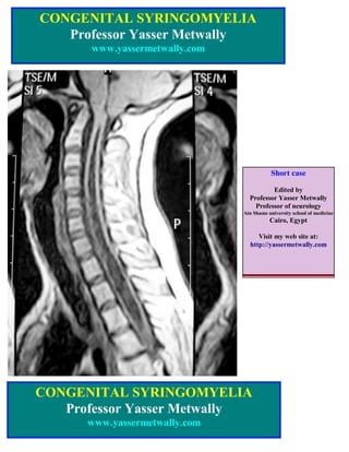
- 2. Figure 1. A case of congenital syringomyelia, MRI T1 images showing a longitudinal multisegmental continuous syringomyelic cavity involving the whole of the cervical spinal cord and extending to the upper dorsal segments with complete absence of transverse band and septations (Type I syringomyelic cavity). Notice herniation of the cerebellar tonsils below the level of the foramen magnum. The intracavitary MRI signal of the syringomyelic slit is identical with that of the CSF signal in the subarachnoid spaces in MRI T1 and T2 images. Figure 2. A case of congenital syringomyelia, MRI T1, T2 images showing a longitudinal multisegmental continuous syringomyelic cavity involving the whole of the cervical spinal cord and extending to the upper dorsal segments with complete absence of transverse band and septations.(Type I syringomyelic cavity). Notice herniation of the cerebellar tonsils below the level of the foramen magnum. The intracavitary MRI signal of the syringomyelic slit is identical with that of the CSF signal in the subarachnoid spaces in MRI T1 and T2 images.
- 3. Figure 3. A case of congenital syringomyelia, MRI T1,T2 images showing a longitudinal multisegmental continuous syringomyelic cavity involving the whole of the cervical spinal cord and extending to the upper dorsal segments with complete absence of transverse band and septations (Type I syringomyelic cavity). Notice herniation of the cerebellar tonsils below the level of the foramen magnum causing marked stenosis at that level. The intracavitary MRI signal of the syringomyelic slit is identical with that of the CSF signal in the subarachnoid spaces in MRI T1 and T2 images. Figure 4. MRI T1,T2 images showing a central syringomyelic cavity representing dilatation of the central canal of the spinal cord, notice the peripheral signal void area (A) that probably represent a CSF flow void sign inside the syringomyelic cavity. The intracavitary MRI signal of the syringomyelic slit is identical with that of the CSF signal in the subarachnoid spaces in MRI T1 and T2 images.
- 4. The case represents a congenital subtype syringomyelia because of the following The presence of Arnold Chiari malformation which represents the aetiopathogenic factor of congenital syringomyelia The central location of the syringomyelic cavity which represents dilation of the central canal of the spinal cord. The syringomyelic cavity is a continuous slit without transverse bands or septations. The involvement of the cervico-dorsal region of the spinal cord which is commonly the site of involvement in congenital syringomyelia. The intracavitary MRI signal of the syringomyelic slit is identical with that of the CSF signal in the subarachnoid spaces in MRI T1 and T2 images. Table 1. Differences between congenital and neoplastic syringomyelia Congenital hydrosyringomyelia Neoplastic syringomyelia The presence of Arnold Chiari malformation Absence of Arnold Chiari malformation which represents the aetiopathogenic factor of congenital syringomyelia The syringomyelic cavity is centrally located Two types of cavities are noted. and represents dilation of the central canal of the spinal cord. (Hydromyelia) 1- A peripheral irregular cavities inside the tumors which represents cystic breakdown of tumor tissue (the cavitations are part of the tumor). 2- Cavitations rostral and caudal to the spinal tumors, these cavitations are not part of the tumors and represents intramedullary cavitations due to CSF flow obstruction. It commonly involves the cervico-dorsal region Any part of the spinal cord can be involved. The syringomyelic cavity is a continuous slit Transverse bands and septations are common. without transverse bands or septations. The intracavitary MRI signal of the The intracavitary MRI signal of the syringomyelic slit is identical with that of the syringomyelic slit is different from that of the CSF signal in the subarachnoid spaces in MRI CSF signal in the subarachnoid spaces in MRI T1 and T2 images. T1 and T2 images. References Metwally, MYM: Textbook of neurimaging, A CD-ROM publication, (Metwally, MYM editor) WEB-CD agency for electronic publishing, version 9.1a January 2008