Short case...brain stem glioma
•
0 gostou•269 visualizações

Shor case...brain stem glioma http://yassermetwally.com http://yassermetwally.net

Recomendados
Recomendados
Mais conteúdo relacionado
Mais procurados
Mais procurados (10)
Destaque
Destaque (20)
Semelhante a Short case...brain stem glioma
Semelhante a Short case...brain stem glioma (20)
Mais de Professor Yasser Metwally
Mais de Professor Yasser Metwally (20)
Último
Último (20)
Short case...brain stem glioma
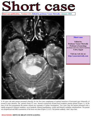
- 1. Short case publication... Version 3.12 | Edited by professor Yasser Metwally | January 2010 Short case Edited by Professor Yasser Metwally Professor of neurology Ain Shams university school of medicine Cairo, Egypt Visit my web site at: http://yassermetwally.com A 50 years old male patient presented clinically for the first time complaining of marked limitation of horizontal gaze bilaterally of around 4 years duration. The patient's brain CT scan showed a posteriorly located focal exophytic pontine glioma (Figure 1). At the time of the first presentation the patient refused any treatment or further investigation. Two years later the patient presented with a rapidly progressive bilateral weakness, sever headache, bilateral papilledema, vertigo and bilateral cerebellar manifestations. The patient was hospitalized and MRI examination of the brain was done (Figure 2,3,4,5). The patient suddenly died 2 days later. DIAGNOSIS: DIFFUSE BRAIN STEM GLIOMA
- 2. Figure 1. Postcontrast CT scan image showing a dorsally exophytic focal pontine glioma. The 4th ventricle is compressed and pushed posteriorly. Notice absence of any significant contrast enhancement. Figure 2. Postcontrast MRI T1 images showing a hypointense nonenhanced posteriorly located brain stem glioma. The tumor is located in the posterior parts of pons, with upward extension to the posterior parts of the midbrain and showed forward extension to the crus cerebri at the midbrain level. The cephalocaudal extension of the tumor is best depicted in the sagittal view in C and is seen as a hypointense broad band in the posterior parts of the brain stem. Mild punctate hypointensities are seen in the basis pontis and probably reflect forward extension of the tumour cells.
- 3. Figure 3. The tumor is seen hyperintense on the MRI T2 images. Notice the punctate hyperintensities that surround the the main bulk of the tumor. Most probably these punctate hyperintensities represent peritumoral satellitosis that occured due to extension of the tumor cells along myelinated and non-myelinated pyramidal tract fibers. Figure 4. The tumor showed upward extension to the crus cerebri and the posterior limbs of the internal capsule, more on the right side. This pattern of spread reflects extension of the tumor cells along myelinated and non-myelinated pyramidal tract fibers.
- 4. Figure 5. MRI T2 (A) mage and postcontrast MRI T1 image (B) at the pontine level. Notice the posteriorly located tumor and the peritumoral satellitosis. References 1. Metwally, MYM: Textbook of neurimaging, A CD-ROM publication, (Metwally, MYM editor) WEB-CD agency for electronic publishing, version 11.1a December 2010 Addendum A new version of short case is uploaded in my web site every week (every Saturday and remains available till Friday.) To download the current version follow the link "http://pdf.yassermetwally.com/short.pdf". You can download the long case version of this short case during the same week from: http://pdf.yassermetwally.com/case.pdf or visit web site: http://pdf.yassermetwally.com To download the software version of the publication (crow.exe) follow the link: http://neurology.yassermetwally.com/crow.zip At the end of each year, all the publications are compiled on a single CD-ROM, please contact the author to know more details. Screen resolution is better set at 1024*768 pixel screen area for optimum display For an archive of the previously reported cases go to www.yassermetwally.net, then under pages in the right panel, scroll down and click on the text entry "downloadable short cases in PDF format" Also to view a list of the previously published case records follow the following link (http://wordpress.com/tag/case- record/) or click on it if it appears as a link in your PDF reader