Case record...Intramedullary cystic spinal cord metastasis
•
2 gostaram•436 visualizações

case record...Intramedullary cystic spinal cord metastasis http://yassermetwally.com http://yassermetwally.net

Recomendados
Recomendados
Mais conteúdo relacionado
Destaque
Destaque (16)
Mais de Professor Yasser Metwally
Mais de Professor Yasser Metwally (20)
Último
Último (20)
Case record...Intramedullary cystic spinal cord metastasis
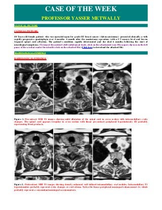
- 1. CLINICAL PICTURE: 48 Years old female patient- who was operated upon for grade III breast cancer (Adenocarcinoma)- presented clinically a with rapidly progressive quadriplegia over 4 months- 6 month after the mastectomy operation- with a C3 sensory level and brown Sequard spinal cord affection. The patient's condition rapidly deteriorated and she died 6 months following the start of neurological symptoms. (To inspect the patient's full radiological study, click on the attachment icon (The paper clip icon in the left pane) of the acrobat reader then double click on the attached file) (Click here to download the attached file) RADIOLOGICAL FINDINGS: Figure 1. Precontrast MRI T1 images showing mild dilatation of the spinal cord in cross section with intramedullary cystic changes. The spinal cord appears irregular in cross section with linear precontrast peripheral hyperintensity (D) probably representing blood products. Figure 2. Postcontrast MRI T1 images showing densely enhanced well defined intramedullary oval nodules. Intramedullary T1 hypointensities probably represent cystic changes or cord edema. Notice the linear peripheral meningeal enhancement (A) which probably represent a concomitant meningeal carcinomatosis. CASE OF THE WEEK PROFESSOR YASSER METWALLY CLINICAL PICTURE RADIOLOGICAL FINDINGS
- 2. Figure 3. Precontrast MRI T1 image (A) and postcontrast MRI T1 images (B,C) showing irregular intramedullary patchy enhancement with central MRI T1 hypointensities which probably represent cord edema or cystic changes. The spinal cord is moderately enlarged from C2-D2. Notice the linear anterior peripheral meningeal enhancement which is -most probably- due to concomitant meningeal carcinomatosis. DIAGNOSIS: INTRAMEDULLARY CYSTIC CERVICAL SPINAL CORD METASTASIS DISCUSSION: Metastasis to the intramedullary spinal cord rarely occurs with systemic cancer. Autopsy series have reported its prevalence in cancer patients as ranging from 0.9 to 2.1% (3,6), whereas clinically significant intramedullary spinal cord metastasis have been reported in 0.1 to 0.4% of cancer patients (5). Lung cancer is the most common source of intramedullary spinal cord metastasis, comprising approximately 50% of reported cases (5,10,13,14,18). Breast carcinoma is another less frequently seen source, accounting for 8 to 14% of intramedullary spinal cord metastasis. Other primary sources for intramedullary spinal cord metastasis include renal cell carcinoma, melanoma, colorectal carcinoma, and lymphoma (5,13,14). Figure 4. Intramedullary cystic metastasis: Notice the asymmetric bulging of the spinal cord with multiple nodular enhancing lesions. DIAGNOSIS: DISCUSSION
- 3. Figure 5. Intramedullary spinal cord metastasis Clinical presentation with rapid progression of symptoms is highly suggestive of a metastatic lesion (2,15). The most common symptom associated with intramedullary spinal cord metastasis is weakness (5,14). In their series of 40 patients with Intramedullary spinal cord metastasis, Schiff and O’Neill (13) reported that 92.5% of patients had motor deficits at the time of diagnosis. In addition, 45% of patients had either a true Brown-Séquard syndrome or a pseudo-Brown-Séquard syndrome. Patients with intramedullary spinal cord metastasis often present with a true Brown-Séquard syndrome, a finding that often portends an intramedullary spinal cord metastasis in cancer patients (11,13). In addition to intramedullary spinal cord metastasis, patients might also have evidence of brain metastases, an occurrence that Schiff and O’Neill (13) observed in 57.5% of patients with intramedullary spinal cord metastasis. Other series have reported even higher rates of brain metastases (10). Although the vast majority of patients with intramedullary spinal cord metastasis often have other locations of systemic spread, only 8 to 11% of patients have been found to have multiple intramedullary lesions (13,14). Neuroimaging of intramedullary metastasis Figure 6. Metastatic involvement of the spine. Intramedullary metastases reach the cord through hematogenous dissemination and grow within the cord parenchyma (1). Leptomeningeal metastases involve the meningeal membranes of the subarachnoid space, which are extramedullary and intradural (2). Epidural metastases usually arise from the highly vascular posterior aspect of the vertebral body and produce compression of the anterior aspect of the spinal cord (3). Epidural compression can also result from paravertebral tumors that invade the vertebral foramina (4) and, less often, from metastases arising in the epidural space itself (5).
- 4. Intramedullary metastasis, in general, produce segmental enlargement of the spinal cord and are often 3-4 spinal segments in length. However, and and unlike primary spinal cord neoplasms, intramedullary spinal cord metastasis produce asymmetric nodular bulging of the spinal cord in cross-section. Intramedullary multiple enhancing nodules might be demonstrated in cross section. Intramedullary metastasis are often cystic and might show hemorrhagic components. Management of Intramedullary spinal cord metastasis As no prospective trials on treatment have been performed, treatment of intramedullary spinal cord metastasis has primarily been based on anecdotal experience. External beam radiation therapy is the most effective treatment option, but its effectiveness depends primarily on the radiosensitivity of the tumor, the duration of symptoms, and the degree of preoperative neurological deficit (17). To avoid the sequelae of external beam radiation therapy, stereotactic radiosurgery can be used as an alternative to treat individual metastatic lesions. However, the current literature only reports effectiveness of this treatment modality with primary vascular tumors (19). Surgical management has led to improved neurological function in patients with rapidly progressive neurological deficits (9). However, as aggressive metastatic lesions typically have unidentifiable margins, the goal of surgery must be to decompress the spinal cord while, at the same time, maintaining residual neurological function (4). Focal radiation can then be used as an adjunct to treat residual disease within the resection cavity (13). Conclusion Intramedullary spinal cord metastasis are rare complications of systemic cancer. Patients with Intramedullary spinal cord metastasis experience the rapid onset of a Brown-Séquard syndrome in the setting of recently treated cancer. A clinical presentation such as this should be considered highly suggestive of an intramedullary spinal cord metastasis. SUMMARY Spinal cord tumors may be either primary or metastatic, and are further categorized by location: extradural, intradural- extramedullary or intramedullary. Intramedullary spinal cord lesions comprise only 1-5% of spinal cord tumors (1, 2) . The majority are primary gliomas, while intramedullary spinal cord metastases (ISCM) account for only 1-3% of all intramedullary neoplasms (1,2,3,4,5). ISCM are associated primarily with lung carcinoma (50%), especially small cell type, breast carcinoma (13%), melanoma (9%), lymphoma (5%) and renal cell cancer (4%) and are usually diagnosed well into the course of primary disease (1,2,4) . As many as 2% of patients in the end stages of disseminated cancer are found to have ISCM. This number is expected to grow with the increasing availability of improved diagnostic imaging, predominantly MRI, which has become the gold standard for diagnosis and evaluation of these rare tumors. A biopsy is often required for definitive diagnosis of the lesion as metastasis versus primary tumor and can help tailor therapeutic approach (5) . Presenting symptoms of ISCM usually consist of weakness, followed in frequency by sensory loss, pain and bowel and bladder disturbance. Weakness and pain present early, as compared to sensory loss, with sphincter disturbance having the latest onset. ISCM are therefore difficult to distinguish clinically from extradural tumors, which often present with a similar constellation of symptoms. Unlike extradural lesions, and exemplified by this case report, intramedullary lesions may first present with asymmetric or unilateral symptoms 6 . ISCM can be distinguished clinically from primary tumors by the rapidity of onset of symptoms. A cause of the sudden onset of motor symptoms may be a vascular event in the tumor bed, such as a venous infarct. Neurological deficits tend to be irreversible, especially if severe motor weakness and bladder/bowel involvement are noted at the time of first presentation. Prognosis of these metastases is guarded, compounded by both treatment modality and tumor type, with lung and breast metastases correlating with the shortest survival (4) . Treatment of choice for ISCM consists of immediate steroid bolus, radiation and chemotherapy with subtotal cytoreductive surgery considered on a case by case basis. High dose dexamethasone may allow for limited and transient neurological improvement, while radiotherapy often results in stasis of the deficit without improvement. However, in combination with chemotherapy, radiation correlates with an increased length of survival. In one review of 177 cases, steroids gave approximately 5 weeks of survival, compared to radiation and radiation with chemotherapy allowing for approximately 15 and 29 weeks of survival, respectively (21) . Cytoreductive surgery is another treatment option for lesions early in the course of neurological deficits, and is associated with a 75% increase in time of “high quality of life” survival (3) . It has not been found to increase the length of survival as compared to other treatment modalities 4 . Our patient presented late in her course, with a high lesion in the cervical cord. Her surgery carried the specific risks of increasing cord edema, resulting in the need for long-term mechanical ventilation and worsening neurological deficits. In the face of these risks and the poor temporal prognosis associated with her breast cancer, it was felt by us that the risks of such surgery were too great. It is likely that the surgical risks would have been minimized had the patient presented with a smaller lesion prior to the precipitous event that caused her such sudden quadriparesis. Such early presentation was precluded in our case due to the patient's perceived exclusion from the healthcare system because of her immigrant status and her lack of insurance. SUMMARY
- 5. Addendum A new version of this PDF file (with a new case) is uploaded in my web site every week (every Saturday and remains available till Friday.) To download the current version follow the link "http://pdf.yassermetwally.com/case.pdf". You can also download the current version from my web site at "http://yassermetwally.com". To download the software version of the publication (crow.exe) follow the link: http://neurology.yassermetwally.com/crow.zip The case is also presented as a short case in PDF format, to download the short case follow the link: http://pdf.yassermetwally.com/short.pdf At the end of each year, all the publications are compiled on a single CD-ROM, please contact the author to know more details. Screen resolution is better set at 1024*768 pixel screen area for optimum display. Also to view a list of the previously published case records follow the following link (http://wordpress.com/tag/case-record/) or click on it if it appears as a link in your PDF reader To inspect the patient's full radiological study, click on the attachment icon (The paper clip icon in the left pane) of the acrobat reader then double click on the attached file. Click here to download the short case version of this case record in PDF format References 1. Anderson TS, Regine WF, Kryscio R, Patchell RA: Neurologic complications of bladder carcinoma: A review of 359 cases. Cancer 97:2267-2272, 2003. 2. Aryan HE, Farin A, Nakaji P, Imbesi SG, Abshire BB: Intramedullary spinal cord metastasis of lung adenocarcinoma presenting as Brown-Sequard syndrome. Surg Neurol 61:72-76, 2004. 3. Chason JL, Walker FB, Landers JW: Metastatic carcinoma in the central nervous system and dorsal root ganglia. A prospective autopsy study. Cancer 16:781-787, 1963. 4. Conill C, Sanchez M, Puig S, Planas I, Castel T: Intramedullary spinal cord metastases of melanoma. Melanoma Res 14:431-433, 2004. 5. Connolly ES Jr, Winfree CJ, McCormick PC, Cruz M, Stein BM: Intramedullary spinal cord metastasis: Reports of three cases and review of the literature. Surg Neurol 46:329-338, 1996. 6. Costigan DA, Winkelman MD: Intramedullary spinal cord metastasis. A clinicopathological study of 13 cases. J Neurosurg 62:227-233, 1985. 7. Edelson RN, Deck MD, Posner JB: Intramedullary spinal cord metastases. Clinical and radiographic findings in nine cases. Neurology 22:1222-1231, 1972. 8. Grem JL, Burgess J, Trump DL: Clinical features and natural history of intramedullary spinal cord metastasis. Cancer 56:2305- 2314, 1985. 9. Kalayci M, Cagavi F, Gul S, Yenidünya S, Açikgöz B: Intramedullary spinal cord metastases: Diagnosis and treatment-An illustrated review. Acta Neurochir (Wien) 146:1347-1354, 2004. 10. Lee SS, Kim MK, Sym SJ, Kim SW, Kim WK, Kim SB, Ahn JH: Intramedullary spinal cord metastases: A single-institution experience. J Neurooncol 84:85-89, 2007. 11. Mut M, Schiff D, Shaffrey ME: Metastasis to nervous system: Spinal epidural and intramedullary metastases. J Neurooncol 75:43-56, 2005. 12. Rades D, Walz J, Schild SE, Veninga T, Dunst J: Do bladder cancer patients with metastatic spinal cord compression benefit from radiotherapy alone? Urology 69:1081-1085, 2007. 13. Schiff D, O’Neill BP: Intramedullary spinal cord metastases: Clinical features and treatment outcome. Neurology 47:906-912, 1996. 14. Schwechheimer K, Lemminger JM: Intramedullary metastases: Report of 4 cases and review of the literature. Clin Neuropathol 4:28-37, 1985. 15. Sutter B, Arthur A, Laurent J, Chadduck J, Friehs G, Clarici G, Pendl G: Treatment options and time course for intramedullary spinal cord metastasis. Report of three cases and review of the literature. Neurosurg Focus 4:e3, 1998. REFERENCES
- 6. 16. Tognetti F, Lanzino G, Calbucci F: Metastases of the spinal cord from remote neoplasms. Study of five cases. Surg Neurol 30:220-227, 1988. 17. Villegas AE, Guthrie TH: Intramedullary spinal cord metastasis in breast cancer: Clinical features, diagnosis, and therapeutic consideration. Breast J 10:532-535, 2004. 18. Watanabe M, Nomura T, Toh E, Sato M, Mochida J: Intramedullary spinal cord metastasis: A clinical and imaging study of seven patients. J Spinal Disord Tech 19:43-47, 2006. 19. Winkelman MD, Adelstein DJ, Karlins NL: Intramedullary spinal cord metastasis. Diagnostic and therapeutic considerations. Arch Neurol 44:526-531, 1987. 20. Metwally, MYM: Textbook of neuroimaging, A CD-ROM publication, (Metwally, MYM editor) WEB-CD agency for electronic publication, version 11.4a October 2010 21. Connolly ES, Winfree CJ, McCormick PC, Cruz M, Stein BM. Intramedullary Spinal Cord Metastasis: Report of Three Cases and Review of the Literature. Surgical Neurology 1996; 46(4): 329-337 (s)