SlideShare uma empresa Scribd logo
1 de 7
Baixar para ler offline
THE NEW ZEALAND
MEDICAL JOURNAL
Journal of the New Zealand Medical Association
NZMJ 3 April 2009, Vol 122 No 1292; ISSN 1175 8716 Page 9
URL: http://www.nzma.org.nz/journal/122-1292/3536/ ©NZMA
Are our medical graduates in New Zealand safe and
accurate in ECG interpretation?
Nigel A Lever, Peter D Larsen, Mathew Dawes, Annie Wong, Scott A Harding
Abstract
Aim We aimed to assess the skills of final year medical students and resident medical
officers in recognising and interpreting important common or life-threatening
abnormalities in the electrocardiogram (ECG).
Methods 102 participants at two study sites (52 of whom were final year medical
students) attempted to determine the heart rate and rhythm and identify and interpret
any abnormalities present in 15 ECGs in a 30-minute time period.
Results Accurate determination of heart rate was poor, ranging from 0% to 89%
correct across the 15 ECGs. Normal sinus rhythm in 8 ECGs was identified 81% to
95% of the time, and ventricular tachycardia was identified by 98% of participants.
Atrial fibrillation (55%), second degree heart block (19%) and ventricular pacing
(9%) were not well identified. Four ECGs showed acute ischaemic ST segment
changes, and these were correctly identified in 87% to 93% of cases, although
interpretation of these abnormalities was less accurate. Long QT interval (7%) and
pre-excitation (WPW pattern, 11%) were not well recognised. Nearly half of the
participants rated their ability to interpret ECGs as less than satisfactory while just
over half rated the ECG teaching they had received as less than satisfactory.
Conclusions Overall study participants did not achieve what we would consider an
adequate standard in recognising and interpreting important common or life-
threatening abnormalities in the ECG. To address this we need to define minimum
standards in ECG interpretation, to improve our teaching to meet these standards, and
to assess our graduates against these.
A century after its introduction into clinical practice, the electrocardiogram (ECG)
remains one of the most commonly used tests for the assessment of cardiac disease.
ECG interpretation is therefore a core skill and medical practitioners are expected to
be proficient at reading ECGs. This is particularly important when dealing with
acutely ill patients where failure to recognise an abnormality may have life-
threatening consequences.1,2
There is increasing concern that ECG teaching and the
ability of medical professionals to interpret ECGs are declining.3
Teaching ECG interpretation is complex, and the best methods for ECG teaching to
recognise and interpret abnormalities remains unclear.4–6
The use of pattern
recognition versus understanding the principles of vector analysis has occupied
commentary, as has the role of computer-aided learning. Whilst there are guidelines
on the competency requirements for practitioners who are in training programmes for
various disciplines (cardiology, emergency medicine7
), guidelines are lacking for
teaching and assessment of medical graduates.
NZMJ 3 April 2009, Vol 122 No 1292; ISSN 1175 8716 Page 10
URL: http://www.nzma.org.nz/journal/122-1292/3536/ ©NZMA
As part of our role in providing undergraduate ECG teaching, we undertook this study
to assess the skills of final year medical students and house officers in recognising and
interpreting important common or life-threatening abnormalities. We were also
interested in how this group rated their own ability to interpret ECGs and the teaching
they had received.
Methods
Final year medical students from University of Auckland and University of Otago, Wellington as well
as first and second year resident medical officers (RMOs) from Auckland and Wellington Hospitals
were invited to participate in the study between March and June 2007 during their regular teaching
programmes. The study was approved by the local institutional ethics committees. Respondents
consented to participate in an anonymised fashion.
Participants were given 15 ECGs (Table 1; Appendix 1—view ECGs at www.nzma.org.nz/journal/122-
1292/3536/appendix.pdf) to interpret over a 30-minute time period. For each ECG they were asked to
determine the rate and rhythm, and to identify any abnormalities present. In the presence of
abnormalities, they were asked to interpret what the abnormality meant. Participants were instructed to
be as specific as possible when describing the rhythm and abnormalities. Following the 30-minute time
period they were then asked to complete a short survey providing demographic details, and answering
questions related to their ECG teaching.
The ECGs used were selected from a pool of departmental teaching ECGs and were considered
representative of important ECG abnormalities. The selection was reviewed with general physicians,
experienced medical educators and cardiologists. The group defined a marking schedule that was
applied to each participant’s answers. For heart rate, a range of ±5 beats per minute for regular rates
below 100, ±10 beats per minute for regular rates between 100 and 150 beats per minute, and ±15 beats
per minute for heart rates greater than 150 beats per minute was allowed. For irregular heart rates the
above ranges were applied to the fastest and slowest heart rates on the ECG rhythm strip.
When assessing the interpretation of rhythm and abnormalities present, only complete answers were
considered correct (for example, atrial fibrillation was considered correct where as irregularly irregular
rhythm was not). Responses for each participant were assessed against the marking schedule by two
physicians.
Statistical analysis—For each participant we calculated the percentage of correct answers and
examined the relationship between this score and self-reported level of confidence using Pearson’s
correlation. We also compared the scores of the final year medical students with those of the house
surgeons, and the scores between the two study sites (Wellington and Auckland) using Mann Whitney
U Test. A p value <0.05 was considered statistically significant. Statistical analysis was performed
using SPSS v11.0 software (SPSS, Chicago, IL).
Results
All 102 subjects approached participated in the study. Of those surveyed, 49 were
male, 52 were final year medical students, and 50 were house officers. The results of
the interpretation of the study ECGs are presented in Table 1. All subjects completed
the first 8 ECGs, but only 73% were able to complete all 15 ECGs in the 30 minutes
allocated.
The ability to determine heart rate was highly variable, with responses ranging from
0% to 89% correct. Where heart rate was inaccurately determined, the tendency was
for it to be underestimated by 10–20 beats per minute. For example, in ECG 4, we
accepted answers between 85 and 95 beats per minute as correct, and the median
answer given was 75 beats per minute; and for ECG 8, we accepted answers in the
range 60–70 beats per minute, and the median answer given 50 beats per minute.
NZMJ 3 April 2009, Vol 122 No 1292; ISSN 1175 8716 Page 11
URL: http://www.nzma.org.nz/journal/122-1292/3536/ ©NZMA
Heart rhythm accuracy ranged from 9% for ventricular pacing to 98% for ventricular
tachycardia. The eight ECGs showing sinus rhythm were identified with accuracy
ranging from 81% to 95%. Atrial fibrillation (55%) and atrio-ventricular conduction
disease (second degree heart block type 1, 19%) were poorly recognised.
Table 1. Description of each ECG, percentage of responders answering each
question, and the percentage of those correctly determining heart rate, rhythm,
abnormality, and interpreting abnormalities other than rhythm correctly if
present
ECG ECG Description Responders
(%)
Rate
(%)
Rhythm
(%)
Abnormality
(%)
Accurate
Interpretation (%)
1 SR, CHB with acute inferior
STEMI
100 85 72 87 54
2 SR with trifascicular block 100 48 90 22 3
3 Ventricular tachycardia 100 89 98 – –
4 SR with LBBB 100 0 87 44 44
5 Atrial flutter 100 79 59 – –
6 SR with acute antero-lateral
STEMI
100 10 93 93 46
7 Ventricular pacing 100 79 9 – –
8 SR with pre-excitation (Wolff-
Parkinson-White pattern)
100 3 95 11 11
9 SR with acute inferior STEMI 92 42 90 93 61
10 SR, 2nd
degree type 1AV block 90 73 19 – –
11 Supraventricular tachycardia 86 5 79 – –
12 SR with Long QT interval and
repolarisation abnormalities
80 11 89 7 7
13 Sinus Rhythm, normal ECG 77 18 81 – –
14 Atrial fibrillation with old
anterior Q wave MI
78 60 55 61 61
15 SR with acute ischaemic
antero-lateral ST depression
73 35 86 92 66
CHB = complete heart block, LBBB = left bundle branch block, MI = myocardial infarction,
SR = sinus rhythm, STEMI = ST elevation myocardial infarction.
There were 4 ECGs showing acute ischaemic ST segment changes (3 with ST
elevation and 1 with ST depression). These changes were identified correctly by
between 87% and 93% of those answering. The interpretation of the ST segment
changes was considerably less accurate, with correct answers in between 46% and
66% of cases. Conduction defects were poorly identified, although a more lenient
marking schedule that accepted any identification of a conduction abnormality would
have seen accuracy increase to approximately 80%.
Prolongation of the QT interval and pre-excitation (WPW pattern) were signs that
were not well detected despite the fact that the examples selected had marked
abnormalities. Also of note is that 34% of those who interpreted the normal ECG
(number 13) reported that an abnormality was present.
The majority (76%) of participants had reported that they had recorded less than 5
ECGs in the previous year. More than half (54%) had examined fewer than 50 ECGs
NZMJ 3 April 2009, Vol 122 No 1292; ISSN 1175 8716 Page 12
URL: http://www.nzma.org.nz/journal/122-1292/3536/ ©NZMA
with 24% reporting that they had interpreted fewer than 10 ECGs in the previous year.
When asked how many ECGs they had discussed with a registrar or consultant, 87%
reported that they had discussed fewer than 50 with 38% reporting that they had
discussed fewer than 10 ECGs in the previous year.
Participant’s ratings of confidence in their ability to interpret ECGs, and of the quality
of their teaching are given in Figure 1. Participants reported that they had received a
mean of 6.3 hours of formal teaching on ECGs during their medical school training.
Figure 1. Participants rating of their own abilities to interpret ECGs (A) and the
quality of the teaching they had received (B)
A. Rating of own abilities in ECG interpretation
0
5
10
15
20
25
30
35
40
45
50
poor less than
satisfactory
satisfactory good excellent
Percentage
B. Rating of quality of ECG teaching
0
5
10
15
20
25
30
35
40
45
50
poor less than
satisfactory
satisfactory good excellent
Percentage
NZMJ 3 April 2009, Vol 122 No 1292; ISSN 1175 8716 Page 13
URL: http://www.nzma.org.nz/journal/122-1292/3536/ ©NZMA
There was a positive correlation between levels of confidence and overall mark
(r=0.242, p=0.03). Final year medical students (median score 52%, range 20–65%)
scored significantly higher than house officers (40%, 18–61%, p=0.001). There was
no difference in score between the two centres in the study (Wellington median score
50%, range 18-65%, Auckland median score 48%, range 21–61%, p=0.67).
Discussion
The major finding of our study is that there are significant deficiencies in the ability
of final year medical students and resident medical officer to interpret common and
important ECG changes. However, the majority of participants were able to recognise
the most important rhythm disturbance, ventricular tachycardia, accurately as well as
identify ischaemic ST segment changes. Furthermore, nearly half of the respondents
rated their ability to interpret ECGs as less than satisfactory while just over half rated
the ECG teaching they had received as less than satisfactory.
Currently there are no clearly defined standards in ECG interpretation for medical
graduates for us to compare the results of this study against. A New York study of
residents in emergency medicine and internal medicine reported 93% accuracy in
identifying VT8
while an Australian study of emergency medicine trainees reported
40% accuracy in identifying VT.9
In this context, the 98% accuracy seen in this New
Zealand study is very good.
With respect to identifying signs of myocardial infarction, previous studies have
reported levels of accuracies of around 90% in registrars8,9
and a retrospective audit of
1684 patients with acute MI in 5 emergency departments in the United States reported
an overall accuracy of 88%.1
The level of accuracy for our medical graduates in
recognising ST segment changes (87–93%) is probably reasonable in comparison.
Clearly, failure to recognise ST segment changes could potentially lead to
inappropriate patient management. Similarly, there are concerns that simple
recognition of ST segment changes without any understanding of what these changes
mean may also lead to inappropriate management decisions being made.10
Given this,
we would have hoped to see considerably better performance in the interpretation of
ST segment changes in this study.
The recognition of pre-excitation and long QT syndrome we observed was poor, but
in Hoyle et al’s study of emergency trainees9
these were identified in only 24% and
43% of cases respectively so it is perhaps not surprising that more junior doctors and
medical students struggle with these ECGs. Again, failure to recognise these
abnormalities has the potential to result in inappropriate patient management.
It is difficult to account for the systematic inaccuracy in calculation of heart rate
observed in the present study. Perhaps heart rate is regarded as clinically unimportant,
and rounding down is viewed by the study participants as a reasonable approximation.
While it is difficult to argue that errors of the magnitude we observed in the current
study are likely to lead to errors in patient management it is none the less
disappointing that a fairly basic skill was not performed to a higher standard.
NZMJ 3 April 2009, Vol 122 No 1292; ISSN 1175 8716 Page 14
URL: http://www.nzma.org.nz/journal/122-1292/3536/ ©NZMA
The time pressure and the lack of a clinical scenario may have detrimentally
influenced the overall level of accuracy. There is good evidence that accuracy is
improved when clinical vignettes are provided in addition to the ECG7
—although
conversely there is evidence that where ECG changes occur in the context of atypical
symptoms, inaccuracy increases.1
Previous studies do not appear to have limited time in any way.6,8,9
We chose to do so
on the basis that 2 minutes per ECG was considered a reasonable time period given
that in clinical practice there are time constraints.
Our medical graduates expressed a lack of confidence in their abilities in ECG
interpretation, and this was related to their accuracy levels. Previous studies of New
Zealand graduates in other areas have identified knowledge gaps and low levels of
confidence similar to those we report.11,12
Perhaps of slightly greater concern to those of us involved in the provision of ECG
teaching is that this was rated as less than satisfactory or poor by more than half the
study participants. It is possible that we have tended to assume that students interpret
and discuss a considerable volume of ECGs in the course of various clinical rotations.
However, our results suggest than more than half the study participants looked at less
than one ECG per week in a clinical context.
The fact that medical students performed better than the residents again suggests that
skills in ECG interpretation gained in medical school are not been built upon by
normal clinical duties or post graduate teaching. The teaching implications are that
more has to be done to assist the students to consolidate the teaching that they get, and
to provide additional teaching opportunities.
While some aspects of performance in the current study were encouraging, overall our
final year medical students and house officers do not achieve what we would consider
an adequate standard in recognising and interpreting important common or life-
threatening abnormalities in the ECG. Consistent with this, the majority of
participants rated both their own ability and the teaching they had received as less
than satisfactory.
To address this problem we need to define minimum standards in ECG interpretation,
improve our teaching to meet these standards, and assess our graduates against them.
Competing interests: None known.
Author information: Nigel A Lever, Senior Lecturer, Department of Medicine,
University of Auckland and Green Lane Cardiovascular Service, Auckland City
Hospital, Auckland; Peter D Larsen, Senior Lecturer, Department of Surgery and
Anaesthesia, University of Otago, Wellington; Mathew Dawes, Senior Lecturer,
Department of Medicine, University of Auckland, Auckland; Annie Wong, House
Surgeon, Wellington Hospital, Wellington; Scott A Harding, Cardiologist, Wellington
Hospital, Wellington
Correspondence: N A Lever, Green Lane Cardiovascular Service, Auckland City
Hospital, Private Bag 92024, Auckland 1030, New Zealand. Fax: +64 (0)9 3078941;
email: nlever@adhb.govt.nz
NZMJ 3 April 2009, Vol 122 No 1292; ISSN 1175 8716 Page 15
URL: http://www.nzma.org.nz/journal/122-1292/3536/ ©NZMA
References:
1. Masoudi FA, Magid DJ, Vinson DR, et al. Implications of the failure to identify high-risk
electrocardiogram findings for the quality of care of patients with acute myocardial infarction:
results of the Emergency Department Quality in Myocardial Infarction (EDQMI) study.
Circulation. 2006;114:1565–71.
2. Todd KH, Hoffman JR, Morgan MT. Effect of cardiologist ECG review on emergency
department practice. Annals of emergency medicine. 1996;27:16–21.
3. Hurst JW. The interpretation of electrocardiograms: pretense or a well-developed skill?
Cardiology Clinics. 2006;24:305-7, vii.
4. Childers R. Teaching electrocardiogram interpretation. Journal of Electrocardiology.
2006;39:426–9.
5. Hurst JW. Thoughts about the abnormalities in the electrocardiogram of patients with acute
myocardial infarction with emphasis on a more accurate method of interpreting ST-segment
displacement: part I. Clinical Cardiology. 2007;30:381–90.
6. Pines JM, Perina DG, Brady WJ. Electrocardiogram interpretation training and competency
assessment in emergency medicine residency programs. Acad Emerg Med. 2004;11:982–4.
7. Salerno SM, Alguire PC, Waxman HS. Competency in interpretation of 12-lead
electrocardiograms: a summary and appraisal of published evidence. Annals of Internal
Medicine. 2003;138:751–60.
8. Berger JS, Eisen L, Nozad V, et al. Competency in electrocardiogram interpretation among
internal medicine and emergency medicine residents. The American Journal of Medicine.
2005;118:873–80.
9. Hoyle RJ, Walker KJ, Thomson G, Bailey M. Accuracy of electrocardiogram interpretation
improves with emergency medicine training. Emerg Med Australas. 2007;19:143–50.
10. Hurst JW. To simply identify "up or down" displacement of the ST segment in the
electrocardiogram can lead to serious errors. Journal of Electrocardiology. 2008 Sep-
Oct;41(5):436–7. Epub 2008 Mar 19.
11. Price CS, Bell SF, Janes SE, Ardagh M. Cardio-pulmonary resuscitation training, knowledge
and attitudes of newly-qualified doctors in New Zealand in 2003. Resuscitation. 2006;68:295–
9.
12. Subramaniam R, Hall T, Chou T, Sheehan D. Radiology knowledge in new medical graduates
in New Zealand. N Z Med J. 2005; 118(1224). http://www.nzmj.com/journal/118-1224/1699

Mais conteúdo relacionado

Mais procurados

Cathet Cardio Intervent - 2019 - Baran - SCAI clinical expert consensus state...
Cathet Cardio Intervent - 2019 - Baran - SCAI clinical expert consensus state...Cathet Cardio Intervent - 2019 - Baran - SCAI clinical expert consensus state...
Cathet Cardio Intervent - 2019 - Baran - SCAI clinical expert consensus state...ssuser36abb7
 
Elevated Tissue Doppler E/E' on Index Admission Can Help Identify Patients at...
Elevated Tissue Doppler E/E' on Index Admission Can Help Identify Patients at...Elevated Tissue Doppler E/E' on Index Admission Can Help Identify Patients at...
Elevated Tissue Doppler E/E' on Index Admission Can Help Identify Patients at...crimsonpublishersOJCHD
 
Murley Meghan Thesis Female Heart Rate Variability - A Pilot Reliability Study
Murley Meghan Thesis Female Heart Rate Variability - A Pilot Reliability StudyMurley Meghan Thesis Female Heart Rate Variability - A Pilot Reliability Study
Murley Meghan Thesis Female Heart Rate Variability - A Pilot Reliability StudyMeghan Murley
 
Cpet in cr in lvad saudi prevent 2019
Cpet in cr in lvad saudi prevent 2019Cpet in cr in lvad saudi prevent 2019
Cpet in cr in lvad saudi prevent 2019asadsoomro1960
 
Pulse Wave Velocity: Theory, Applications, Methods, and Future Directions
Pulse Wave Velocity: Theory, Applications, Methods, and Future DirectionsPulse Wave Velocity: Theory, Applications, Methods, and Future Directions
Pulse Wave Velocity: Theory, Applications, Methods, and Future DirectionsInsideScientific
 
Cerebral oximetry as a real time monitoring tool to assess quality
Cerebral oximetry as a real time monitoring tool to assess qualityCerebral oximetry as a real time monitoring tool to assess quality
Cerebral oximetry as a real time monitoring tool to assess qualityEmeka Onuorah
 
missed myocardial infarction among patients discharged with chest pain in the...
missed myocardial infarction among patients discharged with chest pain in the...missed myocardial infarction among patients discharged with chest pain in the...
missed myocardial infarction among patients discharged with chest pain in the...guest75efe2
 
Terapia de resincronizacion cardiaca
Terapia de resincronizacion cardiacaTerapia de resincronizacion cardiaca
Terapia de resincronizacion cardiacaResidentes1hun
 
216904432 pubmed-prevalencia-dysarthria
216904432 pubmed-prevalencia-dysarthria216904432 pubmed-prevalencia-dysarthria
216904432 pubmed-prevalencia-dysarthriahomeworkping9
 
Tenecteplase before mechanical thrombectomy journal copy
Tenecteplase before mechanical thrombectomy journal   copyTenecteplase before mechanical thrombectomy journal   copy
Tenecteplase before mechanical thrombectomy journal copypradeep3188
 
Reduce the hospitalization
Reduce the hospitalizationReduce the hospitalization
Reduce the hospitalizationAnna Wu
 
Recent evidence for mechanical thrombolysis
Recent evidence for mechanical thrombolysisRecent evidence for mechanical thrombolysis
Recent evidence for mechanical thrombolysisDr Vipul Gupta
 
Experience of Vascular Interventional Procedures of Adana Numune Research and...
Experience of Vascular Interventional Procedures of Adana Numune Research and...Experience of Vascular Interventional Procedures of Adana Numune Research and...
Experience of Vascular Interventional Procedures of Adana Numune Research and...ijtsrd
 
00000542 200710000-00007
00000542 200710000-0000700000542 200710000-00007
00000542 200710000-00007Juan Siri
 
Comparison of clinical, radiological and outcome characteristics of ischemic ...
Comparison of clinical, radiological and outcome characteristics of ischemic ...Comparison of clinical, radiological and outcome characteristics of ischemic ...
Comparison of clinical, radiological and outcome characteristics of ischemic ...MIMS Hospital
 

Mais procurados (20)

Cathet Cardio Intervent - 2019 - Baran - SCAI clinical expert consensus state...
Cathet Cardio Intervent - 2019 - Baran - SCAI clinical expert consensus state...Cathet Cardio Intervent - 2019 - Baran - SCAI clinical expert consensus state...
Cathet Cardio Intervent - 2019 - Baran - SCAI clinical expert consensus state...
 
Elevated Tissue Doppler E/E' on Index Admission Can Help Identify Patients at...
Elevated Tissue Doppler E/E' on Index Admission Can Help Identify Patients at...Elevated Tissue Doppler E/E' on Index Admission Can Help Identify Patients at...
Elevated Tissue Doppler E/E' on Index Admission Can Help Identify Patients at...
 
Factors determining outcomes in grown up patients operated
Factors determining outcomes in grown up patients operatedFactors determining outcomes in grown up patients operated
Factors determining outcomes in grown up patients operated
 
1428931228
14289312281428931228
1428931228
 
Murley Meghan Thesis Female Heart Rate Variability - A Pilot Reliability Study
Murley Meghan Thesis Female Heart Rate Variability - A Pilot Reliability StudyMurley Meghan Thesis Female Heart Rate Variability - A Pilot Reliability Study
Murley Meghan Thesis Female Heart Rate Variability - A Pilot Reliability Study
 
Cpet in cr in lvad saudi prevent 2019
Cpet in cr in lvad saudi prevent 2019Cpet in cr in lvad saudi prevent 2019
Cpet in cr in lvad saudi prevent 2019
 
Pulse Wave Velocity: Theory, Applications, Methods, and Future Directions
Pulse Wave Velocity: Theory, Applications, Methods, and Future DirectionsPulse Wave Velocity: Theory, Applications, Methods, and Future Directions
Pulse Wave Velocity: Theory, Applications, Methods, and Future Directions
 
Cerebral oximetry as a real time monitoring tool to assess quality
Cerebral oximetry as a real time monitoring tool to assess qualityCerebral oximetry as a real time monitoring tool to assess quality
Cerebral oximetry as a real time monitoring tool to assess quality
 
missed myocardial infarction among patients discharged with chest pain in the...
missed myocardial infarction among patients discharged with chest pain in the...missed myocardial infarction among patients discharged with chest pain in the...
missed myocardial infarction among patients discharged with chest pain in the...
 
International Journal of Cardiovascular Diseases & Diagnosis
International Journal of Cardiovascular Diseases & DiagnosisInternational Journal of Cardiovascular Diseases & Diagnosis
International Journal of Cardiovascular Diseases & Diagnosis
 
Terapia de resincronizacion cardiaca
Terapia de resincronizacion cardiacaTerapia de resincronizacion cardiaca
Terapia de resincronizacion cardiaca
 
216904432 pubmed-prevalencia-dysarthria
216904432 pubmed-prevalencia-dysarthria216904432 pubmed-prevalencia-dysarthria
216904432 pubmed-prevalencia-dysarthria
 
Tenecteplase before mechanical thrombectomy journal copy
Tenecteplase before mechanical thrombectomy journal   copyTenecteplase before mechanical thrombectomy journal   copy
Tenecteplase before mechanical thrombectomy journal copy
 
Reduce the hospitalization
Reduce the hospitalizationReduce the hospitalization
Reduce the hospitalization
 
reoperations in complete av canal
reoperations in complete av canalreoperations in complete av canal
reoperations in complete av canal
 
Recent evidence for mechanical thrombolysis
Recent evidence for mechanical thrombolysisRecent evidence for mechanical thrombolysis
Recent evidence for mechanical thrombolysis
 
The Computer Database of 1421 Belarusian Patients with Ischemic Stroke: Desig...
The Computer Database of 1421 Belarusian Patients with Ischemic Stroke: Desig...The Computer Database of 1421 Belarusian Patients with Ischemic Stroke: Desig...
The Computer Database of 1421 Belarusian Patients with Ischemic Stroke: Desig...
 
Experience of Vascular Interventional Procedures of Adana Numune Research and...
Experience of Vascular Interventional Procedures of Adana Numune Research and...Experience of Vascular Interventional Procedures of Adana Numune Research and...
Experience of Vascular Interventional Procedures of Adana Numune Research and...
 
00000542 200710000-00007
00000542 200710000-0000700000542 200710000-00007
00000542 200710000-00007
 
Comparison of clinical, radiological and outcome characteristics of ischemic ...
Comparison of clinical, radiological and outcome characteristics of ischemic ...Comparison of clinical, radiological and outcome characteristics of ischemic ...
Comparison of clinical, radiological and outcome characteristics of ischemic ...
 

Destaque (11)

A Guide to Scalable SharePoint Governance
A Guide to Scalable SharePoint GovernanceA Guide to Scalable SharePoint Governance
A Guide to Scalable SharePoint Governance
 
各醫院健檢套餐
各醫院健檢套餐各醫院健檢套餐
各醫院健檢套餐
 
Apostila De Html
Apostila De HtmlApostila De Html
Apostila De Html
 
Plantilla para Resúmenes de Trabajos de investigación: CIMA Puno 2, 3 y 4 feb...
Plantilla para Resúmenes de Trabajos de investigación: CIMA Puno 2, 3 y 4 feb...Plantilla para Resúmenes de Trabajos de investigación: CIMA Puno 2, 3 y 4 feb...
Plantilla para Resúmenes de Trabajos de investigación: CIMA Puno 2, 3 y 4 feb...
 
Youth HPV Prevention in Brazil
Youth HPV Prevention in BrazilYouth HPV Prevention in Brazil
Youth HPV Prevention in Brazil
 
Manual sindico
Manual sindicoManual sindico
Manual sindico
 
Cbhpm 5 edicao
Cbhpm 5 edicaoCbhpm 5 edicao
Cbhpm 5 edicao
 
FTN COMRA 9º EDICION
FTN COMRA 9º EDICIONFTN COMRA 9º EDICION
FTN COMRA 9º EDICION
 
Kode etik-kedokteran
Kode etik-kedokteranKode etik-kedokteran
Kode etik-kedokteran
 
Krystol Magazine 16.1
Krystol Magazine 16.1Krystol Magazine 16.1
Krystol Magazine 16.1
 
27 48 section32
27 48 section3227 48 section32
27 48 section32
 

Semelhante a Content

The role of Echocardiography In coronary artery disease and Acute Myocardial...
The role of Echocardiography In  coronary artery disease and Acute Myocardial...The role of Echocardiography In  coronary artery disease and Acute Myocardial...
The role of Echocardiography In coronary artery disease and Acute Myocardial...Nizam Uddin
 
Septic cmp poster
Septic cmp poster Septic cmp poster
Septic cmp poster jhammes
 
J clin exp card predictors of ischaemia and outcomes in egyptian patients wit...
J clin exp card predictors of ischaemia and outcomes in egyptian patients wit...J clin exp card predictors of ischaemia and outcomes in egyptian patients wit...
J clin exp card predictors of ischaemia and outcomes in egyptian patients wit...Alexandria University, Egypt
 
Nurses' Performance regarding Patients with Cardiac Arrhythmias
Nurses' Performance regarding Patients with Cardiac ArrhythmiasNurses' Performance regarding Patients with Cardiac Arrhythmias
Nurses' Performance regarding Patients with Cardiac Arrhythmiasmohammed Abd el sayed
 
Abstract world congress
Abstract world congressAbstract world congress
Abstract world congressSergio Pinski
 
Levosimendan articulo
Levosimendan articuloLevosimendan articulo
Levosimendan articulolemaotoya
 
Fragmented QRS Complex is associated with the Left Ventricular Remodeling in ...
Fragmented QRS Complex is associated with the Left Ventricular Remodeling in ...Fragmented QRS Complex is associated with the Left Ventricular Remodeling in ...
Fragmented QRS Complex is associated with the Left Ventricular Remodeling in ...submissionclinmedima
 
FragmentedQRSComplexisassociatedwiththeLeftVentricular Remodeling in Patients...
FragmentedQRSComplexisassociatedwiththeLeftVentricular Remodeling in Patients...FragmentedQRSComplexisassociatedwiththeLeftVentricular Remodeling in Patients...
FragmentedQRSComplexisassociatedwiththeLeftVentricular Remodeling in Patients...semualkaira
 
Non invasive evaluation of arrhythmias
Non invasive evaluation of arrhythmiasNon invasive evaluation of arrhythmias
Non invasive evaluation of arrhythmiasSunil Reddy D
 
ARVC and flecainide case report[EI] Jim.docx.pdf
ARVC and flecainide case report[EI] Jim.docx.pdfARVC and flecainide case report[EI] Jim.docx.pdf
ARVC and flecainide case report[EI] Jim.docx.pdfJim Dowling
 
art%3A10.1007%2Fs00415-014-7621-3
art%3A10.1007%2Fs00415-014-7621-3art%3A10.1007%2Fs00415-014-7621-3
art%3A10.1007%2Fs00415-014-7621-3Bob Oude Velthuis
 
Mangement of chronic heart failure
Mangement of chronic heart failure Mangement of chronic heart failure
Mangement of chronic heart failure Irfan iftekhar
 
Mangement of chronic heart failure 93432-rephrased
Mangement of chronic heart failure 93432-rephrasedMangement of chronic heart failure 93432-rephrased
Mangement of chronic heart failure 93432-rephrasedIrfan iftekhar
 
Copeptin as a Novel Biomarker in the Diagnosis of Acute Myocardial Infarction...
Copeptin as a Novel Biomarker in the Diagnosis of Acute Myocardial Infarction...Copeptin as a Novel Biomarker in the Diagnosis of Acute Myocardial Infarction...
Copeptin as a Novel Biomarker in the Diagnosis of Acute Myocardial Infarction...Premier Publishers
 
Early Detection of Left Ventricular Dysfunction in Type II Diabetic Patients ...
Early Detection of Left Ventricular Dysfunction in Type II Diabetic Patients ...Early Detection of Left Ventricular Dysfunction in Type II Diabetic Patients ...
Early Detection of Left Ventricular Dysfunction in Type II Diabetic Patients ...Premier Publishers
 
Mitral valve separation index
Mitral valve separation indexMitral valve separation index
Mitral valve separation indexRamachandra Barik
 
Higher event rate in patients with known CAD despite a normal myocardial perf...
Higher event rate in patients with known CAD despite a normal myocardial perf...Higher event rate in patients with known CAD despite a normal myocardial perf...
Higher event rate in patients with known CAD despite a normal myocardial perf...Cardiovascular Diagnosis and Therapy (CDT)
 

Semelhante a Content (20)

The role of Echocardiography In coronary artery disease and Acute Myocardial...
The role of Echocardiography In  coronary artery disease and Acute Myocardial...The role of Echocardiography In  coronary artery disease and Acute Myocardial...
The role of Echocardiography In coronary artery disease and Acute Myocardial...
 
CCTGA dobutamine
CCTGA dobutamineCCTGA dobutamine
CCTGA dobutamine
 
Septic cmp poster
Septic cmp poster Septic cmp poster
Septic cmp poster
 
J clin exp card predictors of ischaemia and outcomes in egyptian patients wit...
J clin exp card predictors of ischaemia and outcomes in egyptian patients wit...J clin exp card predictors of ischaemia and outcomes in egyptian patients wit...
J clin exp card predictors of ischaemia and outcomes in egyptian patients wit...
 
Nurses' Performance regarding Patients with Cardiac Arrhythmias
Nurses' Performance regarding Patients with Cardiac ArrhythmiasNurses' Performance regarding Patients with Cardiac Arrhythmias
Nurses' Performance regarding Patients with Cardiac Arrhythmias
 
Abstract world congress
Abstract world congressAbstract world congress
Abstract world congress
 
Murthy2014
Murthy2014Murthy2014
Murthy2014
 
Levosimendan articulo
Levosimendan articuloLevosimendan articulo
Levosimendan articulo
 
Fragmented QRS Complex is associated with the Left Ventricular Remodeling in ...
Fragmented QRS Complex is associated with the Left Ventricular Remodeling in ...Fragmented QRS Complex is associated with the Left Ventricular Remodeling in ...
Fragmented QRS Complex is associated with the Left Ventricular Remodeling in ...
 
FragmentedQRSComplexisassociatedwiththeLeftVentricular Remodeling in Patients...
FragmentedQRSComplexisassociatedwiththeLeftVentricular Remodeling in Patients...FragmentedQRSComplexisassociatedwiththeLeftVentricular Remodeling in Patients...
FragmentedQRSComplexisassociatedwiththeLeftVentricular Remodeling in Patients...
 
Non invasive evaluation of arrhythmias
Non invasive evaluation of arrhythmiasNon invasive evaluation of arrhythmias
Non invasive evaluation of arrhythmias
 
ARVC and flecainide case report[EI] Jim.docx.pdf
ARVC and flecainide case report[EI] Jim.docx.pdfARVC and flecainide case report[EI] Jim.docx.pdf
ARVC and flecainide case report[EI] Jim.docx.pdf
 
art%3A10.1007%2Fs00415-014-7621-3
art%3A10.1007%2Fs00415-014-7621-3art%3A10.1007%2Fs00415-014-7621-3
art%3A10.1007%2Fs00415-014-7621-3
 
Mangement of chronic heart failure
Mangement of chronic heart failure Mangement of chronic heart failure
Mangement of chronic heart failure
 
Mangement of chronic heart failure 93432-rephrased
Mangement of chronic heart failure 93432-rephrasedMangement of chronic heart failure 93432-rephrased
Mangement of chronic heart failure 93432-rephrased
 
Copeptin as a Novel Biomarker in the Diagnosis of Acute Myocardial Infarction...
Copeptin as a Novel Biomarker in the Diagnosis of Acute Myocardial Infarction...Copeptin as a Novel Biomarker in the Diagnosis of Acute Myocardial Infarction...
Copeptin as a Novel Biomarker in the Diagnosis of Acute Myocardial Infarction...
 
Early Detection of Left Ventricular Dysfunction in Type II Diabetic Patients ...
Early Detection of Left Ventricular Dysfunction in Type II Diabetic Patients ...Early Detection of Left Ventricular Dysfunction in Type II Diabetic Patients ...
Early Detection of Left Ventricular Dysfunction in Type II Diabetic Patients ...
 
Mitral valve separation index
Mitral valve separation indexMitral valve separation index
Mitral valve separation index
 
Nstemi
NstemiNstemi
Nstemi
 
Higher event rate in patients with known CAD despite a normal myocardial perf...
Higher event rate in patients with known CAD despite a normal myocardial perf...Higher event rate in patients with known CAD despite a normal myocardial perf...
Higher event rate in patients with known CAD despite a normal myocardial perf...
 

Mais de Yabniel Lit Jingga (20)

Mantri ireng manfaat besar ciplukan
Mantri ireng   manfaat besar ciplukanMantri ireng   manfaat besar ciplukan
Mantri ireng manfaat besar ciplukan
 
Cover
CoverCover
Cover
 
Bab i
Bab iBab i
Bab i
 
Tumor tulang shb
Tumor tulang shbTumor tulang shb
Tumor tulang shb
 
Skoliosis shb
Skoliosis shbSkoliosis shb
Skoliosis shb
 
Rematoid arthritis shb
Rematoid arthritis shbRematoid arthritis shb
Rematoid arthritis shb
 
Perawatan luka
Perawatan lukaPerawatan luka
Perawatan luka
 
Osteoporosis shb
Osteoporosis shbOsteoporosis shb
Osteoporosis shb
 
Osteomalasia pada anak shb
Osteomalasia pada anak shbOsteomalasia pada anak shb
Osteomalasia pada anak shb
 
Osteomalacia dewasa shb
Osteomalacia dewasa shbOsteomalacia dewasa shb
Osteomalacia dewasa shb
 
Lordosis shb
Lordosis shbLordosis shb
Lordosis shb
 
Anatomi fisiologi sistem hematologi
Anatomi fisiologi sistem hematologiAnatomi fisiologi sistem hematologi
Anatomi fisiologi sistem hematologi
 
Anatomi & fisiologi sistem imunologi
Anatomi & fisiologi sistem imunologiAnatomi & fisiologi sistem imunologi
Anatomi & fisiologi sistem imunologi
 
Bahan perkuliahan ke 8
Bahan perkuliahan ke 8Bahan perkuliahan ke 8
Bahan perkuliahan ke 8
 
Bahan perkuliahan ke 6
Bahan perkuliahan ke 6Bahan perkuliahan ke 6
Bahan perkuliahan ke 6
 
Bahan perkuliahan ke 5
Bahan perkuliahan ke 5Bahan perkuliahan ke 5
Bahan perkuliahan ke 5
 
Bahan perkuliahan ke 4
Bahan perkuliahan ke 4Bahan perkuliahan ke 4
Bahan perkuliahan ke 4
 
Bahan perkuliahan ke 3
Bahan perkuliahan ke 3Bahan perkuliahan ke 3
Bahan perkuliahan ke 3
 
Bahan perkuliahan ke 2
Bahan perkuliahan ke 2Bahan perkuliahan ke 2
Bahan perkuliahan ke 2
 
Bahan perkuliahan ke 1
Bahan perkuliahan ke 1Bahan perkuliahan ke 1
Bahan perkuliahan ke 1
 

Content

  • 1. THE NEW ZEALAND MEDICAL JOURNAL Journal of the New Zealand Medical Association NZMJ 3 April 2009, Vol 122 No 1292; ISSN 1175 8716 Page 9 URL: http://www.nzma.org.nz/journal/122-1292/3536/ ©NZMA Are our medical graduates in New Zealand safe and accurate in ECG interpretation? Nigel A Lever, Peter D Larsen, Mathew Dawes, Annie Wong, Scott A Harding Abstract Aim We aimed to assess the skills of final year medical students and resident medical officers in recognising and interpreting important common or life-threatening abnormalities in the electrocardiogram (ECG). Methods 102 participants at two study sites (52 of whom were final year medical students) attempted to determine the heart rate and rhythm and identify and interpret any abnormalities present in 15 ECGs in a 30-minute time period. Results Accurate determination of heart rate was poor, ranging from 0% to 89% correct across the 15 ECGs. Normal sinus rhythm in 8 ECGs was identified 81% to 95% of the time, and ventricular tachycardia was identified by 98% of participants. Atrial fibrillation (55%), second degree heart block (19%) and ventricular pacing (9%) were not well identified. Four ECGs showed acute ischaemic ST segment changes, and these were correctly identified in 87% to 93% of cases, although interpretation of these abnormalities was less accurate. Long QT interval (7%) and pre-excitation (WPW pattern, 11%) were not well recognised. Nearly half of the participants rated their ability to interpret ECGs as less than satisfactory while just over half rated the ECG teaching they had received as less than satisfactory. Conclusions Overall study participants did not achieve what we would consider an adequate standard in recognising and interpreting important common or life- threatening abnormalities in the ECG. To address this we need to define minimum standards in ECG interpretation, to improve our teaching to meet these standards, and to assess our graduates against these. A century after its introduction into clinical practice, the electrocardiogram (ECG) remains one of the most commonly used tests for the assessment of cardiac disease. ECG interpretation is therefore a core skill and medical practitioners are expected to be proficient at reading ECGs. This is particularly important when dealing with acutely ill patients where failure to recognise an abnormality may have life- threatening consequences.1,2 There is increasing concern that ECG teaching and the ability of medical professionals to interpret ECGs are declining.3 Teaching ECG interpretation is complex, and the best methods for ECG teaching to recognise and interpret abnormalities remains unclear.4–6 The use of pattern recognition versus understanding the principles of vector analysis has occupied commentary, as has the role of computer-aided learning. Whilst there are guidelines on the competency requirements for practitioners who are in training programmes for various disciplines (cardiology, emergency medicine7 ), guidelines are lacking for teaching and assessment of medical graduates.
  • 2. NZMJ 3 April 2009, Vol 122 No 1292; ISSN 1175 8716 Page 10 URL: http://www.nzma.org.nz/journal/122-1292/3536/ ©NZMA As part of our role in providing undergraduate ECG teaching, we undertook this study to assess the skills of final year medical students and house officers in recognising and interpreting important common or life-threatening abnormalities. We were also interested in how this group rated their own ability to interpret ECGs and the teaching they had received. Methods Final year medical students from University of Auckland and University of Otago, Wellington as well as first and second year resident medical officers (RMOs) from Auckland and Wellington Hospitals were invited to participate in the study between March and June 2007 during their regular teaching programmes. The study was approved by the local institutional ethics committees. Respondents consented to participate in an anonymised fashion. Participants were given 15 ECGs (Table 1; Appendix 1—view ECGs at www.nzma.org.nz/journal/122- 1292/3536/appendix.pdf) to interpret over a 30-minute time period. For each ECG they were asked to determine the rate and rhythm, and to identify any abnormalities present. In the presence of abnormalities, they were asked to interpret what the abnormality meant. Participants were instructed to be as specific as possible when describing the rhythm and abnormalities. Following the 30-minute time period they were then asked to complete a short survey providing demographic details, and answering questions related to their ECG teaching. The ECGs used were selected from a pool of departmental teaching ECGs and were considered representative of important ECG abnormalities. The selection was reviewed with general physicians, experienced medical educators and cardiologists. The group defined a marking schedule that was applied to each participant’s answers. For heart rate, a range of ±5 beats per minute for regular rates below 100, ±10 beats per minute for regular rates between 100 and 150 beats per minute, and ±15 beats per minute for heart rates greater than 150 beats per minute was allowed. For irregular heart rates the above ranges were applied to the fastest and slowest heart rates on the ECG rhythm strip. When assessing the interpretation of rhythm and abnormalities present, only complete answers were considered correct (for example, atrial fibrillation was considered correct where as irregularly irregular rhythm was not). Responses for each participant were assessed against the marking schedule by two physicians. Statistical analysis—For each participant we calculated the percentage of correct answers and examined the relationship between this score and self-reported level of confidence using Pearson’s correlation. We also compared the scores of the final year medical students with those of the house surgeons, and the scores between the two study sites (Wellington and Auckland) using Mann Whitney U Test. A p value <0.05 was considered statistically significant. Statistical analysis was performed using SPSS v11.0 software (SPSS, Chicago, IL). Results All 102 subjects approached participated in the study. Of those surveyed, 49 were male, 52 were final year medical students, and 50 were house officers. The results of the interpretation of the study ECGs are presented in Table 1. All subjects completed the first 8 ECGs, but only 73% were able to complete all 15 ECGs in the 30 minutes allocated. The ability to determine heart rate was highly variable, with responses ranging from 0% to 89% correct. Where heart rate was inaccurately determined, the tendency was for it to be underestimated by 10–20 beats per minute. For example, in ECG 4, we accepted answers between 85 and 95 beats per minute as correct, and the median answer given was 75 beats per minute; and for ECG 8, we accepted answers in the range 60–70 beats per minute, and the median answer given 50 beats per minute.
  • 3. NZMJ 3 April 2009, Vol 122 No 1292; ISSN 1175 8716 Page 11 URL: http://www.nzma.org.nz/journal/122-1292/3536/ ©NZMA Heart rhythm accuracy ranged from 9% for ventricular pacing to 98% for ventricular tachycardia. The eight ECGs showing sinus rhythm were identified with accuracy ranging from 81% to 95%. Atrial fibrillation (55%) and atrio-ventricular conduction disease (second degree heart block type 1, 19%) were poorly recognised. Table 1. Description of each ECG, percentage of responders answering each question, and the percentage of those correctly determining heart rate, rhythm, abnormality, and interpreting abnormalities other than rhythm correctly if present ECG ECG Description Responders (%) Rate (%) Rhythm (%) Abnormality (%) Accurate Interpretation (%) 1 SR, CHB with acute inferior STEMI 100 85 72 87 54 2 SR with trifascicular block 100 48 90 22 3 3 Ventricular tachycardia 100 89 98 – – 4 SR with LBBB 100 0 87 44 44 5 Atrial flutter 100 79 59 – – 6 SR with acute antero-lateral STEMI 100 10 93 93 46 7 Ventricular pacing 100 79 9 – – 8 SR with pre-excitation (Wolff- Parkinson-White pattern) 100 3 95 11 11 9 SR with acute inferior STEMI 92 42 90 93 61 10 SR, 2nd degree type 1AV block 90 73 19 – – 11 Supraventricular tachycardia 86 5 79 – – 12 SR with Long QT interval and repolarisation abnormalities 80 11 89 7 7 13 Sinus Rhythm, normal ECG 77 18 81 – – 14 Atrial fibrillation with old anterior Q wave MI 78 60 55 61 61 15 SR with acute ischaemic antero-lateral ST depression 73 35 86 92 66 CHB = complete heart block, LBBB = left bundle branch block, MI = myocardial infarction, SR = sinus rhythm, STEMI = ST elevation myocardial infarction. There were 4 ECGs showing acute ischaemic ST segment changes (3 with ST elevation and 1 with ST depression). These changes were identified correctly by between 87% and 93% of those answering. The interpretation of the ST segment changes was considerably less accurate, with correct answers in between 46% and 66% of cases. Conduction defects were poorly identified, although a more lenient marking schedule that accepted any identification of a conduction abnormality would have seen accuracy increase to approximately 80%. Prolongation of the QT interval and pre-excitation (WPW pattern) were signs that were not well detected despite the fact that the examples selected had marked abnormalities. Also of note is that 34% of those who interpreted the normal ECG (number 13) reported that an abnormality was present. The majority (76%) of participants had reported that they had recorded less than 5 ECGs in the previous year. More than half (54%) had examined fewer than 50 ECGs
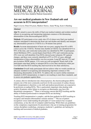
  • 4. NZMJ 3 April 2009, Vol 122 No 1292; ISSN 1175 8716 Page 12 URL: http://www.nzma.org.nz/journal/122-1292/3536/ ©NZMA with 24% reporting that they had interpreted fewer than 10 ECGs in the previous year. When asked how many ECGs they had discussed with a registrar or consultant, 87% reported that they had discussed fewer than 50 with 38% reporting that they had discussed fewer than 10 ECGs in the previous year. Participant’s ratings of confidence in their ability to interpret ECGs, and of the quality of their teaching are given in Figure 1. Participants reported that they had received a mean of 6.3 hours of formal teaching on ECGs during their medical school training. Figure 1. Participants rating of their own abilities to interpret ECGs (A) and the quality of the teaching they had received (B) A. Rating of own abilities in ECG interpretation 0 5 10 15 20 25 30 35 40 45 50 poor less than satisfactory satisfactory good excellent Percentage B. Rating of quality of ECG teaching 0 5 10 15 20 25 30 35 40 45 50 poor less than satisfactory satisfactory good excellent Percentage
  • 5. NZMJ 3 April 2009, Vol 122 No 1292; ISSN 1175 8716 Page 13 URL: http://www.nzma.org.nz/journal/122-1292/3536/ ©NZMA There was a positive correlation between levels of confidence and overall mark (r=0.242, p=0.03). Final year medical students (median score 52%, range 20–65%) scored significantly higher than house officers (40%, 18–61%, p=0.001). There was no difference in score between the two centres in the study (Wellington median score 50%, range 18-65%, Auckland median score 48%, range 21–61%, p=0.67). Discussion The major finding of our study is that there are significant deficiencies in the ability of final year medical students and resident medical officer to interpret common and important ECG changes. However, the majority of participants were able to recognise the most important rhythm disturbance, ventricular tachycardia, accurately as well as identify ischaemic ST segment changes. Furthermore, nearly half of the respondents rated their ability to interpret ECGs as less than satisfactory while just over half rated the ECG teaching they had received as less than satisfactory. Currently there are no clearly defined standards in ECG interpretation for medical graduates for us to compare the results of this study against. A New York study of residents in emergency medicine and internal medicine reported 93% accuracy in identifying VT8 while an Australian study of emergency medicine trainees reported 40% accuracy in identifying VT.9 In this context, the 98% accuracy seen in this New Zealand study is very good. With respect to identifying signs of myocardial infarction, previous studies have reported levels of accuracies of around 90% in registrars8,9 and a retrospective audit of 1684 patients with acute MI in 5 emergency departments in the United States reported an overall accuracy of 88%.1 The level of accuracy for our medical graduates in recognising ST segment changes (87–93%) is probably reasonable in comparison. Clearly, failure to recognise ST segment changes could potentially lead to inappropriate patient management. Similarly, there are concerns that simple recognition of ST segment changes without any understanding of what these changes mean may also lead to inappropriate management decisions being made.10 Given this, we would have hoped to see considerably better performance in the interpretation of ST segment changes in this study. The recognition of pre-excitation and long QT syndrome we observed was poor, but in Hoyle et al’s study of emergency trainees9 these were identified in only 24% and 43% of cases respectively so it is perhaps not surprising that more junior doctors and medical students struggle with these ECGs. Again, failure to recognise these abnormalities has the potential to result in inappropriate patient management. It is difficult to account for the systematic inaccuracy in calculation of heart rate observed in the present study. Perhaps heart rate is regarded as clinically unimportant, and rounding down is viewed by the study participants as a reasonable approximation. While it is difficult to argue that errors of the magnitude we observed in the current study are likely to lead to errors in patient management it is none the less disappointing that a fairly basic skill was not performed to a higher standard.
  • 6. NZMJ 3 April 2009, Vol 122 No 1292; ISSN 1175 8716 Page 14 URL: http://www.nzma.org.nz/journal/122-1292/3536/ ©NZMA The time pressure and the lack of a clinical scenario may have detrimentally influenced the overall level of accuracy. There is good evidence that accuracy is improved when clinical vignettes are provided in addition to the ECG7 —although conversely there is evidence that where ECG changes occur in the context of atypical symptoms, inaccuracy increases.1 Previous studies do not appear to have limited time in any way.6,8,9 We chose to do so on the basis that 2 minutes per ECG was considered a reasonable time period given that in clinical practice there are time constraints. Our medical graduates expressed a lack of confidence in their abilities in ECG interpretation, and this was related to their accuracy levels. Previous studies of New Zealand graduates in other areas have identified knowledge gaps and low levels of confidence similar to those we report.11,12 Perhaps of slightly greater concern to those of us involved in the provision of ECG teaching is that this was rated as less than satisfactory or poor by more than half the study participants. It is possible that we have tended to assume that students interpret and discuss a considerable volume of ECGs in the course of various clinical rotations. However, our results suggest than more than half the study participants looked at less than one ECG per week in a clinical context. The fact that medical students performed better than the residents again suggests that skills in ECG interpretation gained in medical school are not been built upon by normal clinical duties or post graduate teaching. The teaching implications are that more has to be done to assist the students to consolidate the teaching that they get, and to provide additional teaching opportunities. While some aspects of performance in the current study were encouraging, overall our final year medical students and house officers do not achieve what we would consider an adequate standard in recognising and interpreting important common or life- threatening abnormalities in the ECG. Consistent with this, the majority of participants rated both their own ability and the teaching they had received as less than satisfactory. To address this problem we need to define minimum standards in ECG interpretation, improve our teaching to meet these standards, and assess our graduates against them. Competing interests: None known. Author information: Nigel A Lever, Senior Lecturer, Department of Medicine, University of Auckland and Green Lane Cardiovascular Service, Auckland City Hospital, Auckland; Peter D Larsen, Senior Lecturer, Department of Surgery and Anaesthesia, University of Otago, Wellington; Mathew Dawes, Senior Lecturer, Department of Medicine, University of Auckland, Auckland; Annie Wong, House Surgeon, Wellington Hospital, Wellington; Scott A Harding, Cardiologist, Wellington Hospital, Wellington Correspondence: N A Lever, Green Lane Cardiovascular Service, Auckland City Hospital, Private Bag 92024, Auckland 1030, New Zealand. Fax: +64 (0)9 3078941; email: nlever@adhb.govt.nz
  • 7. NZMJ 3 April 2009, Vol 122 No 1292; ISSN 1175 8716 Page 15 URL: http://www.nzma.org.nz/journal/122-1292/3536/ ©NZMA References: 1. Masoudi FA, Magid DJ, Vinson DR, et al. Implications of the failure to identify high-risk electrocardiogram findings for the quality of care of patients with acute myocardial infarction: results of the Emergency Department Quality in Myocardial Infarction (EDQMI) study. Circulation. 2006;114:1565–71. 2. Todd KH, Hoffman JR, Morgan MT. Effect of cardiologist ECG review on emergency department practice. Annals of emergency medicine. 1996;27:16–21. 3. Hurst JW. The interpretation of electrocardiograms: pretense or a well-developed skill? Cardiology Clinics. 2006;24:305-7, vii. 4. Childers R. Teaching electrocardiogram interpretation. Journal of Electrocardiology. 2006;39:426–9. 5. Hurst JW. Thoughts about the abnormalities in the electrocardiogram of patients with acute myocardial infarction with emphasis on a more accurate method of interpreting ST-segment displacement: part I. Clinical Cardiology. 2007;30:381–90. 6. Pines JM, Perina DG, Brady WJ. Electrocardiogram interpretation training and competency assessment in emergency medicine residency programs. Acad Emerg Med. 2004;11:982–4. 7. Salerno SM, Alguire PC, Waxman HS. Competency in interpretation of 12-lead electrocardiograms: a summary and appraisal of published evidence. Annals of Internal Medicine. 2003;138:751–60. 8. Berger JS, Eisen L, Nozad V, et al. Competency in electrocardiogram interpretation among internal medicine and emergency medicine residents. The American Journal of Medicine. 2005;118:873–80. 9. Hoyle RJ, Walker KJ, Thomson G, Bailey M. Accuracy of electrocardiogram interpretation improves with emergency medicine training. Emerg Med Australas. 2007;19:143–50. 10. Hurst JW. To simply identify "up or down" displacement of the ST segment in the electrocardiogram can lead to serious errors. Journal of Electrocardiology. 2008 Sep- Oct;41(5):436–7. Epub 2008 Mar 19. 11. Price CS, Bell SF, Janes SE, Ardagh M. Cardio-pulmonary resuscitation training, knowledge and attitudes of newly-qualified doctors in New Zealand in 2003. Resuscitation. 2006;68:295– 9. 12. Subramaniam R, Hall T, Chou T, Sheehan D. Radiology knowledge in new medical graduates in New Zealand. N Z Med J. 2005; 118(1224). http://www.nzmj.com/journal/118-1224/1699