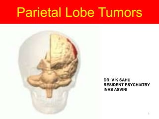
SlideShare uma empresa Scribd logo
1 de 55
Parietal Lobe Tumors
DR V K SAHU
RESIDENT PSYCHIATRY
INHS ASVINI
1
Aim and Main Headings
• Anatomy of Parietal lobe
• Functions
• Tests
• Tumors
• Clinical features
• Investigations
• Management
2
Anatomy
3
Anatomy
• Anterior border - Central Sulcus - parietal lobe & frontal lobe
• Posterior border - Parieto-occipital Sulcus - parietal & occipital
lobes
• Ventral border - Lateral Sulcus (sylvian fissure) is the most
lateral boundary separating it from the temporal lobe
• Medial Longitudinal Fissure divides the two hemispheres.
4
Blood supply
• Anterior parietal artery
• Posterior parietal artery
• Angular artery
• Temporaloccipital: The longest cortical artery.
5
6
7
8
 Body image representation
Body in space
Tactile discrimination
Functions
9
 3 D analysis of body space interactions (body schema)
Visual spatial properties
Visual attention
Motivation and grasping functions.
(parietal lobe lesions - there is ‘self grasping’ of forearm opp. the
lesion )
mediate influence of emotion, attention and motivation
on behavior
 INFERIOR PARIETAL LOBULE
Last to mature anatomically and functionally.
So, the functions are late, to develop b/w 5 and 8 yrs age. ( reading ,
calculations )
Angular gyrus & Supra marginal gyrus - they have
interconnections with visual, auditory, somasthetic, supr. colliculus,
Lateral Geniculate Body and other lobes. 10
Parietal lobe function Impairment of parietal lobe function
Sensory cortex: (represents similar to motor cortex)
---receives afferent pathways for
- appreciation of posture
- touch
- passive movement
Contraleteral disturbances of cortical sensation
- postural sensation disturbed
- sensation of passive movement disturbed
- accurate localization of light touch
disturbed
- 2 point discrimination disturbed
- Asterognosis
Supramarginal angular gyrus: (dominant
hemisphere)
- Wernicke’s language area
- receptive area where auditory , visual
aspect of comprehension are integrated.
Supramarginal angular gyrus: (dominant
hemisphere)
Gerstmann’s syn
-confusion of right & left limb.
-Finger agnosia
-Acalculia
-Agraphia
Supramarginal angular gyrus: (non dominant
hemisphere)
- concept of body image
- awareness of external movement
- skills of handling numbers/calculation
- visual pathway (optic radiation pass through
parietal lobe)
Supramarginal angular gyrus: (non dominant
hemisphere)
-Unaware of opposite limbs
-Anosognosia
-Geographical agnosia
-Constitutional apraxia:
- cannot copy geometric pattern
-Damage of optic radiation: lower homonymous
quadranopia
11
1. Multimodal assimilation – capacity for
organizing , labelling and conceptualizing , using
all senses.
Ex : chair
2. Language capabilities
angular gyrus - anomia
supramarginal gyrus – conduction aphasia
visual cortex to IPL connections – word
blindness
12
3. Agraphia – lt. lobe
Engrams for production and perception
of written language are stored in IPL .
So, misspellings, distorsions, and inversions occur.
4. Temporal sequential functions
IPL is the main track of input and output.
Therefore, information is organized appropriately
into a sequence here.
5. Calculation (Lt.) and computation (Rt.)
13
IPL lesions leads to disruption of visual spatial
functioning and temporal sequencing ability
(apraxia).
i.e either spatial sequential tasks lost - OXOXOX
or sequential grammar relations are lost.
14
Cerebral laterality
15
Either hemisphere
1. Cortical sensations.
2. Integration of sensory , motor and attention signals
(i.e disengage attention - do other activity -
immediately reengage correctly)
3. Optic radiation passes through.
4. Constructional ability – capacity to construct or draw
3D/2D figures or shapes.
Lt. – programming of movements necessary for
constructional activity. (simplification of complex
diagrams)
Rt. – related to spatial relationships or imagery.
(rotation of diagrams)
16
Right hemisphere
1. Constructional skills
2. Dressing apraxia
3. Calculations –
arithmetic concepts of carrying and borrowing
spatial alignment of written calculations.
(computational difficulty – inability to manipulate
no.s in spatial relation, like using decimals,etc –
but he is able to do problems in his head )
4. Perceptual functions (inattention/neglect of lt.
hemispace)
17
Statistics
• Estimated 69,720 new cases of primary malignant and non–
malignant brain and CNS tumors are expected in 2013.
• The incidence rate of all primary malignant and non-
malignant brain and CNS tumors is 20.6 cases per 100,000 (7.3
per 100,000 for malignant tumors and 13.3 per 100,000 for
non–malignant tumors). The rate is higher in females (22.3
per 100,000) than males (18.8 per 100,000).
• Estimated 24,620 new cases of primary malignant brain and
CNS system tumors are expected in 2013
( Central Brain Tumor Registry of the United States ) 18
Primary Brain Tumor Frequency
Tumor Frequency (Percent)
Meningioma 24
Glioblastoma 23
Astrocytoma 12
Pituitary tumors 10
Nerve Sheath tumors & Primary Acoustic Neuroma 7
Medulloblastoma and Pinealomas 5
Anaplastic Astrocytoma and lymphomas 4
Oligodrogliomas 3
All others 12 19
Most Common Brain Tumor by Age Group
Age Range (yr ) Tumor Types
0-9 Primitive Neuroectodermal tumors
medulloblastomas
10-19 Astrocytomas
20-34 Pituitary tumors
35-44 Menigiomas
45-75 Glioblastomas
76 and older Meningiomas
20
Anatomic location of Brain Tumors and
Frequency of Neuropsychiatric symptoms
Anatomic location % of all Brain tumors % with Psychiatric &
Behavioral Symptoms
Frontal lobes 22 As much as 90
Temporal lobes 22 50-55
Parietal lobes 12 As much as 16
Pituitary 10 As much as 60
Occipital 4 As much as 25
Diencephalic Region 2 50 or more
Posterior fossa, Cerebellum
and Brainstem
28 Uncertain,Numerous
neuropsychiatric symptoms
reported
21
General characteristics in Brain Tumors
• Only 18 % of Brain tumor psychiatric
1st manifest with behavioral/
Neuropsychiatric symptoms.
• When mental disturbance most
common pt may first come to psychiatrist
• Many patient with cerebral symptoms
have some psych symptom during illness
• Mental symptoms little guide to
location of tumor Vs Neurological signs.
22
General characteristics in Brain Tumors
• Tumor material proved to be
disappointing for study of cerebral
basis of mental symptoms
• Depressive symptoms- single most
important predictor of quality of
life.
• Slow growing tumor tumor cause
changes of personality , allow
premorbid tendencies to manifest
themselves
• Rapid growing tumors-impairment
of consciousness
23
Neuropsychiatric & Behavior associated
Symptoms in Parietal lobe tumors
• Primarily affective symptoms, depressive > hypomania or
mania
• Psychotic manifestation Paranoid delusions & Cotard’s
syndrome (delusion that they are dead/do not
exist/putrefying/lost blood/internal organs,rarely delusion of
immortality)
24
Neuropsychiatric & Behavior associated
Symptoms (Contd)
• Many have imp lateralizing characteristics
• Results in Contralateral disturbance in
- Two point discrimination
- Joint position sense
- Stereognosis
- Graphesthesia
25
Neuropsychiatric & Behavior associated
Symptoms (Contd)
• Tumors in Dominant Parietal lobe
- difficulties with reading & spelling
- receptive aphasias
- Gerstmann’s syndrome
• Tumors in Non dominant lobe
- visuospatial discrimination
- anosognosia (lack of awareness, denial or complete neglect
of obvious contralateral neurological deficits )
• Various Apraxias
26
Psychiatric & Behavioral Complications of
Medical & Surgical Treatment
• Therapeutic interventions causing abnormalities
• Intraoperative injury to normal brain tissue in
resection/debulking.
e.g. Nonverbal learning disabilities & psychotic
symptoms in children, in frontal lobe –executive dysfunction.
• Radiation induced damage – transient & reversible vs
Permanent
• Chemotherapy causing Delirium
• Treatment of ↑ ICT /Cerebral oedema , Corticosteroid result
in Psychotic and affective symptoms
27
Contributing factors in development of
Neuropsychiatric manifestations
• General Considerations
- Prevalence more in Psychiatric population
- Not commonly the earliest manifestation
• Anatomical localization
- not sole criteria
- lateralization & features not consistent
- symptom far away from location of tumor due to diaschisis
and connection syndrome esp corpus callosum.
- only two mental syndrome consistent – in acute stage
clouding of consciousness & chronic amnesic syndrome in
chronic stage
28
Contributing factors in development of
Neuropsychiatric manifestations (contd)
• Tumor growth
- Rapidity/extent of spread-type/acuity & severity of
symptoms
- Rapid growing– acute, significant neurocognitive impairment
- Slow growing – more vague & subtle behavioral change
- Metastatic lesion & multiple locations
• Tumor type
- more aggressive tumor (high grade gliomas)
- Menigiomas – slow growing & disproportionately in frontal
region cause silent growth & vague/subtle change.
29
Contributing factors in development of
Neuropsychiatric manifestations (contd)
- local effects may be seen e.g. focal cognitive deficits with parietal
lobe tumor & focal amnesic syndrome with diencephalic tumors
- Hallucination – derive from focal lesions of brain
• Intra cranial pressure
- focal & nonfocal neurological symptoms/signs
( - diffuse cognitive impairment,
- changes in attention & concentration
- alteration of level of consciousness
- anxiety,agitation,irritability,depression/apathy )
30
Premorbid Patient characteristics &
Psychosocial factors
• Depression or preexisting psychiatric illness
• Cognitive capacity,coping skills,
adaptive/maladaptive behavioral style
• Psychosocial support
• Challenges by tumor and treatment
31
Diagnostic considerations
• Symptoms and signs
• High index of suspicion & low threshold
for diagnosis in new onset psychiatric
symptoms, esp
- if negative past/personal history
- unexplained personality change
- New neurological/neurocognitive dysfn
• Family History
32
Symptoms suggestive of Brain tumor in
Psychiatric patients
• New onset seizure (focal/partial/generalised) in adult
• Headache - ↑frequency/severity, persistent & nonmigrainous ,
nocturnal, present on awakening, worsened by
position/Valsalva maneuver
• Nausea/Vomiting - esp if nonmigrainous headache
• ↓ Visual acuity, field cuts and double vision
• Unlilateral High Frequency hearing loss,intermittent tinnitus,
vertigo
• Focal weakness
• Focal sensory loss, paresthesias, and dysyesthesias
• Gait disturbances, incoordination, ataxia, and dysarthria
33
Diagnostic studies
• Plain Skull X-ray : pituitary adenomas, craniopharyngiomas,
intracranial calcification, bony metastasis
involving skull ( Bone Scan Preferred )
1. Calcification
- oligodendroglioma
- meningioma
- craniopharyngioma
34
Diagnostic studies
• Signs Of Raised Intracranial Pressure:
-suture separation(diastasis)
- “beaten brass” appreance
• Osteolytic lesion :
primary/secondary bone tumour.
- dermiod/epidermoid
- chordoma
- nasopharyngeal carcinoma
- myeloma
35
Diagnostic studies (contd)
CAT SCAN :
• Calcification
• erosion of bony intracranial structures
• shift in midline cerebral structures
• Abnormalties involving venetricular system : Hydorcephalus
36
Diagnostic studies
• High definition scan:
indication
- pituitary
- orbital
- posterior fossa tumour
- tumour of skull base
Coronal and sagital reconstruction
- diagnosing vertical extent
- relationship with other structure
• IV Contrast : enhance visibility 37
Diagnostic studies (Contd)
• MRI
Indication
- tumours around the skull base/close to bone
- brainstem
Advantage of MRI
- Multiplanar - exact anatomy
- paramagnetic enhancement
- ↑ sensitivity & clarifies the site of origin.
- delineate border b/w tumour & surrounding edema
- more sensitive in identifying
- small tumours ( < 0.5 cms diameter) solid or cystic
- multiple lesions- metastasis 38
Diagnostic studies
• Angiography/ MRA:
reveal
- tumour ‘blush’
- vessel displacement
- preoperative information
- for identifying feeding to
vascular tumours
- tumour involvement and
constriction of major vessels.
39
Diagnostic studies (Contd)
• CT & MRI Cistenography
- evaluation of circulation of CSF
- morphology of ventricular system
- tumor associated hydeocephalus
- CSF leaks
- Intraventricular tumors
• MRI cisternography is better
40
Diagnostic studies (Contd)
• Electroencephalography
- Non specific information, no precise location
- 10-25 % of undiagnosed tumor has no finding/non diagnostic
non specific
- useful for tumor causing seizure
- Findings more in rapidly growing/aggressive
• Lumbar Puncture
- may be useful in leukemias,lymphomas & meningeal
carcinomatosis ( may be missed othervise )
41
Diagnostic studies (Contd)
• Other diagnostic procedures
- Chest x-ray, urinalysis & stool exam
( rule out origination of metastatic tumors)
- PET & SPECT
- tumor recurrence from radiation necrosis
- CNS lymphoma from opportunistic infn
- Magnetoencephalography (MEG)
-may help in phenomenon of disachiasis &
disconnection syndromes 42
Diagnostic studies (Contd)
- Visual field assesment
By Goldman Kinetic perimetry, Humphries static perimetry
- Endocrinological evaluation – hypothalamic-pituitary
axis (for regional involvement & pt treated with radiotherapy.
- Evoked potentials – role in diagnosis & monitoring of
Neurological function during surgical resection.
43
Management of Brain tumors
• Medical Management (Pharmacological)
-Acute treatment/Psychiatric sequelae
• Non pharmacological management
• Management of tumor
- Chemotherapy/Surgery/Radiotherapy
• New Therapeutic Modalities
44
Acute Medical Management
– Stablise patient with peritumoral oedema/↑
ICT/seizure/Delirium.
– Cognitive impairment (slowing of mental performance
,sedation & fatigue )may result from antiepileptics drugs.
– Dexamethasone (low mineralocorticoid activity &
possibly lesser risk of infn & cognitive impairment(but
can cause delirium/psychosis, sleep disturbance &
osteopenia )
– Risk of Opportunistic infn
– Risk of thromboemolism
– Identify hypothalamic-pituitary abnormality
45
Managing Psychiatric Sequalae
• Treatment of Anxiety & Depression
- SSRI drug of choice
• Supportive psychotherapy & CBT recommended
• Cognitive deterioration early marker of progression
(serial neuropsychiatric testing recommended)
• Methylphenidate may improve cognition.
• Palliative care – for disabling neurological symptoms
( e.g. dysphagia)
46
Managing Tumor
• Chemotherapy : Medulloblastoma, lymphomas, oligodendriomas &
germ cell tumor –Highly sensitive
• Neurotoxicity is troublesome s/e esp if intrathecal chemotherapy
• Intrathecal Methotrexate can cause necrotising encephalopathy.
• Cisplatin cause encephalopathy & peripheral neuropathy
• Risk of toxicity increases with associated radiotherapy
47
Managing Tumor
• Radiotherapy
- Cognitive deficits reported in children (RT to brain for acute
leukemia),adults in gliomas, brain metastasis, nasopharyngeal
malignancies, small lung carcinoma.
- Vascular & endothelial damage main features of radiation damage.
- Demyelination may happen subsequently
- Acute radiation encephalopathy (within 2 weeks)
- About 1-6 months after RT may develop radiation encephalopathy
48
Managing Tumor
- Late-delayed encephalopathy is serious & irreversible
- Memory, attention & new learning are sensitive to RT.
- Common neurological sequelae include urinary incontinence,
ataxia, pyramidal as well as extrapyramidal signs.
- Intensity modulated radiation therapy (IMRT) attack from
various angles in 3 dimensional manner.
- Gamma knife uses emitted photons that are precisely directed
- Cyber knife has compact light weight linear accelerator on a
robotic arm 49
New Modalities
(a) Gene therapy – Viral genes to malignant cells
(b) Signal Transduction Inhibitors – aim to reverse the abnormal
activation/suppression responsible for resistance to radiotherapy
(c) Immunotherapy – monoclonal antibody against antigens expressed
by glioma cells
- Interferons also being used
(d) Tamoxifen (modulated Protein Kinase C-involved in cellular signal
transduction) –may have a role.
(e) Stem Cell Therapy- aim to deliver molecules capable of enhancing
antitumor immunity/altering their gene structure
50
Main points
• Disturbance of affect/personality is not specific
to specific portion of brain.
• Fast growing tumors cause acute changes ,
slow growing tumors results in changes in
personality.
• Neurological symptoms/signs has more
localising value.
• Depressive symptoms- single most important
predictor of quality of life.
• Premorbid cognitive capacity/coping skills
important in degree of dysfunction.
51
Main points
• Treatment can also result in Neuropsychiatric
abnormalities.
• Identify patient having suicidal tendencies
• Avoid drugs at risk of inducing seizure in patient
with past h/o of seizure (Bupropion,Lithium
carbonate)
• Adopt active “here & now” therapeutic
psychoeducational approach along with
pharmacotherapy
52
• CANCERS SO LIMITED
It can't cripple love
It can't shatter hope
It can't corrode faith
It can't eat away peace
It can't destroy confidence
It can't kill friendship
It can't shut out memories
It can't silence courage
It can't invade the soul
It can't reduce eternal life
It can't quench the Spirits
It can't lessen the power of the resurrection.
CANCER IS SO LIMITED
It can't cripple love
It can't shatter hope
It can't corrode faith
It can't eat away peace
It can't destroy confidence
It can't kill friendship
It can't shut out memories
It can't silence courage
It can't invade the soul
It can't reduce eternal life
It can't quench the Spirits
It can't lessen the power of the resurrection.
53
54
References
• (http://www.cbtrus.org/factsheet/factsheet.h
tml
( Central Brain Tumor Registry of the United
States )
• Comprehensive Textbook of Psychiatry (Kaplan
& Sadock’s) – Ninth Edition
• Lishman’s Organic Psychiatry – Fourth Edition
55

Mais conteúdo relacionado

Mais procurados

Parietal lobe and its functions
Parietal lobe and its functionsParietal lobe and its functions
Parietal lobe and its functions
drnaveent
 
Limbic system by dr ali
Limbic system by dr aliLimbic system by dr ali
Limbic system by dr ali
OSMAN ALI MD
 
Occipital lobe
Occipital lobeOccipital lobe
Occipital lobe
PS Deb
 
Posterior association cortex
Posterior association cortexPosterior association cortex
Posterior association cortex
PS Deb
 
Parietal lobe
Parietal lobeParietal lobe
Parietal lobe
Arun S
 
Temporal lobe
Temporal lobeTemporal lobe
Temporal lobe
PS Deb
 
Parietal lobe
Parietal lobeParietal lobe
Parietal lobe
PS Deb
 
Frontal lobe 2010
Frontal lobe 2010Frontal lobe 2010
Frontal lobe 2010
PS Deb
 

Mais procurados (20)

Parietal Lobe Anatomy 16 jan 2019 Ashish
Parietal Lobe Anatomy 16 jan 2019 AshishParietal Lobe Anatomy 16 jan 2019 Ashish
Parietal Lobe Anatomy 16 jan 2019 Ashish
 
Parietal lobe and its functions
Parietal lobe and its functionsParietal lobe and its functions
Parietal lobe and its functions
 
Temporal lobe ppt
Temporal lobe pptTemporal lobe ppt
Temporal lobe ppt
 
Seminar Cerebral neurolocalization
Seminar Cerebral neurolocalizationSeminar Cerebral neurolocalization
Seminar Cerebral neurolocalization
 
Frontal lobe
Frontal lobeFrontal lobe
Frontal lobe
 
Frontallobe dr prashant mishra
Frontallobe dr prashant mishra Frontallobe dr prashant mishra
Frontallobe dr prashant mishra
 
The Parietal Lobe
The Parietal LobeThe Parietal Lobe
The Parietal Lobe
 
Frontal lobe epilepsy
Frontal lobe epilepsyFrontal lobe epilepsy
Frontal lobe epilepsy
 
Limbic system by dr ali
Limbic system by dr aliLimbic system by dr ali
Limbic system by dr ali
 
Occipital lobe
Occipital lobeOccipital lobe
Occipital lobe
 
Posterior association cortex
Posterior association cortexPosterior association cortex
Posterior association cortex
 
Cortical areas of brain
Cortical areas of brainCortical areas of brain
Cortical areas of brain
 
Parietal lobe
Parietal lobeParietal lobe
Parietal lobe
 
Frontal lobe
Frontal lobeFrontal lobe
Frontal lobe
 
Temporal lobe
Temporal lobeTemporal lobe
Temporal lobe
 
Parietal lobe
Parietal lobeParietal lobe
Parietal lobe
 
Frontal lobe & subcortical circuits
Frontal lobe & subcortical circuitsFrontal lobe & subcortical circuits
Frontal lobe & subcortical circuits
 
Agnosia ppt
Agnosia pptAgnosia ppt
Agnosia ppt
 
Frontal lobe 2010
Frontal lobe 2010Frontal lobe 2010
Frontal lobe 2010
 
Disconnection syndrome
Disconnection syndromeDisconnection syndrome
Disconnection syndrome
 

Destaque

Parietal lobe 2010
Parietal lobe 2010Parietal lobe 2010
Parietal lobe 2010
PS Deb
 
Lec9 MECH ENG STRucture
Lec9   MECH ENG  STRuctureLec9   MECH ENG  STRucture
Lec9 MECH ENG STRucture
Mohamed Yaser
 

Destaque (14)

Cpqe power point
Cpqe power pointCpqe power point
Cpqe power point
 
Approach to-a-patient-with-chronic-diarrhoea (7)
Approach to-a-patient-with-chronic-diarrhoea (7)Approach to-a-patient-with-chronic-diarrhoea (7)
Approach to-a-patient-with-chronic-diarrhoea (7)
 
Epileptic encephalopathy -EEG
Epileptic encephalopathy -EEGEpileptic encephalopathy -EEG
Epileptic encephalopathy -EEG
 
Basal ganglia – Neuropsychiatric aspect
Basal ganglia  –  Neuropsychiatric  aspectBasal ganglia  –  Neuropsychiatric  aspect
Basal ganglia – Neuropsychiatric aspect
 
Temporal lobe
Temporal lobeTemporal lobe
Temporal lobe
 
Functions of Parietal Lobe
Functions of Parietal Lobe Functions of Parietal Lobe
Functions of Parietal Lobe
 
Case Studies In Stroke
Case Studies In StrokeCase Studies In Stroke
Case Studies In Stroke
 
Brain cortical regions and functions
Brain cortical regions and functionsBrain cortical regions and functions
Brain cortical regions and functions
 
Parietal lobe 2010
Parietal lobe 2010Parietal lobe 2010
Parietal lobe 2010
 
Frontal lobe dr. arpit
Frontal lobe dr. arpitFrontal lobe dr. arpit
Frontal lobe dr. arpit
 
Lec9 MECH ENG STRucture
Lec9   MECH ENG  STRuctureLec9   MECH ENG  STRucture
Lec9 MECH ENG STRucture
 
Harbor UCLA Neuro-Radiology — Case 4
Harbor UCLA Neuro-Radiology — Case 4Harbor UCLA Neuro-Radiology — Case 4
Harbor UCLA Neuro-Radiology — Case 4
 
Functional anatomy of Frontal lobe
Functional anatomy of Frontal lobeFunctional anatomy of Frontal lobe
Functional anatomy of Frontal lobe
 
Limbic system
Limbic systemLimbic system
Limbic system
 

Semelhante a Parietal lobe tumor

Cerebral tumors 1
Cerebral tumors 1Cerebral tumors 1
Cerebral tumors 1
Sabir Ahmed
 
Stroke and cerebrovascular accident
Stroke and cerebrovascular accidentStroke and cerebrovascular accident
Stroke and cerebrovascular accident
Dr. Rubz
 
Dementia- recent updates
Dementia-  recent updatesDementia-  recent updates
Dementia- recent updates
Santanu Ghosh
 
CLINICAL FEATURES, INVESTIGATIONS AND PROGNOSIS OF BRAIN SEMINAR 2.pptx
CLINICAL FEATURES, INVESTIGATIONS AND PROGNOSIS OF BRAIN SEMINAR 2.pptxCLINICAL FEATURES, INVESTIGATIONS AND PROGNOSIS OF BRAIN SEMINAR 2.pptx
CLINICAL FEATURES, INVESTIGATIONS AND PROGNOSIS OF BRAIN SEMINAR 2.pptx
cheshtasharma22
 

Semelhante a Parietal lobe tumor (20)

Cerebral tumors 1
Cerebral tumors 1Cerebral tumors 1
Cerebral tumors 1
 
NeuroLocalisation.ppt
NeuroLocalisation.pptNeuroLocalisation.ppt
NeuroLocalisation.ppt
 
Lecture 4 CNS TUMOR 1 2.ppt
Lecture 4 CNS TUMOR 1 2.pptLecture 4 CNS TUMOR 1 2.ppt
Lecture 4 CNS TUMOR 1 2.ppt
 
Brain tumor
Brain tumorBrain tumor
Brain tumor
 
Brainstem glioma part 1
Brainstem glioma   part 1Brainstem glioma   part 1
Brainstem glioma part 1
 
Supratentorial brain tumours
Supratentorial brain tumoursSupratentorial brain tumours
Supratentorial brain tumours
 
Brain Tumors.ppt
Brain Tumors.pptBrain Tumors.ppt
Brain Tumors.ppt
 
Neuroimaging in Psychiatry
Neuroimaging in PsychiatryNeuroimaging in Psychiatry
Neuroimaging in Psychiatry
 
Stroke and cerebrovascular accident
Stroke and cerebrovascular accidentStroke and cerebrovascular accident
Stroke and cerebrovascular accident
 
Part 1 function of brain and history taking of a neurological patient
Part 1 function of brain and history taking of a neurological patientPart 1 function of brain and history taking of a neurological patient
Part 1 function of brain and history taking of a neurological patient
 
Brain tumors in children with Updates- Pranav
Brain tumors in children with Updates- PranavBrain tumors in children with Updates- Pranav
Brain tumors in children with Updates- Pranav
 
Dementia- recent updates
Dementia-  recent updatesDementia-  recent updates
Dementia- recent updates
 
CP ANGLE TUMOR.pptx
CP ANGLE TUMOR.pptxCP ANGLE TUMOR.pptx
CP ANGLE TUMOR.pptx
 
CLINICAL FEATURES, INVESTIGATIONS AND PROGNOSIS OF BRAIN SEMINAR 2.pptx
CLINICAL FEATURES, INVESTIGATIONS AND PROGNOSIS OF BRAIN SEMINAR 2.pptxCLINICAL FEATURES, INVESTIGATIONS AND PROGNOSIS OF BRAIN SEMINAR 2.pptx
CLINICAL FEATURES, INVESTIGATIONS AND PROGNOSIS OF BRAIN SEMINAR 2.pptx
 
Icsol
IcsolIcsol
Icsol
 
What is Epilepsy?
What is Epilepsy?What is Epilepsy?
What is Epilepsy?
 
NEUROPSYCHIATRIC ASPECTS OF BRAIN TUMORS.pptx
NEUROPSYCHIATRIC ASPECTS OF BRAIN TUMORS.pptxNEUROPSYCHIATRIC ASPECTS OF BRAIN TUMORS.pptx
NEUROPSYCHIATRIC ASPECTS OF BRAIN TUMORS.pptx
 
Surgically Treatable Dementias
Surgically Treatable DementiasSurgically Treatable Dementias
Surgically Treatable Dementias
 
Acoustic neuroma
Acoustic neuromaAcoustic neuroma
Acoustic neuroma
 
Brain tumors
Brain tumorsBrain tumors
Brain tumors
 

Último

Call Girl In Pune 👉 Just CALL ME: 9352988975 💋 Call Out Call Both With High p...
Call Girl In Pune 👉 Just CALL ME: 9352988975 💋 Call Out Call Both With High p...Call Girl In Pune 👉 Just CALL ME: 9352988975 💋 Call Out Call Both With High p...
Call Girl In Pune 👉 Just CALL ME: 9352988975 💋 Call Out Call Both With High p...
chetankumar9855
 

Último (20)

(Low Rate RASHMI ) Rate Of Call Girls Jaipur ❣ 8445551418 ❣ Elite Models & Ce...
(Low Rate RASHMI ) Rate Of Call Girls Jaipur ❣ 8445551418 ❣ Elite Models & Ce...(Low Rate RASHMI ) Rate Of Call Girls Jaipur ❣ 8445551418 ❣ Elite Models & Ce...
(Low Rate RASHMI ) Rate Of Call Girls Jaipur ❣ 8445551418 ❣ Elite Models & Ce...
 
Top Rated Bangalore Call Girls Richmond Circle ⟟ 9332606886 ⟟ Call Me For Ge...
Top Rated Bangalore Call Girls Richmond Circle ⟟  9332606886 ⟟ Call Me For Ge...Top Rated Bangalore Call Girls Richmond Circle ⟟  9332606886 ⟟ Call Me For Ge...
Top Rated Bangalore Call Girls Richmond Circle ⟟ 9332606886 ⟟ Call Me For Ge...
 
Manyata Tech Park ( Call Girls ) Bangalore ✔ 6297143586 ✔ Hot Model With Sexy...
Manyata Tech Park ( Call Girls ) Bangalore ✔ 6297143586 ✔ Hot Model With Sexy...Manyata Tech Park ( Call Girls ) Bangalore ✔ 6297143586 ✔ Hot Model With Sexy...
Manyata Tech Park ( Call Girls ) Bangalore ✔ 6297143586 ✔ Hot Model With Sexy...
 
8980367676 Call Girls In Ahmedabad Escort Service Available 24×7 In Ahmedabad
8980367676 Call Girls In Ahmedabad Escort Service Available 24×7 In Ahmedabad8980367676 Call Girls In Ahmedabad Escort Service Available 24×7 In Ahmedabad
8980367676 Call Girls In Ahmedabad Escort Service Available 24×7 In Ahmedabad
 
Russian Call Girls Service Jaipur {8445551418} ❤️PALLAVI VIP Jaipur Call Gir...
Russian Call Girls Service  Jaipur {8445551418} ❤️PALLAVI VIP Jaipur Call Gir...Russian Call Girls Service  Jaipur {8445551418} ❤️PALLAVI VIP Jaipur Call Gir...
Russian Call Girls Service Jaipur {8445551418} ❤️PALLAVI VIP Jaipur Call Gir...
 
Call Girls Gwalior Just Call 8617370543 Top Class Call Girl Service Available
Call Girls Gwalior Just Call 8617370543 Top Class Call Girl Service AvailableCall Girls Gwalior Just Call 8617370543 Top Class Call Girl Service Available
Call Girls Gwalior Just Call 8617370543 Top Class Call Girl Service Available
 
💕SONAM KUMAR💕Premium Call Girls Jaipur ↘️9257276172 ↙️One Night Stand With Lo...
💕SONAM KUMAR💕Premium Call Girls Jaipur ↘️9257276172 ↙️One Night Stand With Lo...💕SONAM KUMAR💕Premium Call Girls Jaipur ↘️9257276172 ↙️One Night Stand With Lo...
💕SONAM KUMAR💕Premium Call Girls Jaipur ↘️9257276172 ↙️One Night Stand With Lo...
 
Top Rated Bangalore Call Girls Mg Road ⟟ 9332606886 ⟟ Call Me For Genuine S...
Top Rated Bangalore Call Girls Mg Road ⟟   9332606886 ⟟ Call Me For Genuine S...Top Rated Bangalore Call Girls Mg Road ⟟   9332606886 ⟟ Call Me For Genuine S...
Top Rated Bangalore Call Girls Mg Road ⟟ 9332606886 ⟟ Call Me For Genuine S...
 
Call Girl In Pune 👉 Just CALL ME: 9352988975 💋 Call Out Call Both With High p...
Call Girl In Pune 👉 Just CALL ME: 9352988975 💋 Call Out Call Both With High p...Call Girl In Pune 👉 Just CALL ME: 9352988975 💋 Call Out Call Both With High p...
Call Girl In Pune 👉 Just CALL ME: 9352988975 💋 Call Out Call Both With High p...
 
Call Girls Visakhapatnam Just Call 8250077686 Top Class Call Girl Service Ava...
Call Girls Visakhapatnam Just Call 8250077686 Top Class Call Girl Service Ava...Call Girls Visakhapatnam Just Call 8250077686 Top Class Call Girl Service Ava...
Call Girls Visakhapatnam Just Call 8250077686 Top Class Call Girl Service Ava...
 
Call Girls Service Jaipur {8445551418} ❤️VVIP BHAWNA Call Girl in Jaipur Raja...
Call Girls Service Jaipur {8445551418} ❤️VVIP BHAWNA Call Girl in Jaipur Raja...Call Girls Service Jaipur {8445551418} ❤️VVIP BHAWNA Call Girl in Jaipur Raja...
Call Girls Service Jaipur {8445551418} ❤️VVIP BHAWNA Call Girl in Jaipur Raja...
 
Call Girls Vadodara Just Call 8617370543 Top Class Call Girl Service Available
Call Girls Vadodara Just Call 8617370543 Top Class Call Girl Service AvailableCall Girls Vadodara Just Call 8617370543 Top Class Call Girl Service Available
Call Girls Vadodara Just Call 8617370543 Top Class Call Girl Service Available
 
Premium Call Girls In Jaipur {8445551418} ❤️VVIP SEEMA Call Girl in Jaipur Ra...
Premium Call Girls In Jaipur {8445551418} ❤️VVIP SEEMA Call Girl in Jaipur Ra...Premium Call Girls In Jaipur {8445551418} ❤️VVIP SEEMA Call Girl in Jaipur Ra...
Premium Call Girls In Jaipur {8445551418} ❤️VVIP SEEMA Call Girl in Jaipur Ra...
 
Best Rate (Guwahati ) Call Girls Guwahati ⟟ 8617370543 ⟟ High Class Call Girl...
Best Rate (Guwahati ) Call Girls Guwahati ⟟ 8617370543 ⟟ High Class Call Girl...Best Rate (Guwahati ) Call Girls Guwahati ⟟ 8617370543 ⟟ High Class Call Girl...
Best Rate (Guwahati ) Call Girls Guwahati ⟟ 8617370543 ⟟ High Class Call Girl...
 
Call Girls Service Jaipur {9521753030} ❤️VVIP RIDDHI Call Girl in Jaipur Raja...
Call Girls Service Jaipur {9521753030} ❤️VVIP RIDDHI Call Girl in Jaipur Raja...Call Girls Service Jaipur {9521753030} ❤️VVIP RIDDHI Call Girl in Jaipur Raja...
Call Girls Service Jaipur {9521753030} ❤️VVIP RIDDHI Call Girl in Jaipur Raja...
 
Most Beautiful Call Girl in Bangalore Contact on Whatsapp
Most Beautiful Call Girl in Bangalore Contact on WhatsappMost Beautiful Call Girl in Bangalore Contact on Whatsapp
Most Beautiful Call Girl in Bangalore Contact on Whatsapp
 
Call Girls Ahmedabad Just Call 9630942363 Top Class Call Girl Service Available
Call Girls Ahmedabad Just Call 9630942363 Top Class Call Girl Service AvailableCall Girls Ahmedabad Just Call 9630942363 Top Class Call Girl Service Available
Call Girls Ahmedabad Just Call 9630942363 Top Class Call Girl Service Available
 
Top Quality Call Girl Service Kalyanpur 6378878445 Available Call Girls Any Time
Top Quality Call Girl Service Kalyanpur 6378878445 Available Call Girls Any TimeTop Quality Call Girl Service Kalyanpur 6378878445 Available Call Girls Any Time
Top Quality Call Girl Service Kalyanpur 6378878445 Available Call Girls Any Time
 
Model Call Girls In Chennai WhatsApp Booking 7427069034 call girl service 24 ...
Model Call Girls In Chennai WhatsApp Booking 7427069034 call girl service 24 ...Model Call Girls In Chennai WhatsApp Booking 7427069034 call girl service 24 ...
Model Call Girls In Chennai WhatsApp Booking 7427069034 call girl service 24 ...
 
Independent Call Girls In Jaipur { 8445551418 } ✔ ANIKA MEHTA ✔ Get High Prof...
Independent Call Girls In Jaipur { 8445551418 } ✔ ANIKA MEHTA ✔ Get High Prof...Independent Call Girls In Jaipur { 8445551418 } ✔ ANIKA MEHTA ✔ Get High Prof...
Independent Call Girls In Jaipur { 8445551418 } ✔ ANIKA MEHTA ✔ Get High Prof...
 

Parietal lobe tumor

  • 1. Parietal Lobe Tumors DR V K SAHU RESIDENT PSYCHIATRY INHS ASVINI 1
  • 2. Aim and Main Headings • Anatomy of Parietal lobe • Functions • Tests • Tumors • Clinical features • Investigations • Management 2
  • 4. Anatomy • Anterior border - Central Sulcus - parietal lobe & frontal lobe • Posterior border - Parieto-occipital Sulcus - parietal & occipital lobes • Ventral border - Lateral Sulcus (sylvian fissure) is the most lateral boundary separating it from the temporal lobe • Medial Longitudinal Fissure divides the two hemispheres. 4
  • 5. Blood supply • Anterior parietal artery • Posterior parietal artery • Angular artery • Temporaloccipital: The longest cortical artery. 5
  • 6. 6
  • 7. 7
  • 8. 8
  • 9.  Body image representation Body in space Tactile discrimination Functions 9
  • 10.  3 D analysis of body space interactions (body schema) Visual spatial properties Visual attention Motivation and grasping functions. (parietal lobe lesions - there is ‘self grasping’ of forearm opp. the lesion ) mediate influence of emotion, attention and motivation on behavior  INFERIOR PARIETAL LOBULE Last to mature anatomically and functionally. So, the functions are late, to develop b/w 5 and 8 yrs age. ( reading , calculations ) Angular gyrus & Supra marginal gyrus - they have interconnections with visual, auditory, somasthetic, supr. colliculus, Lateral Geniculate Body and other lobes. 10
  • 11. Parietal lobe function Impairment of parietal lobe function Sensory cortex: (represents similar to motor cortex) ---receives afferent pathways for - appreciation of posture - touch - passive movement Contraleteral disturbances of cortical sensation - postural sensation disturbed - sensation of passive movement disturbed - accurate localization of light touch disturbed - 2 point discrimination disturbed - Asterognosis Supramarginal angular gyrus: (dominant hemisphere) - Wernicke’s language area - receptive area where auditory , visual aspect of comprehension are integrated. Supramarginal angular gyrus: (dominant hemisphere) Gerstmann’s syn -confusion of right & left limb. -Finger agnosia -Acalculia -Agraphia Supramarginal angular gyrus: (non dominant hemisphere) - concept of body image - awareness of external movement - skills of handling numbers/calculation - visual pathway (optic radiation pass through parietal lobe) Supramarginal angular gyrus: (non dominant hemisphere) -Unaware of opposite limbs -Anosognosia -Geographical agnosia -Constitutional apraxia: - cannot copy geometric pattern -Damage of optic radiation: lower homonymous quadranopia 11
  • 12. 1. Multimodal assimilation – capacity for organizing , labelling and conceptualizing , using all senses. Ex : chair 2. Language capabilities angular gyrus - anomia supramarginal gyrus – conduction aphasia visual cortex to IPL connections – word blindness 12
  • 13. 3. Agraphia – lt. lobe Engrams for production and perception of written language are stored in IPL . So, misspellings, distorsions, and inversions occur. 4. Temporal sequential functions IPL is the main track of input and output. Therefore, information is organized appropriately into a sequence here. 5. Calculation (Lt.) and computation (Rt.) 13
  • 14. IPL lesions leads to disruption of visual spatial functioning and temporal sequencing ability (apraxia). i.e either spatial sequential tasks lost - OXOXOX or sequential grammar relations are lost. 14
  • 16. Either hemisphere 1. Cortical sensations. 2. Integration of sensory , motor and attention signals (i.e disengage attention - do other activity - immediately reengage correctly) 3. Optic radiation passes through. 4. Constructional ability – capacity to construct or draw 3D/2D figures or shapes. Lt. – programming of movements necessary for constructional activity. (simplification of complex diagrams) Rt. – related to spatial relationships or imagery. (rotation of diagrams) 16
  • 17. Right hemisphere 1. Constructional skills 2. Dressing apraxia 3. Calculations – arithmetic concepts of carrying and borrowing spatial alignment of written calculations. (computational difficulty – inability to manipulate no.s in spatial relation, like using decimals,etc – but he is able to do problems in his head ) 4. Perceptual functions (inattention/neglect of lt. hemispace) 17
  • 18. Statistics • Estimated 69,720 new cases of primary malignant and non– malignant brain and CNS tumors are expected in 2013. • The incidence rate of all primary malignant and non- malignant brain and CNS tumors is 20.6 cases per 100,000 (7.3 per 100,000 for malignant tumors and 13.3 per 100,000 for non–malignant tumors). The rate is higher in females (22.3 per 100,000) than males (18.8 per 100,000). • Estimated 24,620 new cases of primary malignant brain and CNS system tumors are expected in 2013 ( Central Brain Tumor Registry of the United States ) 18
  • 19. Primary Brain Tumor Frequency Tumor Frequency (Percent) Meningioma 24 Glioblastoma 23 Astrocytoma 12 Pituitary tumors 10 Nerve Sheath tumors & Primary Acoustic Neuroma 7 Medulloblastoma and Pinealomas 5 Anaplastic Astrocytoma and lymphomas 4 Oligodrogliomas 3 All others 12 19
  • 20. Most Common Brain Tumor by Age Group Age Range (yr ) Tumor Types 0-9 Primitive Neuroectodermal tumors medulloblastomas 10-19 Astrocytomas 20-34 Pituitary tumors 35-44 Menigiomas 45-75 Glioblastomas 76 and older Meningiomas 20
  • 21. Anatomic location of Brain Tumors and Frequency of Neuropsychiatric symptoms Anatomic location % of all Brain tumors % with Psychiatric & Behavioral Symptoms Frontal lobes 22 As much as 90 Temporal lobes 22 50-55 Parietal lobes 12 As much as 16 Pituitary 10 As much as 60 Occipital 4 As much as 25 Diencephalic Region 2 50 or more Posterior fossa, Cerebellum and Brainstem 28 Uncertain,Numerous neuropsychiatric symptoms reported 21
  • 22. General characteristics in Brain Tumors • Only 18 % of Brain tumor psychiatric 1st manifest with behavioral/ Neuropsychiatric symptoms. • When mental disturbance most common pt may first come to psychiatrist • Many patient with cerebral symptoms have some psych symptom during illness • Mental symptoms little guide to location of tumor Vs Neurological signs. 22
  • 23. General characteristics in Brain Tumors • Tumor material proved to be disappointing for study of cerebral basis of mental symptoms • Depressive symptoms- single most important predictor of quality of life. • Slow growing tumor tumor cause changes of personality , allow premorbid tendencies to manifest themselves • Rapid growing tumors-impairment of consciousness 23
  • 24. Neuropsychiatric & Behavior associated Symptoms in Parietal lobe tumors • Primarily affective symptoms, depressive > hypomania or mania • Psychotic manifestation Paranoid delusions & Cotard’s syndrome (delusion that they are dead/do not exist/putrefying/lost blood/internal organs,rarely delusion of immortality) 24
  • 25. Neuropsychiatric & Behavior associated Symptoms (Contd) • Many have imp lateralizing characteristics • Results in Contralateral disturbance in - Two point discrimination - Joint position sense - Stereognosis - Graphesthesia 25
  • 26. Neuropsychiatric & Behavior associated Symptoms (Contd) • Tumors in Dominant Parietal lobe - difficulties with reading & spelling - receptive aphasias - Gerstmann’s syndrome • Tumors in Non dominant lobe - visuospatial discrimination - anosognosia (lack of awareness, denial or complete neglect of obvious contralateral neurological deficits ) • Various Apraxias 26
  • 27. Psychiatric & Behavioral Complications of Medical & Surgical Treatment • Therapeutic interventions causing abnormalities • Intraoperative injury to normal brain tissue in resection/debulking. e.g. Nonverbal learning disabilities & psychotic symptoms in children, in frontal lobe –executive dysfunction. • Radiation induced damage – transient & reversible vs Permanent • Chemotherapy causing Delirium • Treatment of ↑ ICT /Cerebral oedema , Corticosteroid result in Psychotic and affective symptoms 27
  • 28. Contributing factors in development of Neuropsychiatric manifestations • General Considerations - Prevalence more in Psychiatric population - Not commonly the earliest manifestation • Anatomical localization - not sole criteria - lateralization & features not consistent - symptom far away from location of tumor due to diaschisis and connection syndrome esp corpus callosum. - only two mental syndrome consistent – in acute stage clouding of consciousness & chronic amnesic syndrome in chronic stage 28
  • 29. Contributing factors in development of Neuropsychiatric manifestations (contd) • Tumor growth - Rapidity/extent of spread-type/acuity & severity of symptoms - Rapid growing– acute, significant neurocognitive impairment - Slow growing – more vague & subtle behavioral change - Metastatic lesion & multiple locations • Tumor type - more aggressive tumor (high grade gliomas) - Menigiomas – slow growing & disproportionately in frontal region cause silent growth & vague/subtle change. 29
  • 30. Contributing factors in development of Neuropsychiatric manifestations (contd) - local effects may be seen e.g. focal cognitive deficits with parietal lobe tumor & focal amnesic syndrome with diencephalic tumors - Hallucination – derive from focal lesions of brain • Intra cranial pressure - focal & nonfocal neurological symptoms/signs ( - diffuse cognitive impairment, - changes in attention & concentration - alteration of level of consciousness - anxiety,agitation,irritability,depression/apathy ) 30
  • 31. Premorbid Patient characteristics & Psychosocial factors • Depression or preexisting psychiatric illness • Cognitive capacity,coping skills, adaptive/maladaptive behavioral style • Psychosocial support • Challenges by tumor and treatment 31
  • 32. Diagnostic considerations • Symptoms and signs • High index of suspicion & low threshold for diagnosis in new onset psychiatric symptoms, esp - if negative past/personal history - unexplained personality change - New neurological/neurocognitive dysfn • Family History 32
  • 33. Symptoms suggestive of Brain tumor in Psychiatric patients • New onset seizure (focal/partial/generalised) in adult • Headache - ↑frequency/severity, persistent & nonmigrainous , nocturnal, present on awakening, worsened by position/Valsalva maneuver • Nausea/Vomiting - esp if nonmigrainous headache • ↓ Visual acuity, field cuts and double vision • Unlilateral High Frequency hearing loss,intermittent tinnitus, vertigo • Focal weakness • Focal sensory loss, paresthesias, and dysyesthesias • Gait disturbances, incoordination, ataxia, and dysarthria 33
  • 34. Diagnostic studies • Plain Skull X-ray : pituitary adenomas, craniopharyngiomas, intracranial calcification, bony metastasis involving skull ( Bone Scan Preferred ) 1. Calcification - oligodendroglioma - meningioma - craniopharyngioma 34
  • 35. Diagnostic studies • Signs Of Raised Intracranial Pressure: -suture separation(diastasis) - “beaten brass” appreance • Osteolytic lesion : primary/secondary bone tumour. - dermiod/epidermoid - chordoma - nasopharyngeal carcinoma - myeloma 35
  • 36. Diagnostic studies (contd) CAT SCAN : • Calcification • erosion of bony intracranial structures • shift in midline cerebral structures • Abnormalties involving venetricular system : Hydorcephalus 36
  • 37. Diagnostic studies • High definition scan: indication - pituitary - orbital - posterior fossa tumour - tumour of skull base Coronal and sagital reconstruction - diagnosing vertical extent - relationship with other structure • IV Contrast : enhance visibility 37
  • 38. Diagnostic studies (Contd) • MRI Indication - tumours around the skull base/close to bone - brainstem Advantage of MRI - Multiplanar - exact anatomy - paramagnetic enhancement - ↑ sensitivity & clarifies the site of origin. - delineate border b/w tumour & surrounding edema - more sensitive in identifying - small tumours ( < 0.5 cms diameter) solid or cystic - multiple lesions- metastasis 38
  • 39. Diagnostic studies • Angiography/ MRA: reveal - tumour ‘blush’ - vessel displacement - preoperative information - for identifying feeding to vascular tumours - tumour involvement and constriction of major vessels. 39
  • 40. Diagnostic studies (Contd) • CT & MRI Cistenography - evaluation of circulation of CSF - morphology of ventricular system - tumor associated hydeocephalus - CSF leaks - Intraventricular tumors • MRI cisternography is better 40
  • 41. Diagnostic studies (Contd) • Electroencephalography - Non specific information, no precise location - 10-25 % of undiagnosed tumor has no finding/non diagnostic non specific - useful for tumor causing seizure - Findings more in rapidly growing/aggressive • Lumbar Puncture - may be useful in leukemias,lymphomas & meningeal carcinomatosis ( may be missed othervise ) 41
  • 42. Diagnostic studies (Contd) • Other diagnostic procedures - Chest x-ray, urinalysis & stool exam ( rule out origination of metastatic tumors) - PET & SPECT - tumor recurrence from radiation necrosis - CNS lymphoma from opportunistic infn - Magnetoencephalography (MEG) -may help in phenomenon of disachiasis & disconnection syndromes 42
  • 43. Diagnostic studies (Contd) - Visual field assesment By Goldman Kinetic perimetry, Humphries static perimetry - Endocrinological evaluation – hypothalamic-pituitary axis (for regional involvement & pt treated with radiotherapy. - Evoked potentials – role in diagnosis & monitoring of Neurological function during surgical resection. 43
  • 44. Management of Brain tumors • Medical Management (Pharmacological) -Acute treatment/Psychiatric sequelae • Non pharmacological management • Management of tumor - Chemotherapy/Surgery/Radiotherapy • New Therapeutic Modalities 44
  • 45. Acute Medical Management – Stablise patient with peritumoral oedema/↑ ICT/seizure/Delirium. – Cognitive impairment (slowing of mental performance ,sedation & fatigue )may result from antiepileptics drugs. – Dexamethasone (low mineralocorticoid activity & possibly lesser risk of infn & cognitive impairment(but can cause delirium/psychosis, sleep disturbance & osteopenia ) – Risk of Opportunistic infn – Risk of thromboemolism – Identify hypothalamic-pituitary abnormality 45
  • 46. Managing Psychiatric Sequalae • Treatment of Anxiety & Depression - SSRI drug of choice • Supportive psychotherapy & CBT recommended • Cognitive deterioration early marker of progression (serial neuropsychiatric testing recommended) • Methylphenidate may improve cognition. • Palliative care – for disabling neurological symptoms ( e.g. dysphagia) 46
  • 47. Managing Tumor • Chemotherapy : Medulloblastoma, lymphomas, oligodendriomas & germ cell tumor –Highly sensitive • Neurotoxicity is troublesome s/e esp if intrathecal chemotherapy • Intrathecal Methotrexate can cause necrotising encephalopathy. • Cisplatin cause encephalopathy & peripheral neuropathy • Risk of toxicity increases with associated radiotherapy 47
  • 48. Managing Tumor • Radiotherapy - Cognitive deficits reported in children (RT to brain for acute leukemia),adults in gliomas, brain metastasis, nasopharyngeal malignancies, small lung carcinoma. - Vascular & endothelial damage main features of radiation damage. - Demyelination may happen subsequently - Acute radiation encephalopathy (within 2 weeks) - About 1-6 months after RT may develop radiation encephalopathy 48
  • 49. Managing Tumor - Late-delayed encephalopathy is serious & irreversible - Memory, attention & new learning are sensitive to RT. - Common neurological sequelae include urinary incontinence, ataxia, pyramidal as well as extrapyramidal signs. - Intensity modulated radiation therapy (IMRT) attack from various angles in 3 dimensional manner. - Gamma knife uses emitted photons that are precisely directed - Cyber knife has compact light weight linear accelerator on a robotic arm 49
  • 50. New Modalities (a) Gene therapy – Viral genes to malignant cells (b) Signal Transduction Inhibitors – aim to reverse the abnormal activation/suppression responsible for resistance to radiotherapy (c) Immunotherapy – monoclonal antibody against antigens expressed by glioma cells - Interferons also being used (d) Tamoxifen (modulated Protein Kinase C-involved in cellular signal transduction) –may have a role. (e) Stem Cell Therapy- aim to deliver molecules capable of enhancing antitumor immunity/altering their gene structure 50
  • 51. Main points • Disturbance of affect/personality is not specific to specific portion of brain. • Fast growing tumors cause acute changes , slow growing tumors results in changes in personality. • Neurological symptoms/signs has more localising value. • Depressive symptoms- single most important predictor of quality of life. • Premorbid cognitive capacity/coping skills important in degree of dysfunction. 51
  • 52. Main points • Treatment can also result in Neuropsychiatric abnormalities. • Identify patient having suicidal tendencies • Avoid drugs at risk of inducing seizure in patient with past h/o of seizure (Bupropion,Lithium carbonate) • Adopt active “here & now” therapeutic psychoeducational approach along with pharmacotherapy 52
  • 53. • CANCERS SO LIMITED It can't cripple love It can't shatter hope It can't corrode faith It can't eat away peace It can't destroy confidence It can't kill friendship It can't shut out memories It can't silence courage It can't invade the soul It can't reduce eternal life It can't quench the Spirits It can't lessen the power of the resurrection. CANCER IS SO LIMITED It can't cripple love It can't shatter hope It can't corrode faith It can't eat away peace It can't destroy confidence It can't kill friendship It can't shut out memories It can't silence courage It can't invade the soul It can't reduce eternal life It can't quench the Spirits It can't lessen the power of the resurrection. 53
  • 54. 54
  • 55. References • (http://www.cbtrus.org/factsheet/factsheet.h tml ( Central Brain Tumor Registry of the United States ) • Comprehensive Textbook of Psychiatry (Kaplan & Sadock’s) – Ninth Edition • Lishman’s Organic Psychiatry – Fourth Edition 55

Notas do Editor

  1. Anterior parietal: This artery usually originates from the anterior or middle MCA trunk. In some cases it branches from the rolandic artery or from the posterior parietal artery. Extends the interparietal sulcus and descends slightly posteriorly. Posterior parietal: Emerging from the posterior end of the Sylvian fissure and extends first posteriorly, and then anteriorly along the posterior of the parietal lobe. It also branches to the supramarginal gyrus. Angular: The angular artery is a significant terminal branch of the anterior or middle trunk of the MCA. It emerges from the Sylvian fissure and passes over the anterior transverse temporal gyrus and usually divides into two branches. One to the branches supply the angular gyrus while the other supplies the supramarginal gyrus, posterior superior temporal gyrus, and the parietooccipital arcus (sulcus). Temporaloccipital: The longest cortical artery, it run posteriorly and opposite to operculum centre upon its exit from the Sylvian fissure, it run parallel to the superior temporal sulcus and supplies the superior and inferior occipital gyri. This vessel anastamoses with the posterior cerebral artery and may exist as one or two arteries, 67% or 33% of the time, respectively.
  2. Conduction aphasia, also called associative aphasia, is a relatively rare form of aphasia. An acquired language disorder, it is characterized by intact auditory comprehension, fluent (yet paraphasic) speech production, but poor speech repetition. They are fully capable of understanding what they are hearing but they will have difficulty repeating what was actually said
  3. Calculate comes from the Greek word Κάχληκα or gravel in English because Greeks used gravel for counting. In English, calculation involves numbers and the word usually connotes a simple process, Computation is any type of calculation[1] or use of computing technology in information processing.[2][3] Computation is a process following a well-defined model understood and expressed as, for example, an algorithm, or a protocol. The study of computation is paramount to the discipline of computer science.
  4. Apraxia (from Greek praxis, an act, work, or deed[1]) is the inability to execute learned purposeful movements,[2] despite having the desire and the physical capacity to perform the movements. Apraxia is an acquired disorder of motor planning, but is not caused by incoordination, sensory loss, or failure to comprehend simple commands (which can be tested by asking the person to recognize the correct movement from a series).
  5. Other condition associated with Cotard’s syndrome -Typhoid fever,cerebral infarction,temporal lobe epilepsy,TBI,Migraine,Laurence Moon-Bardet Biedel syndrome,AV Malfromation, Multiple Sclerosis, Parkinsonism
  6. Magnetoencephalography (and fMRI will minimize such complication in future.
  7. Lt side cauing depression and depression , rt side causing euphoria, symptom denial and neglect Diaschisis - sudden loss of function in a portion of the brain connected to a distant, but damaged, brain area.[2] The site of the originally damaged area and of the diaschisis are connected to each other by neurons.[3] The loss of the damaged structure disrupts the function of the remaining intact systems and causes a physiological imbalance. The injury is produced by an acute focal disturbance in an area of the brain. Some function may be restored with gradual readjustment of the intact but suppressed areas. diaschisis is a dysfunction of tissue that is not damaged but for which the blood supply has been altered because of interruption of "up stream". Diaschisis can be related to traumatic brain injury or stroke
  8. Family history of tumors, motor,sensory,gait,equilibrium changes. Seizures
  9. tumor blush in cerebral arteriography, the pooling of contrast material where the blood-brain barrier has been interrupted.
  10. 80 percent of metastatic tumors in brain originate from lung, kidney, breast, GI cancers and Skin Melanomas. Breast and skin examination opportunistic infn such as toxoplasmic encehpalitis in AIDS pt
  11. Seizure presenting feature in 20 % of tumors and present in 62 % pt at some stage of illness. Supratentorial tumors have higher incidence of seizure activity. Younger pt , more aggressive tumor/malignancy are more at risk. Levitracetam or Gabapentin may be preferred bec of less interaction with Cytochrome P450 agents. Avoid drugs at risk of inducing seizure in patient with past h/o of seizure (Bupropion,Lithium carbonate)
  12. Differences in Palliative vs. Hospice Care There are differences between Palliative Care and Hospice Care … and yet, there is a relationship between the two. By definition, Palliative Care focuses on relieving symptoms that are related to chronic illnesses, such as cancer, cardiac disease, respiratory disease, kidney failure, Alzheimer’s and other dementias, AIDS, Amyotrophic Lateral Sclerosis (ALS) and other neurological diseases. Palliative Care can be used at any stage of illness — not just the advanced stages. Hospice Care is palliative by nature. The illness, however, has progressed to a point where curative treatment is no longer desired or beneficial. Hospice Care supports the patient and their families while focusing on relieving symptoms and offering comfort from pain, shortness of breath, fatigue, nausea, anxiety, insomnia, constipation, etc. Treatment Differences: Treatments are not limited with Palliative Care and can range from conservative to aggressive/curative. Hospice Care treatments are limited and focus on palliation of symptoms. The goal is no longer to cure, but to promote comfort. Treatment Timing: Palliative Care can be considered at anytime during the course of a chronic illness. With Hospice Care, Medicare requires that a physician certify that a patient’s condition is terminal. The physician must certify that a patient’s life expectancy is six months or less. Place of Treatment: Both Palliative and Hospice Care can be delivered at any location. Differences in Types of Services: Palliative Care services are typically provided through regular physician and nursing visits. Hospice Care services are more inclusive than Palliative Care services. Hospice Care includes physician services, nursing services, social worker, spiritual care, bereavement care and volunteers. In some cases physical, occupational, speech and dietary therapy services, as well as other counseling services deemed necessary as part of the Hospice Wholistic Care Plan to manage terminal symptoms and provide support for the individual and their family. - See more at: http://www.stcam.com/hospice/palliative-vs-hospice/#sthash.uRLMrDCs.dpuf
  13. Acute radiation encephalopathy (within 2 weeks) due to vasogenic oedema and disruption of Blood brain barrier. Symptoms –headache, somnolence, worsening of neurological symptoms. It improves with corticosteroids. Radiation encephalopathy difficult to diagnose from early tumor progression .Drowsiness, worsening of neurological symptoms & transient cognitive deficits consisting of short term memory and attentional deficits are seen. May resolve completely in 6-12 months.
  14. Late-delayed encephalopathy is serious & irreversible. It may take the form of local radionecrosis or diffuse leukoencephalopathy & cerebral atrophy.
  15. “♥Let no one say that one person's brain tumor experience is more difficult than any other♥ It is not a contest♥