Cancer Staging
•Transferir como PPTX, PDF•
5 gostaram•1,243 visualizações

This PPT includes details of Cancer Staging

Recomendados
Mais conteúdo relacionado
Mais procurados
Mais procurados (20)
Semelhante a Cancer Staging
Semelhante a Cancer Staging (20)
Mais de Prof Vijayraddi
Mais de Prof Vijayraddi (20)
Último
Último (20)
Cancer Staging
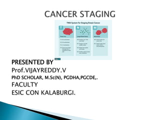
- 1. PRESENTED BY Prof.VIJAYREDDY.V PhD SCHOLAR, M.Sc(N), PGDHA,PGCDE,. FACULTY ESIC CON KALABURGI.
- 2. Cancer is a generic term for a large group of diseases that can affect any part of the body. Other terms used are malignant tumours and neoplasm’s. One defining feature of cancer is the rapid creation of abnormal cells that grow beyond their usual boundaries, and which can then invade adjoining parts of the body and spread to other organs, the latter process is referred to as metastasizing. Metastases are a major cause of death from cancer.
- 3. Cancer is the second leading cause of death globally, and is responsible for an estimated 9.6 million deaths in 2018. Globally, about 1 in 6 deaths is due to cancer. Approximately 70% of deaths from cancer occur in low- and middle-income countries. Around one third of deaths from cancer are due to the 5 leading behavioral and dietary risks: high body mass index, low fruit and vegetable intake, lack of physical activity, tobacco use, and alcohol use. Tobacco use is the most important risk factor for cancer and is responsible for approximately 22% of cancer deaths (2). Cancer causing infections, such as hepatitis and human papilloma virus (HPV), are responsible for up to 25% of cancer cases in low- and middle-income countries (3). Late-stage presentation and inaccessible diagnosis and treatment are common. In 2017, only 26% of low-income countries reported having pathology services generally available in the public sector. More than 90% of high-income countries reported treatment services are available compared to less than 30% of low-income countries. The economic impact of cancer is significant and is increasing. The total annual economic cost of cancer in 2010 was estimated at approximately US$ 1.16 trillion (4). Only 1 in 5 low- and middle-income countries have the necessary data to drive cancer policy (5).
- 5. Cancer arises from the transformation of normal cells into tumour cells in a multistage process that generally progresses from a pre-cancerous lesion to a malignant tumour. These changes are the result of the interaction between a person's genetic factors and 3 categories of external agents, including: physical carcinogens, such as ultraviolet and ionizing radiation; chemical carcinogens, such as asbestos, components of tobacco smoke, aflatoxin (a food contaminant), and arsenic (a drinking water contaminant); and biological carcinogens, such as infections from certain viruses, bacteria, or parasites. WHO, through its cancer research agency, International Agency for Research on Cancer (IARC), maintains a classification of cancer- causing agents.
- 6. Tobacco use, alcohol use, unhealthy diet, and physical inactivity are major cancer risk factors worldwide and are also the 4 shared risk factors for other noncommunicable diseases. Some chronic infections are risk factors for cancer and have major relevance in low- and middle-income countries. Approximately 15% of cancers diagnosed in 2012 were attributed to carcinogenic infections, including Helicobacter pylori, Human papillomavirus (HPV), Hepatitis B virus, Hepatitis C virus, and Epstein-Barr virus3. Hepatitis B and C virus and some types of HPV increase the risk for liver and cervical cancer, respectively. Infection with HIV substantially increases the risk of cancers such as cervical cancer.
- 7. Signs and symptoms caused by cancer will vary depending on what part of the body is affected. Some general signs and symptoms associated with, but not specific to, cancer, include: Fatigue Lump or area of thickening that can be felt under the skin Weight changes, including unintended loss or gain Skin changes, such as yellowing, darkening or redness of the skin, sores that won't heal, or changes to existing moles Changes in bowel or bladder habits Persistent cough or trouble breathing Difficulty swallowing Hoarseness Persistent indigestion or discomfort after eating Persistent, unexplained muscle or joint pain Persistent, unexplained fevers or night sweats Unexplained bleeding or bruising
- 8. Staging is the process of finding out how much cancer is in a person’s body and where it’s located. It’s how the doctor determines the stage of a person’s cancer. Why is cancer staging needed? For most types of cancer, doctors need to know how much cancer there is and where it is (among other things) to help determine the best treatment options. For example, the best treatment for an early-stage cancer may be surgery or radiation, while a more advanced-stage cancer may need treatments that reach all parts of the body, such as chemotherapy, targeted drug therapy, or immunotherapy.
- 9. Aid treatment planning, Provide an indication of prognosis, Assist in the evaluation of treatment results, Facilitate the exchange of information between treatment centres, Contribute to continuing investigations of human malignancies, Support cancer control activities, including through cancer registries.
- 10. What goes into the stage: The TNM system There are different types of systems used to stage cancer, but the most common and useful staging system for most types of cancer is the TNM system. The American Joint Committee on Cancer (AJCC) and the Union for International Cancer Control (UICC) maintain the TNM classification system as a way for doctors to stage many different types of cancer based on certain common standards. In the TNM system, the overall stage is determined after the cancer is assigned a letter or number to describe the tumor (T), node (N), and metastasis (M) categories.
- 11. In the TNM system, the overall stage is determined after the cancer is assigned a letter or number to describe the tumor (T), node (N), and metastasis (M) categories. T describes the original (primary) tumor. N tells whether the cancer has spread to the nearby lymph nodes. M tells whether the cancer has spread (metastasized) to distant parts of the body
- 12. Different types of exams and tests can be used to figure out a cancer’s stage. Depending on where the cancer is located, a physical exam may give some idea as to how much cancer there is. A biopsy often is needed to confirm a cancer diagnosis. Biopsies might also be needed to find out if a lump felt on an exam or if something seen on an imaging test in another part of the body is really from the spread of cancer. During a biopsy, the doctor removes a tumor or pieces of a tumor to be looked at in the lab. Some biopsies are done during surgery. But biopsies can also be done using a thin, hollow needle or through an endoscope. For more on biopsies, see Testing Biopsy and Cytology Specimens for Cancer. Lab tests of cancer cells (from a biopsy or surgery) and blood tests can also be used to help stage some types of cancer.
- 13. Imaging tests like x-rays, CT scans, MRIs, ultrasound, and PET scans may also give information about how much and where cancer is in the body. Endoscopy exams are sometimes used to look for cancer. For these exams, an endoscope, which is a thin, lighted tube (usually with a small video camera on the end) is put inside the body to look for cancer.
- 14. When trying to determine how much and where the cancer is in the body, doctors first look at the primary (main) tumor, which is where the cancer started. The tumor’s size, location, and whether it has grown into nearby areas can all be important. Doctors also check for other nearby tumors. The T category can be assigned a letter or a number: TX means there’s no information about the primary tumor, or it can’t be measured. T0 means there is no evidence of a primary tumor (it cannot be found). Tis means that the cancer cells are only growing in the layer of cells where they started, without growing into deeper layers. This may also be called in situ cancer or pre-cancer.
- 15. Lymph nodes near the primary tumor are usually are checked to find out if cancer has spread into them. Lymph nodes are small, bean-shaped collections of immune cells. Many types of cancer often spread to nearby lymph nodes before they reach other parts of the body. The N category can be assigned a letter or a number: NX means there’s no information about the nearby lymph nodes, or they can’t be assessed. N0 means nearby lymph nodes do not contain cancer. A number after the N (such as N1, N2, or N3) might describe the size, location, and/or the number of nearby lymph nodes affected by cancer. The higher the N number, the greater the cancer spread to nearby lymph nodes.
- 16. Doctors might also look at other parts of the body to see if the cancer has spread. Cancer spread to parts of the body far from the primary tumor is known as metastasis. The M category is assigned a number: M0 means that no distant cancer spread has been found. M1 means that the cancer has been found to have spread to distant organs or tissues. Each cancer type has its own version of the TNM categories, so letters and numbers don’t mean the same thing for every type of cancer. For example, for some types of cancer, the T categories describe the size of the main tumor, while for others they describe how deeply the tumor has grown into the organ it started in, or whether the tumor has grown into nearby structures (regardless of its size).
- 17. Tumour, node and metastasis (TNM) staging is the most common way that doctors stage stomach cancer. Doctors may also use the number staging system. The stage of a cancer tells you how big it is and how far it’s spread. It helps your doctor decide which treatment you need. Your scans will give some information about the stage of your cancer, but your doctor may not be able to tell you the exact stage until you have surgery.
- 18. Tumour describes the size of the tumour. There are 4 main stages of tumour size in stomach cancer. T1 means the tumour has started to grow into the wall of the stomach. It’s divided into T1a and T1b: T1a means the tumour is within the inner layers of the stomach (the mucosa) T1b means the tumour has grown through the mucosa and into a layer of supportive tissue called the submucosa T2 means the tumour has grown into the muscle layer of the stomach T3 means the tumour has grown into the outer lining of the stomach T4 means that the tumour has grown through the outer lining of the stomach. It’s divided into T4a and T4b: T4a means the tumour has broken through the outer lining of the stomach wall T4b means the tumour has grown through the stomach wall and into other organs or body structures nearby such as the liver, food pipe (oeosphagus) or abdominal wall
- 19. Node (N) describes whether the cancer has spread to the lymph nodes. Lymph nodes are a network of glands throughout the body, for example in your armpits, neck and groins. They drain away waste fluid, waste products and damaged cells, and contain cells that fight infection. There are 4 possible stages describing whether cancer cells are in the lymph nodes – N0, N1, N2 and N3: N0 means there are no lymph nodes containing cancer cells. N1 means there are cancer cells in 1 to 2 lymph nodes near to the stomach. N2 means there are cancer cells in 3 to 6 nearby lymph nodes. N3 is split into N3a and N3b: N3a means there are cancer cells in 7 to 15 nearby lymph nodes N3b means there are cancer cells in 16 or more nearby lymph nodes
- 20. Metastasis describes whether the cancer has spread to a different part of the body. There are 2 stages of metastasis: M0 means the cancer has not spread to other organs M1 means the cancer has spread to other parts of the body
- 21. https://www.who.int/ https://www.nccn.org https://www.cancer.org/ https://www.cancerresearchuk.org/ https://www.uicc.org/ https://www.mayoclinic.org/
- 22. Thank you