SlideShare uma empresa Scribd logo
1 de 14
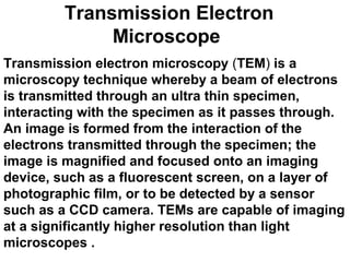
Transmission Electron
Microscope
Transmission electron microscopy (TEM) is a
microscopy technique whereby a beam of electrons
is transmitted through an ultra thin specimen,
interacting with the specimen as it passes through.
An image is formed from the interaction of the
electrons transmitted through the specimen; the
image is magnified and focused onto an imaging
device, such as a fluorescent screen, on a layer of
photographic film, or to be detected by a sensor
such as a CCD camera. TEMs are capable of imaging
at a significantly higher resolution than light
microscopes .
• The first TEM was built by Max Knoll and
Ernst Ruska in 1931, with this group
developing the first TEM with resolving
power greater than that of light in 1933 and
the first commercial TEM in 1939 .
• TEM forms a major analysis method in a range of
scientific fields, in both physical and biological
sciences. It find application in cancer research,
virology, materials science as well as pollution
and semiconductor research.
• TEMs use electrons as "light source" and their
much lower wavelength makes it possible to get a
resolution a thousand times better than with a
light microscope .
• The wavelength of electrons is found by equating
the de Broglie equation to the kinetic energy of an
electron., in a TEM an electron's velocity
approaches the speed of light, c
• where, h is Planck's constant, m0 is the rest mass
of an electron and E is the energy of the
accelerated electron. Electrons are usually
generated in an electron microscope by a process
known as thermionic emission from a filament,
usually tungsten, in the same manner as a light
bulb, or alternatively by field electron emission
.The electrons are then accelerated by an electric
potential (measured in volts) and focused by
electrostatic and electromagnetic lenses onto the
sample. The transmitted beam contains
information about electron density, phase and
periodicity; this beam is used to form an image.
METHODOLOGY
• A "light source" at the top of the microscope emits
the electrons that travel through vacuum in the
column of the microscope. Instead of glass lenses
focusing the light in the light microscope, the TEM
uses electromagnetic lenses to focus the
electrons into a very thin beam. The electron beam
then travels through the specimen you want to
study. Depending on the density of the material
present, some of the electrons are scattered and
disappear from the beam. At the bottom of the
microscope the unscattered electrons hit a
fluorescent screen, which gives rise to a parts
displayed in varied darkness according to their
density. The image can be studied directly by
operator or photographed with a camera.
SAMPLE PREPARATION
• Sample preparation in TEM can be a complex
procedure .The specimens are required to be at
most hundreds of nanometers thick. Preparation
of TEM specimens is specific to the material under
analysis and the desired information to obtain
from the specimen.
• Materials that have dimensions small enough to be
electron transparent, such as powders or
nanotubes, can be quickly prepared by the
deposition of a dilute sample containing the
specimen onto support grids or films .
• Biological specimens can be fixated using either a
negative staining material such as uranyl acetate
or by plastic embedding,or samples may be held
at liquid nitrogen temperatures after embedding in
vitreous ice. By passing samples over a glass or
diamond edge, small, thin sections can be readily
obtained using a semi-automated method or using
microtome. This method is used to obtain thin,
minimally deformed samples that allow for the
observation of tissue samples To prevent charge
build-up at the sample surface, tissue samples
need to be coated with a thin layer of conducting
material .
• TEM sample placing mesh.
SAMPLE STAINING
• TEM samples of biological tissues can utilize high
atomic number stains to enhance contrast. The
stain absorbs electrons or scatters part of the
electron beam which otherwise is projected onto
the imaging system. Compounds of heavy metals
such as osmium, lead, or uranium may be used
prior to TEM observation to selectively deposit
electron dense atoms in or on the sample in
desired cellular or protein regions, requiring an
understanding of how heavy metals bind to
biological tissues.
• Mechanical milling: Polishing needs to be done to
a high quality, to ensure constant sample
thickness across the region of interest .
• Chemical etching :Certain samples may be
prepared by chemical etching, particularly metallic
specimens
• More recently focussed ion beam methods have
been used to prepare samples. FIB is a relatively
new technique to prepare thin samples for TEM
examination from larger specimens.
LIMITATIONS
• There are a number of drawbacks to the TEM
technique. Many materials require extensive
sample preparation to produce a sample thin
enough to be electron transparent, which makes
TEM analysis a relatively time consuming process
with a low throughput of samples. The structure of
the sample may also be changed during the
preparation process. Also the field of view is
relatively small, raising the possibility that the
region analysed may not be characteristic of the
whole sample. There is potential that the sample
may be damaged by the electron beam,
particularly in the case of biological materials.

Mais conteúdo relacionado

Mais procurados

transmission Electron Microscopy (Tem)
transmission Electron Microscopy (Tem)transmission Electron Microscopy (Tem)
transmission Electron Microscopy (Tem)Sivasangari Shanmugam
 
Transmission Electron microscopy
Transmission Electron microscopyTransmission Electron microscopy
Transmission Electron microscopyAditya Singh
 
Scanning Electron microscopy
Scanning Electron microscopy Scanning Electron microscopy
Scanning Electron microscopy sagar sen sen
 
Transmission electron microscope
Transmission electron microscopeTransmission electron microscope
Transmission electron microscopeMonika Uma Shankar
 
Transmission electron microscope
Transmission electron microscopeTransmission electron microscope
Transmission electron microscopeSuganyaPaulraj
 
Transmission electron microscope
Transmission electron microscopeTransmission electron microscope
Transmission electron microscopeSizan Thapa
 
Electron microscope - SEM and TEM
Electron microscope - SEM and TEMElectron microscope - SEM and TEM
Electron microscope - SEM and TEMgaurav raja
 
Scanning and transmission electron microscopy
Scanning and transmission electron microscopyScanning and transmission electron microscopy
Scanning and transmission electron microscopygohil sanjay bhagvanji
 
transmission electron microscopy
transmission electron microscopytransmission electron microscopy
transmission electron microscopyJessa Ariño
 
Transmission Electron Microscope
Transmission Electron MicroscopeTransmission Electron Microscope
Transmission Electron MicroscopeManoranjan Ghosh
 
Scanning Electron Microscope
Scanning Electron Microscope Scanning Electron Microscope
Scanning Electron Microscope VrushankSalimath
 
Transmission electron microscope
Transmission electron microscopeTransmission electron microscope
Transmission electron microscopeSenthil Arasan
 
Transmission electron microscope (TEM)
Transmission electron microscope (TEM)Transmission electron microscope (TEM)
Transmission electron microscope (TEM)Nano Encryption
 

Mais procurados (20)

transmission Electron Microscopy (Tem)
transmission Electron Microscopy (Tem)transmission Electron Microscopy (Tem)
transmission Electron Microscopy (Tem)
 
Transmission Electron microscopy
Transmission Electron microscopyTransmission Electron microscopy
Transmission Electron microscopy
 
SEM- scanning electron microscope
SEM- scanning electron microscope SEM- scanning electron microscope
SEM- scanning electron microscope
 
Scanning Electron microscopy
Scanning Electron microscopy Scanning Electron microscopy
Scanning Electron microscopy
 
Tem ppt
Tem ppt Tem ppt
Tem ppt
 
Atomic Force Microscopy
Atomic Force MicroscopyAtomic Force Microscopy
Atomic Force Microscopy
 
Transmission electron microscope
Transmission electron microscopeTransmission electron microscope
Transmission electron microscope
 
Electron microscope
Electron microscopeElectron microscope
Electron microscope
 
Transmission electron microscope
Transmission electron microscopeTransmission electron microscope
Transmission electron microscope
 
Transmission electron microscope
Transmission electron microscopeTransmission electron microscope
Transmission electron microscope
 
Electron microscope - SEM and TEM
Electron microscope - SEM and TEMElectron microscope - SEM and TEM
Electron microscope - SEM and TEM
 
SEM & TEM
SEM & TEMSEM & TEM
SEM & TEM
 
Scanning and transmission electron microscopy
Scanning and transmission electron microscopyScanning and transmission electron microscopy
Scanning and transmission electron microscopy
 
transmission electron microscopy
transmission electron microscopytransmission electron microscopy
transmission electron microscopy
 
Transmission Electron Microscope
Transmission Electron MicroscopeTransmission Electron Microscope
Transmission Electron Microscope
 
Electron microscope
Electron microscopeElectron microscope
Electron microscope
 
Scanning Electron Microscope
Scanning Electron Microscope Scanning Electron Microscope
Scanning Electron Microscope
 
Transmission electron microscope
Transmission electron microscopeTransmission electron microscope
Transmission electron microscope
 
Transmission electron microscope (TEM)
Transmission electron microscope (TEM)Transmission electron microscope (TEM)
Transmission electron microscope (TEM)
 
Tem
TemTem
Tem
 

Destaque

Dna fingerprinting powerpoint
Dna fingerprinting powerpointDna fingerprinting powerpoint
Dna fingerprinting powerpointGenevia Vincent
 
Transmission Electron Microscope (TEM) for research (Full version)
Transmission Electron Microscope (TEM) for research (Full version)Transmission Electron Microscope (TEM) for research (Full version)
Transmission Electron Microscope (TEM) for research (Full version)Mohit Rajput
 
Types of electron microscope
Types of electron microscopeTypes of electron microscope
Types of electron microscopeNancy Bansal
 
Transmission electron microscopy (1) (1)
Transmission electron microscopy (1) (1)Transmission electron microscopy (1) (1)
Transmission electron microscopy (1) (1)Adnan Murad Bhayo
 
Tissue Management & Impression Technique
Tissue Management & Impression TechniqueTissue Management & Impression Technique
Tissue Management & Impression TechniquePacific University
 
Carpool Effects on Air Quality in Los Angeles
Carpool Effects on Air Quality in Los AngelesCarpool Effects on Air Quality in Los Angeles
Carpool Effects on Air Quality in Los AngelesJikun Lian EIT
 
2017 Ford Fusion near Wilmington | DE Ford ^
2017 Ford Fusion near Wilmington | DE Ford ^2017 Ford Fusion near Wilmington | DE Ford ^
2017 Ford Fusion near Wilmington | DE Ford ^Carman Ford
 
Wakhi language presentation
Wakhi language presentationWakhi language presentation
Wakhi language presentationBaig Ali
 
Herramientas WEB 2.0
Herramientas WEB 2.0Herramientas WEB 2.0
Herramientas WEB 2.0Jesus Lopez
 
NIDITE-Acuerdos-Acta-29-[04-junio-2015]
NIDITE-Acuerdos-Acta-29-[04-junio-2015]NIDITE-Acuerdos-Acta-29-[04-junio-2015]
NIDITE-Acuerdos-Acta-29-[04-junio-2015]NIDITE UPEL-IPRGR
 
Mechanical Engineer having 5+ years experience in Erection & Commissioning En...
Mechanical Engineer having 5+ years experience in Erection & Commissioning En...Mechanical Engineer having 5+ years experience in Erection & Commissioning En...
Mechanical Engineer having 5+ years experience in Erection & Commissioning En...Qamar Ahmad
 
Sequences pavagesposttchequie
Sequences pavagesposttchequieSequences pavagesposttchequie
Sequences pavagesposttchequiemassillonprimaire
 

Destaque (19)

Aox
AoxAox
Aox
 
Photoprotection in plants
Photoprotection in plantsPhotoprotection in plants
Photoprotection in plants
 
Dna fingerprinting powerpoint
Dna fingerprinting powerpointDna fingerprinting powerpoint
Dna fingerprinting powerpoint
 
Transmission Electron Microscope (TEM) for research (Full version)
Transmission Electron Microscope (TEM) for research (Full version)Transmission Electron Microscope (TEM) for research (Full version)
Transmission Electron Microscope (TEM) for research (Full version)
 
Types of electron microscope
Types of electron microscopeTypes of electron microscope
Types of electron microscope
 
Transmission electron microscopy (1) (1)
Transmission electron microscopy (1) (1)Transmission electron microscopy (1) (1)
Transmission electron microscopy (1) (1)
 
Tem
TemTem
Tem
 
Tissue Management & Impression Technique
Tissue Management & Impression TechniqueTissue Management & Impression Technique
Tissue Management & Impression Technique
 
Journal poupée turquie
Journal poupée turquieJournal poupée turquie
Journal poupée turquie
 
Carpool Effects on Air Quality in Los Angeles
Carpool Effects on Air Quality in Los AngelesCarpool Effects on Air Quality in Los Angeles
Carpool Effects on Air Quality in Los Angeles
 
2017 Ford Fusion near Wilmington | DE Ford ^
2017 Ford Fusion near Wilmington | DE Ford ^2017 Ford Fusion near Wilmington | DE Ford ^
2017 Ford Fusion near Wilmington | DE Ford ^
 
Wakhi language presentation
Wakhi language presentationWakhi language presentation
Wakhi language presentation
 
Herramientas WEB 2.0
Herramientas WEB 2.0Herramientas WEB 2.0
Herramientas WEB 2.0
 
Biosensorppt2
Biosensorppt2Biosensorppt2
Biosensorppt2
 
NIDITE-Acuerdos-Acta-29-[04-junio-2015]
NIDITE-Acuerdos-Acta-29-[04-junio-2015]NIDITE-Acuerdos-Acta-29-[04-junio-2015]
NIDITE-Acuerdos-Acta-29-[04-junio-2015]
 
Mechanical Engineer having 5+ years experience in Erection & Commissioning En...
Mechanical Engineer having 5+ years experience in Erection & Commissioning En...Mechanical Engineer having 5+ years experience in Erection & Commissioning En...
Mechanical Engineer having 5+ years experience in Erection & Commissioning En...
 
Chloroplastgee1
Chloroplastgee1Chloroplastgee1
Chloroplastgee1
 
ECE 468 Lab Project 1
ECE 468 Lab Project 1ECE 468 Lab Project 1
ECE 468 Lab Project 1
 
Sequences pavagesposttchequie
Sequences pavagesposttchequieSequences pavagesposttchequie
Sequences pavagesposttchequie
 

Semelhante a Transmission electron microscopeppt

Powerpoint presentation on electron microscopy
Powerpoint presentation on electron microscopyPowerpoint presentation on electron microscopy
Powerpoint presentation on electron microscopykumar virbhadra
 
The Transmission electron microscope .pdf
The Transmission electron microscope .pdfThe Transmission electron microscope .pdf
The Transmission electron microscope .pdfbuckbobby123
 
Electron microscopy ameena
Electron microscopy ameenaElectron microscopy ameena
Electron microscopy ameenaAnnakurian9
 
Lec 3; Microscopy ALI.pptx
Lec 3; Microscopy ALI.pptxLec 3; Microscopy ALI.pptx
Lec 3; Microscopy ALI.pptxalihaider64675
 
Electron microscope
Electron microscopeElectron microscope
Electron microscopeReena Rai
 
Scanning and transmission electroscopy.pptx
Scanning and transmission electroscopy.pptxScanning and transmission electroscopy.pptx
Scanning and transmission electroscopy.pptxNirmal P J
 
ELECTRON MICROSCOPE AND ITS APPLICATION.pptx
ELECTRON MICROSCOPE AND ITS APPLICATION.pptxELECTRON MICROSCOPE AND ITS APPLICATION.pptx
ELECTRON MICROSCOPE AND ITS APPLICATION.pptxPallaviKumari112
 
10. The Transmission Electron Microscope (TEM).pptx
10. The Transmission Electron Microscope (TEM).pptx10. The Transmission Electron Microscope (TEM).pptx
10. The Transmission Electron Microscope (TEM).pptxRENERGISTICS
 
Transmission Electron Microscopy (TEM)
Transmission Electron Microscopy (TEM) Transmission Electron Microscopy (TEM)
Transmission Electron Microscopy (TEM) Zohaib HUSSAIN
 
Electron microscope, principle and application
Electron microscope, principle and applicationElectron microscope, principle and application
Electron microscope, principle and applicationKAUSHAL SAHU
 
Electron microscopy by nishi kumari
Electron microscopy  by nishi kumariElectron microscopy  by nishi kumari
Electron microscopy by nishi kumariNishi Sharma
 
Scanning electron microscopy
Scanning electron microscopyScanning electron microscopy
Scanning electron microscopyJessa Ariño
 
ELECTRON MICROSCOPE TYPES: SEM & TEM NOTES PPT
ELECTRON MICROSCOPE TYPES: SEM & TEM  NOTES PPTELECTRON MICROSCOPE TYPES: SEM & TEM  NOTES PPT
ELECTRON MICROSCOPE TYPES: SEM & TEM NOTES PPTSruthyPB3
 
ect 292 nano electronics
ect 292 nano electronicsect 292 nano electronics
ect 292 nano electronicsRenjithThomas13
 

Semelhante a Transmission electron microscopeppt (20)

Powerpoint presentation on electron microscopy
Powerpoint presentation on electron microscopyPowerpoint presentation on electron microscopy
Powerpoint presentation on electron microscopy
 
The Transmission electron microscope .pdf
The Transmission electron microscope .pdfThe Transmission electron microscope .pdf
The Transmission electron microscope .pdf
 
Electron microscopy ameena
Electron microscopy ameenaElectron microscopy ameena
Electron microscopy ameena
 
Lec 3; Microscopy ALI.pptx
Lec 3; Microscopy ALI.pptxLec 3; Microscopy ALI.pptx
Lec 3; Microscopy ALI.pptx
 
Electron microscope
Electron microscopeElectron microscope
Electron microscope
 
Scanning and transmission electroscopy.pptx
Scanning and transmission electroscopy.pptxScanning and transmission electroscopy.pptx
Scanning and transmission electroscopy.pptx
 
ELECTRON MICROSCOPE AND ITS APPLICATION.pptx
ELECTRON MICROSCOPE AND ITS APPLICATION.pptxELECTRON MICROSCOPE AND ITS APPLICATION.pptx
ELECTRON MICROSCOPE AND ITS APPLICATION.pptx
 
10. The Transmission Electron Microscope (TEM).pptx
10. The Transmission Electron Microscope (TEM).pptx10. The Transmission Electron Microscope (TEM).pptx
10. The Transmission Electron Microscope (TEM).pptx
 
Transmission Electron Microscopy (TEM)
Transmission Electron Microscopy (TEM) Transmission Electron Microscopy (TEM)
Transmission Electron Microscopy (TEM)
 
Electron microscope, principle and application
Electron microscope, principle and applicationElectron microscope, principle and application
Electron microscope, principle and application
 
Electron microscope
Electron microscopeElectron microscope
Electron microscope
 
Sem n tem
Sem n temSem n tem
Sem n tem
 
Dr nh mat.char notes
Dr nh   mat.char  notesDr nh   mat.char  notes
Dr nh mat.char notes
 
Electron microscopy by nishi kumari
Electron microscopy  by nishi kumariElectron microscopy  by nishi kumari
Electron microscopy by nishi kumari
 
Transmission Electron Microscopy
Transmission Electron MicroscopyTransmission Electron Microscopy
Transmission Electron Microscopy
 
Scanning and Transmission Electron Microscope
Scanning and Transmission Electron MicroscopeScanning and Transmission Electron Microscope
Scanning and Transmission Electron Microscope
 
Scanning electron microscopy
Scanning electron microscopyScanning electron microscopy
Scanning electron microscopy
 
ELECTRON MICROSCOPE TYPES: SEM & TEM NOTES PPT
ELECTRON MICROSCOPE TYPES: SEM & TEM  NOTES PPTELECTRON MICROSCOPE TYPES: SEM & TEM  NOTES PPT
ELECTRON MICROSCOPE TYPES: SEM & TEM NOTES PPT
 
ect 292 nano electronics
ect 292 nano electronicsect 292 nano electronics
ect 292 nano electronics
 
5,21 (TEM).pptx
5,21 (TEM).pptx5,21 (TEM).pptx
5,21 (TEM).pptx
 

Mais de Genevia Vincent (20)

Spinal muscular atrophyppt
Spinal muscular atrophypptSpinal muscular atrophyppt
Spinal muscular atrophyppt
 
Sexlinked1
Sexlinked1Sexlinked1
Sexlinked1
 
Scanning electron microscopyppt
Scanning electron microscopypptScanning electron microscopyppt
Scanning electron microscopyppt
 
Rotenoneppt
RotenonepptRotenoneppt
Rotenoneppt
 
Rotenoneppt
RotenonepptRotenoneppt
Rotenoneppt
 
Retinoblastomappt
RetinoblastomapptRetinoblastomappt
Retinoblastomappt
 
Pyrethroids
PyrethroidsPyrethroids
Pyrethroids
 
Presentation1immbol
Presentation1immbolPresentation1immbol
Presentation1immbol
 
Presentation1gee3
Presentation1gee3Presentation1gee3
Presentation1gee3
 
Pptgee
PptgeePptgee
Pptgee
 
Ppt toxicology
Ppt toxicologyPpt toxicology
Ppt toxicology
 
Ppt idc fam
Ppt idc famPpt idc fam
Ppt idc fam
 
Poliomyelitis1
Poliomyelitis1Poliomyelitis1
Poliomyelitis1
 
Poliomyelitis1
Poliomyelitis1Poliomyelitis1
Poliomyelitis1
 
Nucleusppt
NucleuspptNucleusppt
Nucleusppt
 
Nuclear ppt3
Nuclear ppt3Nuclear ppt3
Nuclear ppt3
 
Mitodiseaseppt
MitodiseasepptMitodiseaseppt
Mitodiseaseppt
 
Microscopes.ppt(1)
Microscopes.ppt(1)Microscopes.ppt(1)
Microscopes.ppt(1)
 
Microscope3
Microscope3Microscope3
Microscope3
 
Microscope(222)
Microscope(222)Microscope(222)
Microscope(222)
 

Último

module for grade 9 for distance learning
module for grade 9 for distance learningmodule for grade 9 for distance learning
module for grade 9 for distance learninglevieagacer
 
biology HL practice questions IB BIOLOGY
biology HL practice questions IB BIOLOGYbiology HL practice questions IB BIOLOGY
biology HL practice questions IB BIOLOGY1301aanya
 
Gwalior ❤CALL GIRL 84099*07087 ❤CALL GIRLS IN Gwalior ESCORT SERVICE❤CALL GIRL
Gwalior ❤CALL GIRL 84099*07087 ❤CALL GIRLS IN Gwalior ESCORT SERVICE❤CALL GIRLGwalior ❤CALL GIRL 84099*07087 ❤CALL GIRLS IN Gwalior ESCORT SERVICE❤CALL GIRL
Gwalior ❤CALL GIRL 84099*07087 ❤CALL GIRLS IN Gwalior ESCORT SERVICE❤CALL GIRLkantirani197
 
Porella : features, morphology, anatomy, reproduction etc.
Porella : features, morphology, anatomy, reproduction etc.Porella : features, morphology, anatomy, reproduction etc.
Porella : features, morphology, anatomy, reproduction etc.Silpa
 
Grade 7 - Lesson 1 - Microscope and Its Functions
Grade 7 - Lesson 1 - Microscope and Its FunctionsGrade 7 - Lesson 1 - Microscope and Its Functions
Grade 7 - Lesson 1 - Microscope and Its FunctionsOrtegaSyrineMay
 
Molecular markers- RFLP, RAPD, AFLP, SNP etc.
Molecular markers- RFLP, RAPD, AFLP, SNP etc.Molecular markers- RFLP, RAPD, AFLP, SNP etc.
Molecular markers- RFLP, RAPD, AFLP, SNP etc.Silpa
 
300003-World Science Day For Peace And Development.pptx
300003-World Science Day For Peace And Development.pptx300003-World Science Day For Peace And Development.pptx
300003-World Science Day For Peace And Development.pptxryanrooker
 
Cyathodium bryophyte: morphology, anatomy, reproduction etc.
Cyathodium bryophyte: morphology, anatomy, reproduction etc.Cyathodium bryophyte: morphology, anatomy, reproduction etc.
Cyathodium bryophyte: morphology, anatomy, reproduction etc.Silpa
 
Factory Acceptance Test( FAT).pptx .
Factory Acceptance Test( FAT).pptx       .Factory Acceptance Test( FAT).pptx       .
Factory Acceptance Test( FAT).pptx .Poonam Aher Patil
 
(May 9, 2024) Enhanced Ultrafast Vector Flow Imaging (VFI) Using Multi-Angle ...
(May 9, 2024) Enhanced Ultrafast Vector Flow Imaging (VFI) Using Multi-Angle ...(May 9, 2024) Enhanced Ultrafast Vector Flow Imaging (VFI) Using Multi-Angle ...
(May 9, 2024) Enhanced Ultrafast Vector Flow Imaging (VFI) Using Multi-Angle ...Scintica Instrumentation
 
Dr. E. Muralinath_ Blood indices_clinical aspects
Dr. E. Muralinath_ Blood indices_clinical  aspectsDr. E. Muralinath_ Blood indices_clinical  aspects
Dr. E. Muralinath_ Blood indices_clinical aspectsmuralinath2
 
Digital Dentistry.Digital Dentistryvv.pptx
Digital Dentistry.Digital Dentistryvv.pptxDigital Dentistry.Digital Dentistryvv.pptx
Digital Dentistry.Digital Dentistryvv.pptxMohamedFarag457087
 
Module for Grade 9 for Asynchronous/Distance learning
Module for Grade 9 for Asynchronous/Distance learningModule for Grade 9 for Asynchronous/Distance learning
Module for Grade 9 for Asynchronous/Distance learninglevieagacer
 
CURRENT SCENARIO OF POULTRY PRODUCTION IN INDIA
CURRENT SCENARIO OF POULTRY PRODUCTION IN INDIACURRENT SCENARIO OF POULTRY PRODUCTION IN INDIA
CURRENT SCENARIO OF POULTRY PRODUCTION IN INDIADr. TATHAGAT KHOBRAGADE
 
Phenolics: types, biosynthesis and functions.
Phenolics: types, biosynthesis and functions.Phenolics: types, biosynthesis and functions.
Phenolics: types, biosynthesis and functions.Silpa
 
Thyroid Physiology_Dr.E. Muralinath_ Associate Professor
Thyroid Physiology_Dr.E. Muralinath_ Associate ProfessorThyroid Physiology_Dr.E. Muralinath_ Associate Professor
Thyroid Physiology_Dr.E. Muralinath_ Associate Professormuralinath2
 
LUNULARIA -features, morphology, anatomy ,reproduction etc.
LUNULARIA -features, morphology, anatomy ,reproduction etc.LUNULARIA -features, morphology, anatomy ,reproduction etc.
LUNULARIA -features, morphology, anatomy ,reproduction etc.Silpa
 
Climate Change Impacts on Terrestrial and Aquatic Ecosystems.pptx
Climate Change Impacts on Terrestrial and Aquatic Ecosystems.pptxClimate Change Impacts on Terrestrial and Aquatic Ecosystems.pptx
Climate Change Impacts on Terrestrial and Aquatic Ecosystems.pptxDiariAli
 

Último (20)

module for grade 9 for distance learning
module for grade 9 for distance learningmodule for grade 9 for distance learning
module for grade 9 for distance learning
 
biology HL practice questions IB BIOLOGY
biology HL practice questions IB BIOLOGYbiology HL practice questions IB BIOLOGY
biology HL practice questions IB BIOLOGY
 
Gwalior ❤CALL GIRL 84099*07087 ❤CALL GIRLS IN Gwalior ESCORT SERVICE❤CALL GIRL
Gwalior ❤CALL GIRL 84099*07087 ❤CALL GIRLS IN Gwalior ESCORT SERVICE❤CALL GIRLGwalior ❤CALL GIRL 84099*07087 ❤CALL GIRLS IN Gwalior ESCORT SERVICE❤CALL GIRL
Gwalior ❤CALL GIRL 84099*07087 ❤CALL GIRLS IN Gwalior ESCORT SERVICE❤CALL GIRL
 
Porella : features, morphology, anatomy, reproduction etc.
Porella : features, morphology, anatomy, reproduction etc.Porella : features, morphology, anatomy, reproduction etc.
Porella : features, morphology, anatomy, reproduction etc.
 
Grade 7 - Lesson 1 - Microscope and Its Functions
Grade 7 - Lesson 1 - Microscope and Its FunctionsGrade 7 - Lesson 1 - Microscope and Its Functions
Grade 7 - Lesson 1 - Microscope and Its Functions
 
Molecular markers- RFLP, RAPD, AFLP, SNP etc.
Molecular markers- RFLP, RAPD, AFLP, SNP etc.Molecular markers- RFLP, RAPD, AFLP, SNP etc.
Molecular markers- RFLP, RAPD, AFLP, SNP etc.
 
300003-World Science Day For Peace And Development.pptx
300003-World Science Day For Peace And Development.pptx300003-World Science Day For Peace And Development.pptx
300003-World Science Day For Peace And Development.pptx
 
Cyathodium bryophyte: morphology, anatomy, reproduction etc.
Cyathodium bryophyte: morphology, anatomy, reproduction etc.Cyathodium bryophyte: morphology, anatomy, reproduction etc.
Cyathodium bryophyte: morphology, anatomy, reproduction etc.
 
Factory Acceptance Test( FAT).pptx .
Factory Acceptance Test( FAT).pptx       .Factory Acceptance Test( FAT).pptx       .
Factory Acceptance Test( FAT).pptx .
 
PATNA CALL GIRLS 8617370543 LOW PRICE ESCORT SERVICE
PATNA CALL GIRLS 8617370543 LOW PRICE ESCORT SERVICEPATNA CALL GIRLS 8617370543 LOW PRICE ESCORT SERVICE
PATNA CALL GIRLS 8617370543 LOW PRICE ESCORT SERVICE
 
(May 9, 2024) Enhanced Ultrafast Vector Flow Imaging (VFI) Using Multi-Angle ...
(May 9, 2024) Enhanced Ultrafast Vector Flow Imaging (VFI) Using Multi-Angle ...(May 9, 2024) Enhanced Ultrafast Vector Flow Imaging (VFI) Using Multi-Angle ...
(May 9, 2024) Enhanced Ultrafast Vector Flow Imaging (VFI) Using Multi-Angle ...
 
Dr. E. Muralinath_ Blood indices_clinical aspects
Dr. E. Muralinath_ Blood indices_clinical  aspectsDr. E. Muralinath_ Blood indices_clinical  aspects
Dr. E. Muralinath_ Blood indices_clinical aspects
 
Digital Dentistry.Digital Dentistryvv.pptx
Digital Dentistry.Digital Dentistryvv.pptxDigital Dentistry.Digital Dentistryvv.pptx
Digital Dentistry.Digital Dentistryvv.pptx
 
Module for Grade 9 for Asynchronous/Distance learning
Module for Grade 9 for Asynchronous/Distance learningModule for Grade 9 for Asynchronous/Distance learning
Module for Grade 9 for Asynchronous/Distance learning
 
CURRENT SCENARIO OF POULTRY PRODUCTION IN INDIA
CURRENT SCENARIO OF POULTRY PRODUCTION IN INDIACURRENT SCENARIO OF POULTRY PRODUCTION IN INDIA
CURRENT SCENARIO OF POULTRY PRODUCTION IN INDIA
 
Phenolics: types, biosynthesis and functions.
Phenolics: types, biosynthesis and functions.Phenolics: types, biosynthesis and functions.
Phenolics: types, biosynthesis and functions.
 
Site Acceptance Test .
Site Acceptance Test                    .Site Acceptance Test                    .
Site Acceptance Test .
 
Thyroid Physiology_Dr.E. Muralinath_ Associate Professor
Thyroid Physiology_Dr.E. Muralinath_ Associate ProfessorThyroid Physiology_Dr.E. Muralinath_ Associate Professor
Thyroid Physiology_Dr.E. Muralinath_ Associate Professor
 
LUNULARIA -features, morphology, anatomy ,reproduction etc.
LUNULARIA -features, morphology, anatomy ,reproduction etc.LUNULARIA -features, morphology, anatomy ,reproduction etc.
LUNULARIA -features, morphology, anatomy ,reproduction etc.
 
Climate Change Impacts on Terrestrial and Aquatic Ecosystems.pptx
Climate Change Impacts on Terrestrial and Aquatic Ecosystems.pptxClimate Change Impacts on Terrestrial and Aquatic Ecosystems.pptx
Climate Change Impacts on Terrestrial and Aquatic Ecosystems.pptx
 

Transmission electron microscopeppt

  • 1. Transmission Electron Microscope Transmission electron microscopy (TEM) is a microscopy technique whereby a beam of electrons is transmitted through an ultra thin specimen, interacting with the specimen as it passes through. An image is formed from the interaction of the electrons transmitted through the specimen; the image is magnified and focused onto an imaging device, such as a fluorescent screen, on a layer of photographic film, or to be detected by a sensor such as a CCD camera. TEMs are capable of imaging at a significantly higher resolution than light microscopes .
  • 2.
  • 3. • The first TEM was built by Max Knoll and Ernst Ruska in 1931, with this group developing the first TEM with resolving power greater than that of light in 1933 and the first commercial TEM in 1939 .
  • 4.
  • 5. • TEM forms a major analysis method in a range of scientific fields, in both physical and biological sciences. It find application in cancer research, virology, materials science as well as pollution and semiconductor research. • TEMs use electrons as "light source" and their much lower wavelength makes it possible to get a resolution a thousand times better than with a light microscope .
  • 6. • The wavelength of electrons is found by equating the de Broglie equation to the kinetic energy of an electron., in a TEM an electron's velocity approaches the speed of light, c • where, h is Planck's constant, m0 is the rest mass of an electron and E is the energy of the accelerated electron. Electrons are usually generated in an electron microscope by a process known as thermionic emission from a filament, usually tungsten, in the same manner as a light bulb, or alternatively by field electron emission .The electrons are then accelerated by an electric potential (measured in volts) and focused by electrostatic and electromagnetic lenses onto the sample. The transmitted beam contains information about electron density, phase and periodicity; this beam is used to form an image.
  • 7.
  • 8. METHODOLOGY • A "light source" at the top of the microscope emits the electrons that travel through vacuum in the column of the microscope. Instead of glass lenses focusing the light in the light microscope, the TEM uses electromagnetic lenses to focus the electrons into a very thin beam. The electron beam then travels through the specimen you want to study. Depending on the density of the material present, some of the electrons are scattered and disappear from the beam. At the bottom of the microscope the unscattered electrons hit a fluorescent screen, which gives rise to a parts displayed in varied darkness according to their density. The image can be studied directly by operator or photographed with a camera.
  • 9. SAMPLE PREPARATION • Sample preparation in TEM can be a complex procedure .The specimens are required to be at most hundreds of nanometers thick. Preparation of TEM specimens is specific to the material under analysis and the desired information to obtain from the specimen. • Materials that have dimensions small enough to be electron transparent, such as powders or nanotubes, can be quickly prepared by the deposition of a dilute sample containing the specimen onto support grids or films .
  • 10. • Biological specimens can be fixated using either a negative staining material such as uranyl acetate or by plastic embedding,or samples may be held at liquid nitrogen temperatures after embedding in vitreous ice. By passing samples over a glass or diamond edge, small, thin sections can be readily obtained using a semi-automated method or using microtome. This method is used to obtain thin, minimally deformed samples that allow for the observation of tissue samples To prevent charge build-up at the sample surface, tissue samples need to be coated with a thin layer of conducting material .
  • 11. • TEM sample placing mesh.
  • 12. SAMPLE STAINING • TEM samples of biological tissues can utilize high atomic number stains to enhance contrast. The stain absorbs electrons or scatters part of the electron beam which otherwise is projected onto the imaging system. Compounds of heavy metals such as osmium, lead, or uranium may be used prior to TEM observation to selectively deposit electron dense atoms in or on the sample in desired cellular or protein regions, requiring an understanding of how heavy metals bind to biological tissues.
  • 13. • Mechanical milling: Polishing needs to be done to a high quality, to ensure constant sample thickness across the region of interest . • Chemical etching :Certain samples may be prepared by chemical etching, particularly metallic specimens • More recently focussed ion beam methods have been used to prepare samples. FIB is a relatively new technique to prepare thin samples for TEM examination from larger specimens.
  • 14. LIMITATIONS • There are a number of drawbacks to the TEM technique. Many materials require extensive sample preparation to produce a sample thin enough to be electron transparent, which makes TEM analysis a relatively time consuming process with a low throughput of samples. The structure of the sample may also be changed during the preparation process. Also the field of view is relatively small, raising the possibility that the region analysed may not be characteristic of the whole sample. There is potential that the sample may be damaged by the electron beam, particularly in the case of biological materials.