

Recomendados
Mais conteúdo relacionado
Mais procurados
Mais procurados (20)
Semelhante a Opthalmology
Semelhante a Opthalmology (20)
Mais de Verdah Sabih
Último
Último (20)
Opthalmology
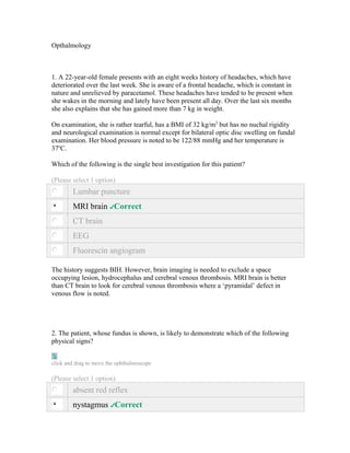
- 1. Opthalmology 1. A 22-year-old female presents with an eight weeks history of headaches, which have deteriorated over the last week. She is aware of a frontal headache, which is constant in nature and unrelieved by paracetamol. These headaches have tended to be present when she wakes in the morning and lately have been present all day. Over the last six months she also explains that she has gained more than 7 kg in weight. On examination, she is rather tearful, has a BMI of 32 kg/m2 but has no nuchal rigidity and neurological examination is normal except for bilateral optic disc swelling on fundal examination. Her blood pressure is noted to be 122/88 mmHg and her temperature is 37oC. Which of the following is the single best investigation for this patient? (Please select 1 option) Lumbar puncture MRI brain Correct CT brain EEG Fluorescin angiogram The history suggests BIH. However, brain imaging is needed to exclude a space occupying lesion, hydrocephalus and cerebral venous thrombosis. MRI brain is better than CT brain to look for cerebral venous thrombosis where a ‘pyramidal’ defect in venous flow is noted. 2. The patient, whose fundus is shown, is likely to demonstrate which of the following physical signs? click and drag to move the ophthalmoscope (Please select 1 option) absent red reflex nystagmus Correct
- 2. ptosis rubeosis iridis xanthelasma This is an albino fundus - there is no retinal pigmentation and all the blood vessels can be clearly seen. Nystagmus and photophobia are common findings in albinos. 3. These are ophthalmoscopic appearances of a 62-year-old male who has recently been diagnosed with diabetes mellitus. He has been referred by his optician after noting the visual acuity in his left eye is 6/36 and 6/12 in his right. There is no correction with pin hole. What is the likely cause of his fundal appearance? (Please select 1 option) Glaucoma Correct Hypertensive retinopathy Macular degeneration Proliferative diabetic retinopathy Retinitis pigmentosa This patient has optic atrophy as revealed by a particularly pale disc. Causes include Glaucoma, external compression of the optic nerves eg pituitary tumour and Multiple sclerosis. 4. This 74-year-old man presents with poor vision. click and drag to move the ophthalmoscope What is the diagnosis? (Please select 1 option) diabetic retinopathy
- 3. glaucoma hypertensive retinopathy macular degeneration Correct pan-retinal photocoagulation The fundus shows small pale dots over the macular area typical of Drusen. This is macular degneration and one of the commonest causes of blindness. 5. With what diseases is this appearance associated?
- 4. (Please select 3 options) Acromegaly Correct Poorly controlled diabetes mellitus Pseudoxanthoma elasticum Correct Chronic lymphocytic leukaemia Hypertriglyceridaemia Paget's disease of bone Correct Acquired Immune Deficiency Syndrome Retinitis pigmentosa Cytomegalovirus infection
- 5. Closed angle glaucoma The slide shows angioid streaks, caused by breaks in Bruch's membrane. Angioid streaks may be associated with pseudoxanthoma elasticum, Ehlers-Danlos syndrome, sickle cell disease, Acromegaly (and other pituitary disorders), and Paget's disease. 6. These are the ophthalmoscopic appearances of a 74-year-old female who presents with a long history of deterioration in her vision. Her vision is 6/36 in both eyes and uncorrected with a pinhole. What is the most likely diagnosis? (Please select 1 option) Glaucoma Correct Macular degeneration Optic atrophy Papilloedema Retinitis pigmentosa This fundus shows cupping of the optic disc which is typical of glaucoma. Almost invariably there is an increase in pressure which is sufficient to cause damage to the optic nerve head and causes changes in the visual field. The mean value for intraocular pressure is 15-16 mm Hg with a standard deviation of +/- 2.5. The upper limit of normal is considered to be 21 mm Hg. 7. A 35 year-old woman with sickle cell disease was admitted to hospital with abdominal pain. This appearance was noted on fundoscopy.
- 6. What is the diagnosis? (Please select 1 option) Branch retinal artery occlusion Angioid streaks Correct Papilloedema Central retinal vein occlusion Lipaemia reinalis The slide shows angioid streaks, caused by breaks in Bruch's membrane.
- 7. 8. This 42-year-old lady has a history of repeated bone fractures. What is the diagnosis? (Please select 1 option) Familial hypercholesterolaemia Gaucher disease Correct Sarcoidosis Phenylketonuria Wilson's disease The slide shows yellow papules (pingeculae) in the cornea; these are characteristic of Gaucher disease. Gaucher disease is inherited as an autosomal recessive disease. Disease is caused by a deficiency of the enzyme glucocerbrosidase, essentail for the metabolism of glycolipids. There are three types of Gaucher disease: 1. Type 1 (chronic non-neuropathic; adult Gaucher disease) 2. Type 2 (acute neuropathic; infentile Gaucher disease) 3. Type 3 (subacute neuropathic; juvenile Gaucher disease) Patients with all types of disease have hepatosplenomegaly and large glucocerebroside- rich cells (Gaucher cells) infiltrating the bone marrow. Type 2 (infentile Gaucher disease) carries the worst prognosis, with children seldom surviving beyond 2 years. Type 1 disease is the commonest, usually presenting in childhood with hepatosplenomegaly, but not uncommonly in middle- or old-age. Bone marrow replacement and hypersplenism result in anaemia and thrombocytopoenia. Pathological bone fractures and avascular necrosis of the femoral heads are not uncommon. Bony disease may be confined to the distal ends of the femurs, with formation of characteristic 'Erlenmeyer flask' shaped cysts. The skin may show a grey-brown discolouration, especially around the forehead, hands and pre-tibial regions. Characteristic yellow or yellow-brown papules (pingeculae) develop at the sclerocorneal junctions.
- 8. 9. A 76-year-old male presents with visual loss. Examination reveals a blood pressure of 168/102 mmHg and fundoscopy shows an embolus to right superior temporal branch of the retinal artery. Carotid dopplers are arranged and these show normal left sided carotids, but 90% Right External Carotid Artery stenosis with approximately 50% Right Internal Carotid Artery stenosis. What would be the most appropriate treatment? (Please select 1 option) Aspirin Correct Dipyridamole Right External Carotid Artery endarterectomy Right Internal Carotid Artery endarterectomy Warfarin Endarterectomy is only beneficial if internal carotid artery stenosis is greater than 70%. If less than that aspirin and control of vascular risk factors is the treatment of choice. In this case hypertensive control is advised. 10. A 24-year-old female presents with an eight weeks history of headaches, which have deteriorated over the last week. She is aware of a frontal headache, which is constant in nature and unrelieved by paracetamol. These headaches have tended to be present when she wakes in the morning and lately have been present all day. Over the last six months she also explains that she has gained more than 7 kg in weight. On examination, she is rather tearful, has a BMI of 32 kg/m2 but has no nuchal rigidity and neurological examination is normal except for bilateral optic disc swelling on fundal examination. Her blood pressure is noted to be 122/88 mmHg and her temperature is 37oC. An MRI scan of her brain is normal and LP reveals an opening pressure of 30 cm H2O but normal CSF analysis. What treatment would you offer this patient if her vision were threatened? (Please select 1 option) IV dexamethasone Acetazolamide Lumbo-peritoneal shunt Correct
- 9. IV mannitol Furosemide Visual loss is the single threatening complication of BIH. Urgent LP shunt is the treatment of choice Optic nerve fenestration is an alternative. There are no comparative studies between the two interventions. 11. A 64-year-old lady presented to the Accident and Emergency Unit with orbital pain and swelling (shown below). She had been under review in the endocrinology clinic and had been started on some new treatment four weeks previously. Thyroid function four weeks ago showed: T4 99 µg/dL (5-12) TSH <0.04 mU/L (0.4-6.0) Thyroid function in A&E shows: T4 2 µg/dL (5-12) TSH 28 mU/L (0.4-6.0)
- 10. What treatment has she received? (Please select 1 option) Carbimazole Prednisolone Propranolol Propylthiouracil Radioiodine Correct The slide shows malignant exophthalmos: malignant refers to the rapidity of onset and threat to eyesight rather than association with malignancy. Radioiodine therapy can worsen thyroid-associated ophthalmopathy patients with thyroid eye disease are generally treated with steroids for 1-2 weeks prior to starting radioiodine therapy. Treatment for malignant exophthalmos is rapid administration of steroids. Where sight is threatened, orbital decompression may be necessary.
- 11. 12. A 35 year-old man with HIV disease presents with sudden loss of vision in his right eye. He is not curently taking antiretroviral therapy. A recent CD4 count was measured at 20 cells/mm3. What is the most appropriate therapy to initiate? (Please select 1 option) High-dose intravenous aciclovir Intravenous ganciclovir Correct Zidovudine + lamivudine + nevirapine Sulfadiazine + pyrimethamine Amphotericin B + flucytosine The slide shows the typical 'cottage cheese and tomato ketchup' or 'pizza' appearance of CMV retinitis in a patient with HIV disease. Intravenous ganciclovir is currently the treatment of choice. Available forms of ganciclovir include intravenous and oral preparations as well as an ocular implant. The principal disadvantages of the latter two are: the poor bioavailability of the oral preparation; the implant is effective at clearing ocular disease, but CMV is disseminated at this stage of AIDS and the implant does not address systemic infection. Pyrimethamine + sulfadiazine are used to treat toxoplams retinitis; zidovudine + lamivudine + nelfinavir constitutes highly active antiretrovral ('combination') therapy (HAART) for HIV disease. Initiation of HAART has been shown to worsen active CMV eye disease and should be witheld until CMV is in remission. 13. A 62-year-old woman presented acutely to Casualty Department with sudden visual loss of the right eye. She had been reading at the time and suddenly noticed that she could not see the print. She denied any headache preceding the symptoms and had not noticed any weakness in her arms or legs. She had a past medical history of hypertension and took regular atenolol. She was a smoker of 20 cigarettes per day and did not drink any alcohol. On examination she was anxious and distressed. Her blood pressure was 145/85 mmHg, pulse was 89/min and irregular and temperature was 36.7oC. On auscultation of the heart there was a mitral regurgitation murmur. On examination of the eyes the pupils were equal and reactive to light and ocular movements were full. Visual field analysis revealed a defect in the right inferior nasal field. The rest of the cranial nerves appeared normal
- 12. and no abnormalities could be found in the peripheral nervous system. She had bilateral non-tender pulsatile temporal arteries. Investigations revealed: Haemoglobin 12.3 g/dl (13.0 – 18.0 g/dL) White cell count 7.8 x 10⁹/L (4 – 11 x 109/L) Platelets 345 x 109/L (150 – 400 x 109/L) ESR (Westergren) 30 mm/1st hour (0 – 30 mm/1st hour) Serum sodium 134 mmol/L (137 – 144 mmol/L) Serum potassium 4.6 mmol/L (3.5 – 4.9 mmol/L) Serum urea 6.7 mmol/L (2.5 – 7.5 mmol/L) Serum creatinine 120 umol/L ( 60 – 110 umol/L) Serum C-reactive protein 9 IU/L (<5 IU/L) Serum cholesterol 8.6 mmol/L (< 5.2 mmol/L) The electrocardiogram showed atrial fibrillation with a ventricular rate of 88 beats per minute. The appearance of the retina on fundoscopy is shown below: What is the most likely diagnosis in this patient? (Please select 1 option) Cholesterol embolus of the retinal artery Correct Factitious disorder Infective endocarditis Parietal lobe infarct Temporal arteritis This patient presents with a monocular visual disturbance. This places the lesion from the retina to the optic tract. She has a very specific visual disturbance affecting the inferior nasal portion of the right visual field. Fundoscopy shows an embolus to the right superior temporal branch of the retinal artery, which would explain the clinical findings. Temporal arteritis presents with a history of a curtain descending over the eye and, given a normal
- 13. ESR and non-tender arteries, this diagnosis is unlikely. Infective endocarditis is unlikely given normal inflammatory markers and haematology. 14. A 60-year-old man was seen in Casualty Department having developed transient painless loss of vision in the right eye lasting several minutes. The loss of vision had been sudden in onset and appeared to descend affecting the entire field of vision. He had no associated symptoms of headache or any weakness of the face or limbs. He had had an episode 2 months ago of left facial weakness, which had lasted approximately 20 minutes. He had a history of migraines and had also recently attended his GP with worsening dyspnoea and occasional palpitations. He took Pizotifen and Imigran as required. He was a non smoker and did not drink any alcohol. On examination there were some purpuric skin lesions over the arms and his face appeared flushed. His blood pressure was 138/76 mmHg, pulse was 88 beats per minute and irregular and temperature was 36.7oC. Auscultation of the heart revealed a mid- diastolic murmur heard at the left sternal edge and there were bilateral basal crackles heard in the chest. There were no carotid bruits. Fundoscopy did not reveal any abnormalities, pupils were intact and peripheral nervous system examination did not reveal any abnormalities. Investigations revealed: Haemoglobin 13.4 g/dL (13.0-18.0 g/dL) White cell count 8.0 x10⁹/L (4-11 x109/L) Platelets 285 x109/L (150-400 x109/L) ESR (Westergren) 5 mm/1st hour (0-15 mm/1st hour) Serum sodium 136 mmol/L (137-144 mmol/L) Serum potassium 3.8 mmol/L (3.5-4.9 mmol/L) Serum urea 4.5 mmol/L (2.5-7.5 mmol/L) Serum creatinine 88 µmol/L (60-110) Serum C-reactive protein 4 U/L (<5 U/L) Fasting plasma glucose 5.7 mmol/L (3-6 mmol/L) Serum cholesterol 4.7 mmol/L (< 5.2 mmol/L) A lumbar puncture was performed and yielded the following data: Opening pressure 14 cmH2O (6-18 cm H2O) CSF protein 0.35 g/L (0.15-0.45 g/L) CSF white cell count 4 cells per mL (<5 per mL) CSF red cell count 1 cell per mL (<5 per mL) What is the most likely diagnosis?
- 14. (Please select 1 option) Cardiac embolus Correct Carotid artery embolus Cerebral vasculitis Complex migraine Temporal arteritis The patient is describing amaurosis fugax, which is unilateral transient loss of vision that develops over seconds, remains for maximal for up to 5 minutes and resolves over 10 - 20 minutes. The only feature that differentiates the middle cerebral artery syndrome from the carotid artery syndrome is amaurosis fugax. The most likely cause in this patient is a cardiac embolus secondary to mitral stenosis as evidenced by malar flush, purpuric embolic skin lesions, signs of left heart failure and a mid-diastolic murmur. Emboli secondary to atheroma is less likely, given the lack of additional risk factors. There are no features in the history suggestive of temporal arteritis and there was no history of headache at the onset of symptoms. Cerebral vasculitis can present with a myriad of symptoms, but is unlikely given the normal inflammatory markers and normal cerebro-spinal fluid analysis. 15.
- 15. What is the diagnosis? (Please select 1 option) Anterior uveitis Dislocation of the lens Hyphaema Correct Hypopyon Malignant melanoma of the iris The slide shows hyphaema, blood in the anterior chamber. Usually caused by trauma – often small objects (champagne corks, squash balls) hitting the eye. Aspiration may be required to prevent loss of vision. 16.
- 16. What is the most likely visual field defect? (Please select 1 option) Arcuate Central Correct Concentric Lower quadrantic Temporal The slide shows the typical appearance of toxocara retinitis with a lesion at the macula. In retinitis due to Toxocara canis, there is usually only a single, well demarcated lesion. 17. A 44-year-old woman complains of gradual loss of night vision. She has had increasing difficulty driving in the dark and recently stumbled on steps when leaving a
- 17. restaurant. Clinical examination revealed loss of peripheral visual fields. Her fundoscopy is shown below. What is the diagnosis? click and drag to move the ophthalmoscope (Please select 1 option) Angioid streaks Branch retinal vein occlusion Central retinal artery occlusion Panretinal photocoagulation scarring Retinitis pigmentosa Correct Loss of night vision and peripheral vision are classic features of Retinitis Pigmentosa. The fundi shows the characteristic 'bone spicule' areas of pigmentation in the periphery of the retina. 18. A 29-year-old man presents with a 6 months history of nasal congestion. He was seen by an ENT consultant who diagnosed sinusitis. He presents now with a two day history of right periorbital swelling and diplopia.
- 18. Examination reveals him to be unwell, with no neck stiffness or photophobia and his temperature is 37.4°C. He has marked drooping of the right eyelid with the right eye congested and deviated right with an enlarged right pupil. Left eye appears normal. Fundoscopy is normal. There is also loss of sensation of the right forehead. What is the likely diagnosis? (Please select 1 option) Cavernous sinus thrombosis Correct Epidural abscess Meningitis Pituitary adenoma Tolosa Hunt syndrome The history is typical for cavernous sinus thrombosis possibly secondary to sinusitis. This is a life threatening condition. Urgent CT brain and antibiotics are needed. 19. A 35 year-old man with HIV disease presents with sudden loss of vision in his right eye. He is not curently taking antiretroviral therapy. A recent CD4 count was measured at 20 cells/mm3. What is the most likely causative agent? (Please select 1 option) Cytomegalovirus Correct Toxocara canis Toxoplasma gondii Cryptococcus neoformans Human immundeficiency virus The slide shows the typical 'cottage cheese and tomato ketchup' or 'pizza' appearance of CMV retinitis in a patient with HIV disease. 20. A 23-year-old obese woman presented to Casualty Department with a 4 day history of progressively worsening generalised headache associated with a buzzing in her ears. In the last few days she had noticed some blurring of her vision and reduction in her visual field. She had started to feel nauseated particularly early in the morning and on the
- 19. morning of admission had vomited several times. She had a history of severe acne which was treated with long-term oral doxycycline and smoked 35 cigarettes a day. On examination she was orientated with a Glasgow coma scale of 15/15. Visual acuity was recorded as 6/18 in both eyes. There was reduction in temporal visual fields bilaterally and enlargement of the blind spot bilaterally. The rest of the neurological examination was entirely normal. The fundoscopic appearance is shown below: An MRI scan of the brain was normal. A lumbar puncture was performed and yielded the following data: Opening pressure 33 cm H2O (6 – 18 cm H2O) CSF protein 0.42 g/L (0.15 – 0.45 g/L) CSF white cell count 2 cells per mL (<5 per mL) CSF red cell count 2 cells per mL (<5 per mL) CSF oligoclonal bands Negative Given the above clinical account, what is the likely cause for this patient’s visual disturbance? (Please select 1 option) Benign intracranial hypertension Correct Central retinal vein occlusion Optic nerve meningioma Optic papillitis Pseudopapilloedema The slide shows papilloedema. Management of optic disc oedema begins with a correct diagnosis. Most importantly, it is crucial to distinguish between papilloedema and the many other forms of optic disc oedema, including 'masqueraders' such as buried optic disc drusen. Consider the acuity, visual fields, ophthalmoscopy findings and especially the laterality of presentation carefully in the initial work-up." 21. A 24-year-old woman presents with an eight weeks history of headaches, which has deteriorated markedly over the last two days and has resulted in her admission. These headaches are distressing her have been problematical in the morning but she has found some relief from paracetamol. However, the headache has deteriorated quite markedly
- 20. over the last two days being constant and intolerable. The only other information is that she has gained 6 kg in weight the last 6 months. On examination she is noted to be obese with a BMI of 32 kg/m2 and a blood pressure of 122/76 mmHg. Fundoscopy reveals bilateral swelling of both optic discs with loss of venous pulsation but otherwise neurological examination is normal. Investigations reveal normal MRI appearances of the brain and a lumbar puncture reveals an opening pressure of 30 cm H2O but CSF analysis is normal. What is the most likely diagnosis? (Please select 1 option) Benign Intracranial Hypertension Correct Subarachnoid Hemorrhage Migraine Tension type headache Multiple sclerosis This is a typical history of BIH. An obese female patient with headaches and papilloedema, normal Brain imaging, CSF analysis and high pressure. The other possible differential diagnosis would be cerebral/sagittal vein thrombosis but one might expect to pick up evidence for this on the MRI.