

Recomendados
Mais conteúdo relacionado
Mais procurados
Mais procurados (20)
Destaque
Destaque (19)
Semelhante a Trigeminal Nerve
Semelhante a Trigeminal Nerve (20)
Mais de Dr. Vartika Srivastava
Último
Último (20)
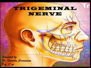
Trigeminal Nerve
- 2. It is V and Largest cranial nerve Mixed -- Small motor root Large sensory root Nerve of the first pharyngeal arch 2
- 3. Exteroceptive from Skin of the face & forehead; Mucous membrane of the nasal cavity; Oral cavity; Nasal sinus; Floor of mouth, teeth; Anterior 2/3 of tongue; Cranial dura Proprioception from Teeth; Periodontium; Hard palate; TMJ 3
- 4. Attached to lateral part of pons Sensory root (portio major) Motor root (portio minor) 4
- 5. 5
- 6. Fibers arise from Semilunar Ganglion Semilunar ganglion – Develops from neural crest – Crescent shaped – Unipolar neurons – Location- Meckel’s cavity; superior to petrous part of temporal bone 6
- 7. 7
- 8. 8
- 9. Afferent station Afferent fibers accompany fibers of motor root Proprioception from TMJ, periodontal membrane, teeth, hard palate Afferent impulses from stretch receptors in the muscles of mastication 9
- 10. 10
- 11. 11
- 12. Located at midpontine level Medial to main sensory nucleus Fibres distribute to muscles of mastication, mylohyoid, anterior belly of digastric, tensor tympani, tensor veli palatini. 12
- 13. Location – midpons Forms dorsal trigeminothalamic tract Ascending fibers terminate in this nucleus Convey light touch, tactile discrimination, sense of position and passive movements 13
- 14. True sensory ganglion Contains cells that are structurally and functionally ganglion cells Convey impulses from the muscles innervated by the trigeminal nerve and the extraocular muscles, as well as from the periodontal ligament of the teeth 14
- 15. Largest nucleus Extends caudally from main nucleus to level C3 of spinal cord Forms ventral trigeminothalamic tract Conveys pain and temperature 15
- 16. extends to the pontomedullary junction inferiorly pontomedullary junction to obex Tactile sense Obex(medulla) to C3 level of spinal cord Pain and temperature 16
- 17. 17
- 18. 18
- 19. Smallest division From anterior medial part of semilunar ganglion lateral wall of cavernous sinus Sensory fibres from Scalp, skin of forehead, upper eyelid lining frontal sinus, conjunctiva of eyeball, lacrimal gland, skin of the lateral angle of eyeball & lining of ethmoid cell 19
- 22. LACRIMAL NERVE (n. lacrimalis) Smallest branch. Enters the orbit through the narrowest part of the superior orbital fissure Runs along the upper part of the lateral rectus Communicates with zygomatic branch of maxillary nerve. Enters the lacrimal gland – gives of several filaments Finally pierces the orbital septum & ends in supplying the skin of upper eyelid. 22
- 23. FRONTAL NERVE (n. frontalis) Largest branch. Enters the orbit through the superior orbital fissure. Runs forward between levator palpebrae superioris and periosteum Divides into two branches in the midway between the apex and base of the orbit Supratrochlear Supraorbital 23
- 24. • Supraorbital Smaller than supratrochlear. Gives filament to join the infratrochlear. Supplies – Skin over the lower forehead. Conjunctiva Skin of the upper eyelid. Supratrochlear Passes through the supraorbital foramen. Branches into medial & lateral. Supplies – Conjunctiva. Skin of the upper eyelid. Twigs to pericranium. 24
- 25. Nasociliary Nerve (n. nasociliaris) Intermediate in size and more deeply placed. Enters the orbit between the two heads of rectus lateralis. Further passes through anterior ethmoidal foramen. Supplies internal nasal branches to mucous menbrane. Emerges as external nasal branch supplying the skin of the ala and apex of the nose. 25
- 26. Sensory From lower eyelid, side of the nose, upper lip; All maxillary teeth & gingivae, mucous membrane of most of nasal cavity, hard and soft palate; Tonsillar region and region of pharynx 26
- 27. 27
- 28. 28
- 30. 30
- 31. Zygomaticotemporal: Exits the zygomatic bone on its medial surface Pierces the temporal fascia to supply skin over temple Receives a branch from lacrimal nerve Communicates with facial nerve and auriculotemporal branch Zygomaticofacial : Leaves zygomatic bone on its lateral surface Supplies skin over malar prominence Communicate with facial nerve & with inferior palpebral branch of maxillary 31
- 32. Posterior superior alveolar nerves • Arise in the pterygopalatine fossa • Leaves maxillary nerve in pterygopalatine fossa • Enters the posterior alveolar canals Branches to Sinus lining Three twigs to Molars Gingiva Adjoining part cheek 32
- 33. Middle superior alveolar nerves • Arise from the nerve in the posterior part of the infraorbital canal • Runs in infraorbital groove on the lateral wall max sinus • Supply premolars, gingiva & adjoining part cheek • Forms a superior dental plexus with anterior and posterior superior alveolar branches 33
- 34. NTERIOR SUPERIOR ALVEOLAR NERVES • Given off just before exiting from the infraorbital foramen. • Supplies the incisor and canine teeth. • Descends in canalis sinosus in anterior wall of maxillary sinus. • Gives off a nasal branch to supply the mucous membrane of the anterior part of inferior meatus and nasal floor. • Communicates with the nasal branches of the sphenopalatine ganglion. 34
- 35. INNERVATION OF HARD PALATE Sensory innervations Greater palatine Nasopalatine These are branches of maxillary N passing through Pterygopalatine ganglion Lesser Palatine N ( middle & posterior ) – Uvula, tonsil, soft palate Pterygopalatine ganglion Greater palatine nerve Lesser palatine nerve 35
- 36. 36
- 37. 37
- 38. 38
- 39. 39
- 40. 40
- 41. GANGLIONS 41
- 42. Ciliary ganglion • Suspended from nasociliary nerve • Anatomically belongs to trigeminal nerve • Functionally belongs to oculomotor nerve • Carries parasympathetic motor fibres from Edinger – Westphal nucleus 42
- 43. OTIC GANGLION • Suspended from mandibular nerve • Anatomically belongs to trigeminal nerve but functionally belongs to glossopharyngeal nerve • Carries the secretomotor ( nucleus is superior salivary )fibres & distributed via lesser superficial petrosal nerve to Parotid glands. 43
- 44. Suspended from lingual nerve Anatomically belongs to trigeminal nerve but functionally belongs to facial nerve Carries the secretomotor ( nucleus is superior salivary ) fibres & distributed via chorda tymoani nerve to submandibular & sublingual salivary glands. 44
- 45. PTERYGOPALATINE GANGLION • Suspended from maxillary nerve • Anatomically belongs to trigeminal nerve but functionally belongs to facial nerve • Carries secretomotor fibres & distributed via the great superficial petrosal nerve ( Nucleus is inferior salivary ) to the Lacrimal glands & the glands of the palate 45
- 46. 46
- 47. 47
- 48. 48
- 49. 49
- 50. 50
- 51. Surgical removal of third molars(Von Arx and Simpson, 1989; Rood, 1992) Osteotomies (Walter and Gregg, 1979; Yoshida et al, 1989) Trauma (De Man and Bax, 1988) Tooth extractions (Strassburg, 1967; Hansen, 1980) Pulpectomy (Holland, 1994) Experimental Trigeminal Nerve Injury G.R. Holland CROBM 1996 7: 237 51
- 52. Implant placement Hydroxypatite ridge augmentation Endodontic surgeries Tumour resection Salivary gland and duct surgery Vestibuloplasty Biopsy procedures 52
- 53. • Inferior alveolar nerve injury – 0.41-7.5% • Lingual nerve injury – 0.06-11.5% 53
- 54. 54
- 55. Risk factors for nerve injury 55
- 56. Local anesthetic toxicity Formation of epineural hematoma Needle-barb mechanism of injury Chemical injury 56
- 57. Sagittal split osteotomy Mandibular osteotomies. advancement procedure Intraoral vertical ramus osteotomy (IVRO) Genioplasty procedures 57
- 58. 58
- 59. Fracture of mandibular body and ramus LeFort I & II fractures Fracture of condylar segment medially Mandibular angle, body and symphysis fracture Inadvertent placement of screws 59
- 60. 60
- 61. 61
- 62. Overinstrumentation Chemical injury Direct trauma from apicoecotomy 62
- 63. TMJ exposures by preauricular approach Damage is minimized by incision and dissecting in close apposition to cartilagenous portion of external auditory meatus Fracture of neck of condyle 63
- 64. Trigeminal neuralgia is defined as sudden, usually unilateral, severe, brief, stabbing, lancinating type of pain in the distribution of one or more branches of 5th cranial nerve Specific etiology unknown 64
- 65. Sudden, unilateral, intermittent paroxysmal, sharp, shooting, lancinating, like pain. Pain is elicited by slight touching superficial ‘Trigger points’ Common triggers include touch, talking, eating, drinking, chewing, tooth brushing, etc. 65
- 66. MEDICAL TREATMENT DRUGS CURENTLY USED:1.Carbamazepine is used as a standard drug , adult dose 200mg TDS & can be increased upto 1600 mg/day in divided doses initially started as small dose & gradually increased to prevent side effects adverse effects include dizziness, ataxia, vertigo, skin rashes . ,etc bone marrow suppression is rare but requires routine . monitoring 66
- 67. 2.Phenytoin sodium usually used in combination of carbamazepine dose 100-400mg/day side effects: gum hyperplasia 3. Gabapentin dose is 1200-3600mg/day used with caution in patients with renal & hapatic disease 4. Gaba agonist these drugs reduce the central projection of painful impulses eg. Baclofen , adult dose being 10-30 mg TDS 67
- 68. Surgical treatment • Peripheral nerve block procedure: It involves blocking of peripheral nerve by long acting LA. • Alcohol block : 0.5 -2 ml of 95% alcohol can be used for blocking of peripheral nerve. • Blocking of gasserian ganglion: more effective but it has hazards in sense that alcohol escape into the surrounding subarchanoid space & may cause palsy of adjacent cranial nerve. 68
- 69. • Post ganglionic sectioning: peripheral neurectomy in which peripheral branch of nerve is avulsed. • Cryotherapy: peripheral branches are subjected to application of extreme cold by using cryoprobes In this the nerve is not sectioned but destroyed • Percutaneous procedure: Percutaneus procedures involes mechanically or chemically damaging parts of trigeminal groove 69
- 70. • Radiation therapy: Gamma knife has been used which consists of multiple rays of high energy photon concentrated on trigeminal nerve root. Can be used to destroy specific components of nerve Source of radiation is Co 60 70
- 71. Head neck and brain- bd chaurasia Gray’s anatomy Monheim’s local anesthesia and pain control in dental practice Handbook of local anesthesia - malamed Peterson’s principles of oral & maxillofacial surgery Experimental trigeminal nerve injury g.R. Holland crobm 1996 7: 237 71
Notas do Editor
- 1-orbital; 2-posterior superior lateral nasal; 3-nasopalatine ; 4-pharyngeal; 5-greater palatine; 6-lesser palatine