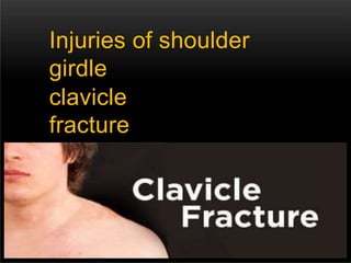
2. CLAVICE
:Is an S-shape long, curved ,tubular bone , lies
horizontally a cross the root of neck .
It articulate with sternum medially to form
sternoclavicular joint.
Also articulate with acromion process of
scapula at acromioclavicular joint and
acromioclavicular ligament .
the muscles inserting on clavicle are :
sternocleidomastoid, And subclavius muscles
.
8. Mechanism of
injury :
Direct traumatic impact or fall on the
shoulder
87%
.07% .
06%
.
Direct impact to
clavicle Fall on
outstretched hand
From fall on the side
.
Vigorous muscle contraction , seizures
[rare] . Pathological
fracture [rare] .
11. Allman classification : according
to site of fracture :
group 1: Fracture mostly
occur in the middle
one third of clavicle 80% .
group 2: The fractures of outer
third is 15% . Fractures
involving the acromioclavicular joint 28%
.
12. Why does the fracture occur in
middle third more ?
It is the thinnest part of the bone .
It is the junction of the tow main curves
of shaft . Site of entrance of nutrient
artery .
13.
14. common pattern of
fractures
of clavicle
are :
1 - Green stick
fracture :
Common at the junction
between middle and
outer third .
Common in children .
17. 4 - With greater
displacement
:
Thereis over lapping and
shortening . •
18. Clinical
presentatio
n :
pain and tenderness at site of injury .
Obvious deformity and
swelling sometimes occur .
Patient come support his injured limb
with other hand and head tilted
toward injured
side . Local
bruising .
19. vascular compilication are rare , but we
must look for it by : check pulse , gently
palpate root of neck
.
Outer third # are easily
missed for
acromioclavicular joint .
20. Diagnosi
s
:
- Clinical picture
examination .
investigation :
x-ray[AP view ] :
# is usually in middle third, outer
fragment below the inner .
#of outer third may be missed .
CT scan : useful for non union
21.
22. Treatmen
t
:
The aim is to provide support for the
weight of the arm .
Fracture of clavicle unite with or without
treatment . Healing occurs usually in 3-6
weeks .
It may be :
conservative or surgical .
24. Rehabilitati
on :The patient should be instructed
regarding hand wrist and elbow
exercises during immobilization .
And regarding shoulder exercises once
fracture healed .
25.
26.
27.
28. Surgical
treatment :Rarely indicated ,
except in :
- lateral one third
fracture .
- presence of neurovascular
injury .
- non union cases .
Internal fixation plate .
29.
30.
31. Complicati
on:late :
Malunion .
Ununion : treated by internal fixation and bone
grafting . Neurovascular injury [rare] . .
Stiffness of shoulder in
elderly . Ulnar
neuropathy .
Refracture .
Early : [subclavian or carotid artery injury
35. Scapul
a :Is a flat triangular bone that lies on the posterior
thorax wall between 2-7 rib.
It envelope by :
supraspinatus
muscle
infraspinatus
muscle
subscapularis
muscle
Attached to clavicle at acromioclavicular joint
,secured by acromioclavicular ligament .
36.
37.
38.
39. Fracture of
scapula :Fractures of scapula are uncommon
because of scapula location and
surrounding muscles whitch protect it .
Fractures of
scapula -
are result of high
energy
trauma with high
40. Associated life threatening injuries with
scapula # : pneumothorax
pulmonary
contusion
arterial injury
abdominal injury
head injury
splenic or liver
laceration brachial
plexus injury
41. Fractures of scapula are
classified according to
location :
body
fracture
neck
fracture
50 % .
5-30
% .glenoid fracture 10
% . Coracoid
fracture 8 % .
Acromion fracture 7
% .
42. Mechanism of
injury :
# of body : from sever direct trauma
- fall from height with direct landing on posterior
aspect of trunk .
- motor vehicle crush .
# of neck : direct blow to shoulder
- fall on shoulder .
- fall on outstretched hand .
# of glenoid : direct blow to lateral aspect of shoulder .
or impaction of humeral head in to glenoid
fossa .
43. # of coracoid process :
direct blow or shoulder
dislocation .
# of acromion :
direct down ward blow to
shoulder .
44. Clinical
picture :Sight > swelling
deformi
ty
ecchymo
sis
erosio
n .
Touch >
pain
tenderne
ss
crepitatio
n .
Pain exacerbated by
movment .
45. Clinical
picture :Brusing over scapula or chest
area . - Pain in
movement . -
Swelling around back of
shoulder . -
Tenderness at site of # .
-
Arm is held immobile .
46. Diagnosi
s :After initial assessment , according to
advanced trauma life support [ATLS]
principles , radiograghic evaluation is
indicated as soon as possible as patient stable
.
X – ray :
Anteroposterior view lateral axillary view .
C T scan :is useful in glenoid or body
47.
48.
49.
50. Treatme
nt :
Reduction is usually unnecessary .
Patient wears a sling for comfort and
from start movement.
Check repeatedly for dislocation of the
shoulder .
51. # of body by :
conservatively by analgesics and
simple sling to rest shoulder for
2-3 weeks .
# of acromion process :
Un displaced :
sling for 3-4 weeks for rest
shoulder. displaced :
52. # of coracoid :
conservatively in major , using a
sling for 2-3 weeks.
Vigorous exercises should be prohibited
for 2 m . If there is marked displacement
> open reduction .
# of neck and glenoid :
- sling for 2-3 weeks
- if there is displacement > shoulder spica after
reduction .
-open reduction > indicated if there is isolated
53.
54.
55.
56.
57.
58. Complicatio
n :Malunion non union >
rare Glenohumeral
arthritis .
Limitation in range of
motion . After surgery :
local
dyscomfort
infection
nerve injuries
post traumatic
59. Notes
:Scapular fracture should alert the
surgeon to presence of other
injuries .
Sever chest injury should also raise
suspicion of possible scapular injury
.