

Recomendados
Mais conteúdo relacionado
Mais procurados
Mais procurados (19)
Semelhante a SINH LÝ NGHE BÌNH THƯỜNG
Semelhante a SINH LÝ NGHE BÌNH THƯỜNG (20)
Mais de SoM
Mais de SoM (20)
Último
Último (20)
SINH LÝ NGHE BÌNH THƯỜNG
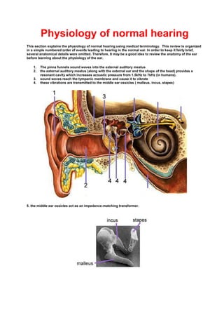
- 1. Physiology of normal hearing This section explains the physiology of normal hearing using medical terminology. This review is organized in a simple numbered order of events leading to hearing in the normal ear. In order to keep it fairly brief, several anatomical details were omitted. Therefore, It may be a good idea to review the anatomy of the ear before learning about the physiology of the ear. 1. The pinna funnels sound waves into the external auditory meatus 2. the external auditory meatus (along with the external ear and the shape of the head) provides a resonant cavity which increases acoustic pressure from 1.5kHz to 7kHz (in humans). 3. sound waves reach the tympanic membrane and cause it to vibrate 4. these vibrations are transmitted to the middle ear ossicles ( malleus, incus, stapes) 5. the middle ear ossicles act as an impedance-matching transformer.
- 2. IMPEDANCE: The impedance of a medium is the opposition it offers to the transmission of acoustic energery. As a general rule, denser materials offer more impedance to sound waves. If sound waves in air (low acoustic impedance) were to directly hit the oval window and the fluid in the cochlea (high acoustic impedance), then most of the acoustic energy would be reflected away, and very little would be transmitted (approximately 3%) to the fluid in the cochlea. The middle ear manages to get around this problem by using two membranes and a system of three ossicles which act as an impedance-matching transformer. As a result, 60-65% of the original sound is transmitted to the cochlea. There are 3 main factors that contribute to this matching of impedance between air and fluid: The tympanic membrane is large and the stapes footplate that lies over the oval window is small. The ratio is approximately 35:1. This concentrates, and therefore increases the amount of sound pressure exerted on the oval window The tympanic membrane is pulled inward by the handle of the malleus. This also serves to increase the force being exerted on the oval window. There is a difference in the effective lever lengths of the malleus and the incus. This also increases the force at the stapes foot plate. The effect of different lever lengths can be illustrated by placing a board unevenly on a fulcrum. Since the lengths on either side of the fulcrum are of different lengths, any pressure (Force) exerted on one side is amplified on the other. The overall pressure increase at the oval window is 23 times more than than what it would have been if sound waves in air directly contacted the oval window. This amounts to an increase in approximately 25 dB. 6. the middle ear muscles affect the vibrations being transmitted through the middle ear.
- 3. MIDDLE EAR MUSCLES: The tensor tympani and stapedius muscles increase the stiffness of the ossicles to attenuate mainly low frequency sounds. This reflex serves several main functions: Protection of the inner ear from intense sounds. This reflex, however, is delayed by approximately 25-35 ms because it requires at least a 3-4 neuron pathway in the brainstem. As a result, it provides little protection from impulse noise. Extends the dynamic range of the ear. Normally, low frequency sounds mask high frequency sounds. However, when low frequency sounds are attenuated, higher frequency sounds are no longer masked, and are therefore more audible. Prevents overstimulation of the cochlea during self-produced sounds (eg. speaking). 7. the inner ear lies in the petrous temporal bone. 8. This is an anterior view of the inner ear. The base of the stapes lies over the oval window. As a result, its vibrations are conducted to the perilymph of the vestibule of the inner ear. Since perilymph and endolymph are incompressible fluids and the vestibular and basilar membranes are slack-flexible, vibrations are easily transmitted from one fluid chamber to the next. 9. The aim of these diagrams is to orient you to a cross section of the cochlea. The pressure waves pass into the scala vestibuli (vestibule) of the cochlea, through the vestibular (Reissner's) membrane into the endolymph of the scala media. They are then transmitted to the perilymph of the scala tympani, and are dissipated at the secondary tympanic membrane that covers the round window.
- 4. 10. These pressure changes induce vibrations in the basilar membrane. The makeup of the basilar membrane varies from its base to its apex, thus allowing for different frequencies to stimulate different areas. Base Apex stiffness stiffer less stiff membrane width small wide fiber arrangement densely packed looser arrangement membrane thickness thicker thinner frequencies sensed high low hair cell length shorter longer number of stereocilia on hair cell more less pure tone stimuli cause pressure variations that establish a travelling wave in the basilar membrane. the wave propagates from base to apex the envelope of the wave reaches a peak and then diminishes rapidly. This peak is determined by the frequency of the stimulus. Stimuli with high frequencies peak near the base of the basilar membrane, and stimuli with lower frequencies peak near the apex.
- 5. Reproduced from Robert Harrison and the Auditory Science Laboratory with permission. 11. Below is a detailed diagram of the organ of Corti. The organ of Corti rests on the basilar membrane, and therefore senses its vibrations.
- 6. 12. the organ of Corti contains sensory (hair) cells. These hair cells have two ends: a tip, which has specialized microvilli called stereocilia a base, which is supported by phalangeal(Deiters) cells and pillar cells that rest on the basilar membrane Reproduced from the Auditory Science Laboratory with permission. there are two sets of hair cells: o inner sensory (hair) cells (1 row) innervated by a high proportion of the afferent nerve fibers, and are the most important transducer sending signals to afferent cochlear neurons and subsequently to the brain for the sensation of hearing o outer sensory (hair) cells (3-5 rows) these have tips that are embedded in the tectorial membrane, and are probably more involved in elecromechanical feedback to the basilar membrane.
- 7. This is an electron micrograph of a right cochlea. Note how it spirals downwards in a clockwise direction. This is an enlarged view of the organ of Corti. If you look closely, you can see the hair cells. This is an enlarged view of the hair cells of the organ of Corti. Note how there is 1 row of inner hair cells, and 3 rows of outer hair cells. Sensorineural hearing loss results from damage to these hair cells. 13. displacement of the basilar membrane causes shearing stresses on the hair cell stereocilia. the tips of inner sensory (hair) cells are free, and are deflected by the flow of endolymph across their apical surface. the tips of the outermost row of outer sensory (hair) cells are embedded in a part of the tectorial membrane called Hardesty's membrane. As a result, any movement of the basilar membrane directly causes deflection of these cell tips. 14. the stereocilia make up a bundle, and are ranked by height with the tallest lying next to the kinocilium (this disappears shortly after birth). The kinocilium is a tall cilium that lacks independent motility. Deflection of the stereocilia towards the kinocilium depolarizes the hair cell and increases the number of action potentials that are generated and sent to the vestibular nerve. (Deflection of the stereocilia away from the kinocilium hyperpolarizes the hair cell and decreases the number of action potentials generated). The greatest hair cell receptor potentials are achieved when endolymph flows across the hair cell tips in a radial direction in the cochlea.
- 8. Reproduced from Robert Harrison and the Auditory Science Laboratory with permission. 15. the different compartments of the cochlea have different compositions and resting potentials. IONIC COMPOSITIONS AND RESTING POTENTIALS Fluid filling the region Conc. of Na Conc. of K Electrical Potential Scala Vestibuli perilymph high (140mM) low (7mM) 0 mV Scala Media endolymph low (2mM) high (150mM) +80mV * this is maintained by the stria vascularis Scala Tympani perilymph high (140mM) low (7mM) 0 mV the +80mV potential in the scala media along with a -40 mV potential in the hair cells of the cochlea results in a 120 mV difference across the apical membrane of the hair cell. 16. when stereocilia are deflected toward the kinocilium, there is an increased flow of positive ions into the hair cell, causing depolarization towards its resting potential of 0 mV. Why the temporal aspect of sound is lost above 3-4 kHz For any given deflection, depolarization changes are greater than hyperpolarization changes. As a result, sinusoidal inputs (such as sound waves) create both a net depolarization and an alternating voltage change in hair cell receptors. Low frequencies of sound produce an alternating voltage change, which allows for temporal coding. At higher frequencies (i.e. above 3-4kHz), the capacitance of the hair cell wall shunts these alternating changes to ground, thus leaving only net depolarization. Thus, temporal aspects of sound are lost above 3-4 kHz. 17. the base of the hair cell rests in a groove at the top of a phalangeal cell. Both afferent and efferent nerve fibers synapse with the base of the hair cell.
- 9. This is a scanning E.M. of the outer hair cells being supported by phalangeal (Deiter's) cells. The stereocilia are at the top of the image and the efferent nerve endings are at the bottom. 18. when the hair cell is depolarized, depolarization of the presynaptic region causes an influx of Ca, which results in exocytosis of synaptic vesicles that contain neurotransmitter. even in the absence of sound stimulation, there is some release of neurotransmitter that results in spontaneous activity in cochlear nerve fibers. the rate of release of neurotransmitter decreases with time as neurotransmitter is depleted, thus accounting for adaptation in nerve fiber responses 19. the INNER hair cells send afferent neurons to the modiolus, which congregate to form the cochlear nerve. The cochlear nerve conserves the cochleotopic (tonotopic) arrangement of hair cells of the organ of Corti. 20. primary afferent neurons innervate three parts of the cochlear nucleus: AVCN - anteroventral cochlear nucleus o receives neurons from the apex of the cochlea (low frequency) PVCN - posteroventral cochlear nucleus DCN - dorsal cochlear nucleus o receives neurons from the base of cochlea (high freq.)
- 10. 21. axons from the AVCN and PVCN pass to the superior olivary complex on both ipsilateral and contralateral sides. The superior olivary complex has 3 nuclei that are important in the ascending auditory pathway: LSO - lateral superior olivary nucleus MSO-medial superior olivary nucleus MTB-medial nucleus of the trapezoid body They are the first to receive information from both ears, and are therefore important in sound localization o axons from the superior olivary complex then ascend in the lateral lemniscus to the central nucleus of the inferior colliculus. axons from the DCN pass mainly to the central nucleus of the contralateral inferior colliculus. 22. There are two connections between both sides of the brainstem. Commissures of Probst - at the lemniscal level Commissures of the inferior Colliculi - at collicular levels 23. axons from the inferior colliculi project to 3 nuclei of the medial geniculate body (MGB). They are: ventral nucleus (VN) o purely auditory o projects to primary auditory cortex medial nucleus (MN) o handles many senses o projects to all areas of the auditory cortex dorsal nucles (DN) o handle many senses o projects to insulo-temporal region, posterior ectosylvian gyrus and to the secondary auditory cortex. 24. the main auditory area in the cortex lies in the sylvian fissure. Even though there are 6 layers in the primary audiory cortex, the geniculate afferents terminate mainly in layer IV (and partly in layer III).
- 11. Reproduced from Robert Harrison and the Auditory Science Laboratory with permission. 25. the cells in layer IV are arranged in vertical columns. There are two types of columns: summation columns (70%): are excited more by stimulation of both ears than by one ear alone. suppression columns (30%): are excited by one ear, and inhibited by the other. 26. There is tonotopic arrangement in the primary auditory cortex. posterior part- codes low frequencies anterior part - codes high frequencies Summary table demonstrating conservation of tonotopicity: basilar membrane cochlear nucleus primary auditory cortex high frequencies base (DCN) dorsal cochlear nucleus anterior part low frequencies apex (AVCN) anteroventral cochlear nucleus posterior part 27. There are further projections to the secondary auditory cortex, and to Wernicke's area, which is an auditory association area usually found on the left side. Sound localization occurs primarily at the superior olivary complex there are several cues used in sound localization: o Interaural time delay - the two ears are far apart, therefore sounds arrive at both ears at different times. Onset time and phase differences of sounds are important. o Interaural sound intensity differences- the head has a shadowing effect, and attenuates sounds farthest from their source. High frequencies are attenuated most effectively The pinna and external auditory meatus cause sound to bounce around, thus modifying it. As a result, sounds can be localized as coming from in front of, or behind the head. This cue is not coded in the superior olivary complex.