
Recomendados
Mais conteúdo relacionado
Mais procurados
Mais procurados (20)
Semelhante a TRIANGLES OF NECK
Semelhante a TRIANGLES OF NECK (20)
Último
Último (20)
TRIANGLES OF NECK
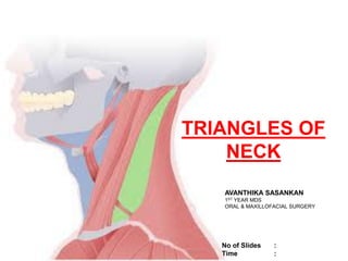
- 1. TRIANGLES OF NECK AVANTHIKA SASANKAN 1ST YEAR MDS ORAL & MAXILLOFACIAL SURGERY No of Slides : Time :
- 2. 1. INTRODUCTION 2. CLASSIFICATION 3. ANTERIOR TRIANGLE 4. SUBMENTAL TRIANGLE 5. SUBMANDIBULAR TRIANGLE 6. CAROTID TRIANGLE 7. MUSCULAR TRIANGLE 8. POSTERIOR TRIANGLE 9. OCCIPITAL TRIANGLE 10. SUBCLAVICULAR TRIANGLE 11. CONCLUSION CONTENTS
- 3. INTRODUCTION • The neck is a geometric region that can be studied and operated using anatomical triangles. • Neck is limited , Superiorly - inferior border of mandible Anteriorly – midline Inferiorly - superior border of clavicle Posteriorly – anterior margin of trapezius • Anatomical triangles reported and depicted are classified within the broader anterior and posterior cervical triangles. • Triangles contain nerves , vessels and other anatomical structures.
- 4. ANTERIOR TRIANGLE SUPERIOR –inferior border of mandible MEDIAL - midline of neck LATERAL - anterior border of sternocleidomastoid SUBDIVISIONS • MUSCULAR TRIANGLE • CAROTID TRIANGLE • SUBMANDIBULAR TRIANGLE • SUBMENTAL TRIANGLE CLASSIFICATION
- 5. POSTERIOR TRIANGLE ANTERIOR - posterior margin of sternocleidomastoid POSTERIOR - anterior margin of trapezius INFERIOR - middle one -third of clavicle SUBDIVISIONS • OCCIPITAL TRIANGLE • SUPRACLAVICULAR TRIANGLE
- 6. ANTERIOR TRIANGLE The anterior triangle refer to bilateral anatomic subdivisions of the neck comprising the anterior surface of the neck, deep to the superficial cervical fascia and platysma muscle.
- 7. SUBMENTAL TRIANGLE Median triangle. • On each side - anterior belly of the digastric muscles. • Base - body of the hyoid bone. • Apex - lies at the chin. • Floor -right and left mylohyoid muscles and the median raphe uniting them .
- 8. CONTENTS OF SUBMENTAL TRIANGLE 2-4 small submental lymph nodes are situated in the superficial fascia between the anterior bellies of the digastric muscles. They drain: • Superficial tissues below the chin. • Central part of the lower lip. • The adjoining gums. • Anterior part of the floor of the mouth. • The tip of the tongue. Their efferent pass to the submandibular nodes. 2 Small submental veins join to form the anterior jugular vein.
- 9. CLINICAL SIGNIFICANCE • Tumors and cysts develop within its limits, and abscesses from other regions extend to the area. • Odontogenic infection from the lower central and lateral incisor teeth can spread into the submental space , may require external incision and drainage. • For head and neck cancer staging , the neck is divided into 6 anatomic lymph node levels . The submental lymph nodes are classified as level Ia.
- 10. ❖ Submandibular triangle. • Anteroinferiorly -Anterior belly of digastric. • Posteroinferiorly -Posterior belly of digastric and the stylohyoid. • Superiorly or base -Base of the mandible and a line joining the angle of the mandible to the mastoid process . DIGASTRIC TRIANGLE
- 11. ROOF • Skin. • Superficial fascia, a. The platysma. b. The cervical branch of the facial nerve. c. The ascending branch of the transverse or anterior cutaneous nerve of the neck. • Deep fascia, which splits to enclose the submandibular salivary gland FLOOR • Anteriorly -Mylohyoid muscle • Posteriorly -Hyoglossus. • A small part of the middle constrictor muscle of the pharynx
- 12. CONTENTS OF DIGASTRIC TRIANGLE 1 .ANTERIOR PART 2. POSTERIOR PART ANTERIOR PART superficial to mylohyoid • Superficial part of the submandibular salivary gland • The facial vein and the submandibular lymph nodes • Facial artery is deep • Submental artery. • Mylohyoid nerve and vessels • The hypoglossal nerve.
- 13. POSTERIOR PART Superficial • Lower part of the parotid gland. • The external carotid artery before it enters the parotid gland. Deep structures, passing between the external and internal carotid arteries • styloglossus. • stylopharyngeus. • glossopharyngeal nerve • pharyngeal branch of the vagus nerve. • styloid process • A part of the parotid gland.
- 14. Deepest structures Internal carotid artery. Internal jugular vein. Vagus nerve .
- 15. CLINICAL SIGNIFICANCE • Neoplasms , infectious and immunologic pathologies can arise from the submandibular triangle. • Sialadenitis – inflammation of the submandibular salivary gland . Most common pathology seen in submandibular triangle. • Pleomorphic adenoma – the most common benign neoplasm ,followed by adenolymphoma ( Warthin’s tumour). • Adenoid cystic carcinoma –the most common malignant neoplasm ,followed by mucoepidermoid carcinoma.
- 16. • Anterosuperiorly : Posterior belly of the digastric muscle and the stylohyoid • Anterioinferiorly: Superior belly of the omohyoid • Posteriorly : Anterior border of the sternocleidomastoid ROOF • Skin. • Superficial fascia o The platysma. o The cervical branch of the facial nerve. o The transverse cutaneous nerve of the neck. • Investing layer of deep cervical fascia. CAROTID TRIANGLE
- 17. FLOOR • The middle constrictor of pharynx. • The inferior constrictor of the pharynx . • Thyrohyoid membrane.
- 18. CONTENTS OF CAROTID TRIANGLE • ARTERIES • VEINS • NERVE
- 19. ARTERIES • The common carotid artery - carotid sinus & carotid body • Internal carotid artery • The external carotid artery
- 20. VEINS • Internal jugular vein. • The common facial vein internal jugular vein. • A pharyngeal vein internal jugular vein. • The lingual vein internal jugular vein.
- 21. NERVES • Vagus • Superior laryngeal branch of the vagus, o external laryngeal nerve o internal laryngeal nerve • Spinal accessory nerve • Hypoglossal nerve . o upper root of the Ansa cervicalis or descendens hyoglossi, • Branch to the thyrohyoid • Sympathetic chain of 344
- 22. LYMPH NODES • The deep cervical lymph nodes • jugulodigastric node • jugulo-omohyoid node
- 23. CLINICAL SIGNIFICANCE • Distention of the jugular vein can be palpated and assessed. • Internal jugular vein is usually utilized to establish central venous access for parenteral nutrition , dialysis and administration of chemotherapy drugs. • Carotid sinus is a baroreceptor that regulates blood pressure . Supraventricular tachycardia may be controlled by carotid sinus massage ,due to inhibitory effects of vagus nerve on the heart. • The carotid sinus is richly supplied by nerves. In some persons ,the sinus may be hyper-sensitive. In such persons , sudden rotation of the head may cause slowing of heart . This condition is called carotid sinus syndrome.
- 24. ANTERIORLY - Anterior median line of the neck from the hyoid bone to the sternum. POSTEROSUPERIORLY - Superior belly of the omohyoid muscle POSTEROINFERIORLY -Lower part of anterior border of the sternocleidomastoid muscle . MUSCULAR TRIANGLE
- 25. CONTENTS INFRAHYOID MUSCLES • Sternohyoid • Sternothyroid • Thyrohyoid • Omohyoid. SUPERFICIAL • Sternohyoid • Omohyoid DEEP • Sternothyroid • Thyrohyoid
- 26. • Superior thyroid artery • Anterior jugular vein • Inferior thyroid vein • Ansa cervicalis LYMPHATICS • Anterior cervical • Infrahyoid • Pretracheal • Paratracheal MEDIAL PART • Esophagus • Trachea • Thyroid gland • Lower part of sternum
- 27. CLINICAL SIGNIFICANCE • Tracheostomy and thyroidectomy – surgically accessed through the muscular triangle. • A potential risk of both is damage to the recurrent laryngeal nerve. Other than the cricothyroid muscle, it supplies all the intrinsic muscles of the larynx , including posterior cricoarytenoid ;the only muscle that abducts the vocal cords. Injury can cause hoarseness, aphonia and dyspnoea. • Injury of the superior thyroid artery can result in bleeding during surgery.
- 28. POSTERIOR TRIANGLE The posterior triangle is a space on the side of the neck situated behind the sternocleidomastoid muscle. • ANTERIOR -Posterior border of sternocleidomastoid • POSTERIOR -Anterior border of trapezius. • INFERIOR OR BASE -Middle one-third of clavicle. • APEX -Lies on the superior nuchal line where the trapezius and sternocleidomastoid meet.
- 29. ROOF • Investing layer of deep cervical fascia. • The superficial fascia o platysma. o external jugular and posterior external jugular veins. o Parts of the supraclavicular, great auricular, transverse cutaneous and lesser occipital nerves . o Unnamed arteries derived from the occipital, transverse cervical and suprascapular arteries. o Lymph vessels which pierce the deep fascia to end in the supraclavicular nodes. The external jugular vein: It lies deep to the platysma
- 30. FLOOR • prevertebral layer of deep cervical fascia, o Splenius capitis. o Levator scapulae. o Scalenus medius o Semispinalis capitis DIVISION OF POSTERIOR TRIANGLE subdivided by the inferior belly of omohyoid into: A larger upper part, called the occipital triangle. A smaller lower part, called the supraclavicular or the subclavian triangle .
- 31. • ANTERIOR - posterior border of sternocleidomastoid • POSTERIOR - anterior border of trapezius • INFERIOR - inferior belly of omohyoid • FLOOR - Levator scapulae splenius capitis middle and posterior scalene muscles. OCCIPITAL TRIANGLE
- 32. CONTENTS OF OCCIPITAL TRIANGLE NERVES • Spinal accessory nerve • Four cutaneous branches of cervical plexus o Lesser occipital (C2) o Great auricular (C2, C3) o Anterior cutaneous nerve of neck (C2, C3) o Supraclavicular nerves (C3, C4) • Muscular branches: o Two small branches to the levator scapulae (C3, C4) o Two small branches to the trapezius (C3, C4) o Nerve to rhomboideus (proprioceptive) (C5)
- 33. VESSELES • Transverse cervical artery and vein • Occipital artery • LYMPH NODES • Along the posterior border of the sternocleidomastoid, o more in the lower part -supraclavicular nodes o and a few at the upper angle-occipital nodes
- 34. CLINICAL SIGNIFICANCE • The most common swelling in the posterior triangle is due to enlargement of the supraclavicular lymph nodes . • While doing biopsy of the lymph node ,one must be careful in preserving the accessory nerve which may get entangled amongst enlarged lymph nodes. • Frequent surgical intervention include radical neck dissection of lymph nodes along the sternocleidomastoid after removal of head and neck squamous cell carcinoma. • Lymph node in this area can cause damage to the accessory nerve and subsequently trapezius innervation , leading to inability of the patient to shrug their shoulder. • Supraclavicular lymph nodes are commonly enlarged in tuberculosis , Hodgkin’s disease and in malignant growths of the breast ,arm or chest • Torticollis or wry neck is a deformity in which the head is bent to one side and the chin points to other side . This is a result of spasm or contracture of the muscles supplied by the spinal accessory nerve , ie, sternocleidomastoid and trapezius.
- 35. ❖SUPRACLAVICULAR TRIANGLE ANTERIOR -posterior border of sternocleidomastoid SUPERIOR -inferior belly of omohyoid INFERIOR -middle one third of clavicle FLOOR -first digitation of serratus anterior , middle scalene and the first rib SUBCLAVIAN TRIANGLE
- 36. CONTENTS OF SUBCLAVIAN TRIANGLE NERVES Roots and trunks of brachial plexus Nerve to serratus anterior (long thoracic, C5-C7) Nerve to subclavius (C5, C6) Suprascapular nerve (C5, C6)
- 37. VESSELS • Third part of subclavian artery and subclavian vein • Suprascapular artery and vein • Commencement of transverse cervical artery and termination of the corresponding vein • Lower part of external jugular vein LYMPH NODES • A few members of the supraclavicular chain
- 38. CLINICAL SIGNIFICANCE A cervical rib may compress the second part of subclavian artery . In this cases, blood supply to upper limb reaches via anastomoses around the scapula . Dysphagia caused by compression of the esophagus by an abnormal subclavian artery is called dysphagia lusoria. In blalock’s operation for fallot’s tetralogy ,the right subclavian artery is anastomosed end to side to short circuit the pulmonary stenosis. External jugular vein is clinically important . The distention of this vein is visible in raised venous pressure due to congestive heart failure. The height of the column above the clavicle provides a rough guide to the increase in the venous pressure. This is called jugular venous pressure. The left supraclavicular lymph node or Virchow’s node is near the junction of the thoracic duct and the left subclavian vein. Enlargement of this node can indicate metastasis of gastrointestinal malignancy.
- 39. Anatomical landmarks can be useful during surgery of the neck. The triangles of neck contain nerves ,arteries ,veins, lymph vessels and other important structures. Assessing neck lumps and understanding the surgical approaches for pathological lesions requires a sound understanding of neck anatomy A better understanding of the triangles of the neck make surgery more efficient and avoid intraoperative complications. Anatomy of these triangles of the neck could help to minimize surgical injuries and make surgical dissections more efficient. Also , clinicians can easily identify some of these triangles and their contents with palpation ,which provides valuable assistance as surface landmarks for clinical examination. The use of the divisions described as the triangles of the neck permit the effective communication of the location of palpable masses located in the neck between health care professionals. CONCLUSION
- 40. • B.D Chaurasia - BD Chaurasia‘s Human Anatomy • Frank H. Netter - Netter Atlas of Human Anatomy REFERNCES