SlideShare uma empresa Scribd logo
1 de 135
Unit One : Cell Biology
Human cheek cells stained with methylene blue, 400x
http://microscopy4kids.org/Comparing_Plant_and_Animal_Cells
1.1 Introduction to Cells
Below are some of the oldest fossilized cells on Earth,
Prokaryotes from 3.0 to 3.5 billion years ago
• Essential idea: The evolution of multicellular organisms
allowed cell specialization and cell replacement.
Cell theory states that:
1. All living things are composed of cells (or cell products)
2. The cell is the smallest unit of life
3. Cells only arise from pre-existing cells
1.1 U.1 According to the cell theory, living organisms are composed of cells.
1. All living things are composed of cells (or cell products)
Living organisms are composed of cells or cell products
Volvox, a type of green algae. It forms
spherical colonies of up to 50,000 cells
2. The cell is the smallest unit of life
Specialized structures within cells (organelles) carry out different functions.
Organelles cannot survive alone.
3. Cells only arise from
pre-existing cells:
• Cells multiply through
division
• All life evolved from
simpler ancestors
• Mitosis results in
genetically identical diploid
daughter cells
• Meiosis generates haploid
gametes (sex cells)
http://cx.aos.ask.com/question/aq/700px-
700px/why-do-cells-divide_462b38a6-0721-4835-
9cbd-e4b9e1a0fb59.jpg
1.1 A.1 Questioning the cell theory using atypical examples, including striated muscle,
giant algae and aseptate fungal hyphae.
Exceptions to the Cell Theory
Example 1: striated muscle
• Challenges the idea that a cell has one nucleus are multi-nucleated. This
does not conform to the standard view of a small single nuclei within a cell
• Muscle cells have more than one nucleus per cell
• Muscle Cells called fibers can be very long (300mm)
Exceptions to the Cell Theory
Example 2: Acetabularia
• Acetabularia is a giant algae is a single-celled organism that challenges both the
idea that cells must be simple in structure and small in size.
• As a comparison in size a human blood cell (the most abundant cell in a human is
about 100 micrometers. cells average between 5000 – 100,000 micrometers.
1.1 A.1 Questioning the cell theory using atypical examples, including striated muscle,
giant algae and aseptate fungal hyphae.
Exceptions to the Cell Theory
Example 3: fungal hyphae
• challenges the idea that a cell
is a single unit.
• Fungal hyphae are again
very large with many nuclei
and a continuous cytoplasm
• The tubular system of hyphae
form dense networks called
mycelium
1.1 A.1 Questioning the cell theory using atypical examples, including striated muscle,
giant algae and aseptate fungal hyphae.
Exceptions to the Cell Theory
Example 4: Bone:
•There are some cells that secrete material outside of the cell membrane
•The secretions solidify and dominate the structure
1.1 A.1 Questioning the cell theory using atypical examples, including striated muscle,
giant algae and aseptate fungal hyphae.
Exceptions to the Cell Theory
Example 5: Red Blood Cells
• Mammalian erythrocytes are
unique among the vertebrates as
they are non-nucleated cells in
their mature form.
• The lack of a nucleus provide
more space for hemoglobin.
• In mammals, erythrocytes also
lose all other
cellular organelles such as
their mitochondria, Golgi
apparatus and endoplasmic
reticulum.
1.1 A.1 Questioning the cell theory using atypical examples, including striated muscle,
giant algae and aseptate fungal hyphae.
1.1 U.2 Organisms consisting of only one cell carry out all functions of life in that cell.
You probably know:
• Movement
• Reproduction
• Homeostasis
• Growth
• Respiration
• Excretion
• Nutrition
• Transport
• Repair
In this course the functions are refined:
• Metabolism - the web of all the enzyme-
catalyzed reactions in a cell or organism,
e.g. respiration
• Response - Living things can respond to
and interact with the environment
• Homeostasis - The maintenance and
regulation of internal cell conditions, e.g.
water and pH
• Growth - Living things can grow or change
size / shape
• Reproduction - Living things produce
offspring, either sexually or asexually
• Excretion – the removal of metabolic waste
• Nutrition – feeding by either the synthesis
of organic molecules (e.g. photosynthesis)
or the absorption of organic matter
An easy way to remember Metabolism, Response, Homeostasis, Growth,
Reproduction, Excretion and Nutrition is: MR H GREN
How does this paramecium show the functions of life?
1.1 U.2 Organisms consisting of only one cell carry out all functions of life in that cell.
http://umanitoba.ca/Biology/BIOL1030/Lab1/biolab1_3.html#Ciliophora
Homeostasis – contractile vacuole fill
up with water and expel it through
the plasma membrane to manage the
water content
Reproduction – The
nucleus can divide to
support cell division by
mitosis, reproduction is
often asexual
Metabolism
– most
metabolic
pathways
happen in
the
cytoplasm
Growth – after
consuming and
assimilating biomass
from food the
paramecium will get
larger until it divides.
Response –
the wave
action of the
cilia moves
the
paramecium
in response
to changes in
the
environment,
e.g. towards
food.
Excretion – the plasma
membrane control the entry
and exit of substances
including expulsion of
metabolic waste
Nutrition – food
vacuoles contain
organisms the
paramecium has
consumed
How does this photosynthetic algae show the functions of life?
1.1 U.2 Organisms consisting of only one cell carry out all functions of life in that cell.
Homeostasis –
contractile
vacuole fill up
with water and
expel it
through the
plasma
membrane to
manage the
water content
Reproduction – The nucleus can
divide to support cell division, by
mitosis (these cells are undergoing
cytokinesis)
Metabolism
– most
metabolic
pathways
happen in
the
cytoplasm
Growth – after consuming and assimilating
biomass from food the algae will get larger
until it divides.
Response –
the wave
action of the
cilia moves
the algae in
response to
changes in
the
environment,
e.g. towards
light.
Excretion – the plasma
membrane control the
entry and exit of
substances including
the diffusion out of
waste oxygen
Nutrition –
photosynthesis
happens inside
the
chloroplasts to
provide the
algae with food
• All organisms need to exchange substances such as food, waste, gases and
heat with their surroundings. These substances must be exchanged between
the organism and its surroundings.
• As the size of a structure increases the surface area to volume ratio
decreases. Therefore the rate of exchange (diffusion/radiation) decreases.
This is true for organelles, cells, tissues, organs and organisms.
• As cells become larger they may not be able to take in essential
nutrients need for survival.
• The rate of exchange of substances therefore depends on the organism's surface
area that is in contact with the surroundings.
• The exchange depends on the volume of the organism, so the ability to meet the
requirements depends on , which is known as the surface area to volume ratio.
All organisms need to have a large surface to volume ration.
• As organisms get bigger their volume and surface area both get bigger, but not
by the same amount. This can be seen by performing some simple calculations
concerning different-sized organisms.
1.1 U.3 Surface area to volume ratio is important in the limitation of cell size.
1
2
3
1 cm 10 cm 100 cm
Assume we have
3 cubes:
With sizes:
What will happen to ratio between V and S.A. as their size increases?
1.1 U.3 Surface area to volume ratio is important in the limitation of cell size.
Ratio of Surface Area to Volume ration of the 3 cells
Cube Side
Length
Volume
(x3)
S.A. (6x2) Ratio
(S.A./V)
1 1 cm
2 10 cm
3 100 cm
1 cm3
1 000 cm3
1 000 000
cm3
6 cm2
600 cm2
60 000 cm2
6
0.6
0.06
1.1 U.3 Surface area to volume ratio is important in the limitation of cell size.
1.1 U.5 Specialized tissues can develop by cell differentiation in multicellular organisms.
• In humans 220 distinct
highly specialized cell
types have been
recognized
• All specialized cells and
the organs constructed
from them have
developed as a result of
differentiation
Emergent properties arise from the interaction of component parts.
The whole is greater than the sum of its parts. Multicellular
organisms are capable of completing functions that individual cells
could not undertake - this is due to the interaction between cells
producing new functions.
1.1 U.4 Multicellular organisms have properties that emerge from the interaction of their cellular
components.
The advantage is that when these functions work
together they can benefit the organism as a whole
1.1 U.4 Multicellular organisms have properties that emerge from the interaction of their cellular
components.
When studying the individual parts of a
living thing, there are many times
when the part alone do not allow the
prediction of the properties of the
whole living thing.
As an example: the toaster parts to the
right. Only when we combine them to
form the toaster can these properties
be determined. There is nothing
supernatural about the emergent
properties rather it is simply the
combination of the parts that results in
new properties emerging.
As a model consider a toaster. The
toaster is the system and is composed
of 400 different parts. We can study
the parts individually how they function
and the properties they posses. Some
of these would be the properties of :
• Steele
• Mica
• Plastic
Toaster Project
1.1 U.6 Differentiation involves the expression of some genes and not others in a cell’s
genome.
• All (diploid) cells of an
individual organisms share
an identical genome - each
cell contains the entire set of
genetic instructions for that
organism
• BUT not all genes are
expressed (activated) in all
cells
• In (totipotent) embryonic
stem cells the entire
genome is active
• Newly formed cells
receive signals which
deactivate (or more rarely
activate) genes, e.g. a skin
cell does not need to be able
to produce hemoglobin (the
pigment in red blood cells
that carries oxygen)
1.1 U.7 The capacity of stem cells to divide and differentiate along different pathways is
necessary in embryonic development and also makes stem cells suitable for therapeutic
uses.
Stem cells are unspecialized cells
that can:
• Can continuously divide and
replicate
• Have the capacity to differentiate
into specialized cell types
Totipotent Can differentiate
into any type of cell.
Pluripotent Can differentiate
into many types of cell.
Multipotent Can differentiate
into a few closely-related
types of cell.
Unipotent Can regenerate but
can only differentiate into their
associated cell type (e.g. liver
stem cells can only make liver
cells).
Potency
Learn about stem cells using the tutorials
1.1 U.7 The capacity of stem cells to divide and differentiate along different pathways is
necessary in embryonic development and also makes stem cells suitable for therapeutic
uses.
1.1 A.4 Ethics of the therapeutic use of stem cells from specially created embryos, from
the umbilical cord blood of a new-born baby and from an adult’s own tissues.
Comparison of stem cell sources
Embryo Cord blood Adult
Differentiation Can differentiate into
any cell type
Limited capacity to
differentiate
(without
inducement only
naturally divide
into blood cells)
Limited capacity to
differentiate
(dependent on the
source tissue)
Genetic damage Less chance of genetic damage than adult
cells
Due to
accumulation of
mutations through
the life of the adult
genetic damage
can occur
Compatibility Stem cells are not
genetically identical to
the patient
Fully compatible with the patient as the
stem cells are genetically identical
Comparison of stem cell sources
Embryo Cord blood Adult
Ease of extraction Can be obtained
from excess
embryos generated
by IVF programs.
Easily obtained
and stored.
Though limited
quantities available
Difficult to obtain
as there are very
few and are buried
deep in tissues
Ethics of the
extraction
Can only be
obtained by
destruction of an
embryo
Umbilical cord is
removed at birth
and discarded
whether or not
stem cells are
harvested
Adult patient can
give permission for
cells to be
extracted
Growth potential Almost unlimited Reduced potential (compared to
embryonic cells)
Tumor risk Higher risk of
development
Lower risk of development
1.1 A.4 Ethics of the therapeutic use of stem cells from specially created embryos, from
the umbilical cord blood of a new-born baby and from an adult’s own tissues.
1.1 A.4 Ethics of the therapeutic use of stem cells from specially created embryos, from
the umbilical cord blood of a new-born baby and from an adult’s own tissues.
1.1 U.7 Use of stem cells to treat Stargardt’s disease and one other named condition.
Photo has been
altered to show
how someone
with Macular
Degeneration
might see the
Mona Lisa.
1.1 U.7 Use of stem cells to treat Stargardt’s disease and one other named condition.
The
problem
• Affects around one in 10,000 children
• Recessive genetic (inherited) condition
• The mutation causes an active transport protein on
photoreceptor cells to malfunction
• The photoreceptor cells degenerate
• the production of a dysfunctional protein that cannot
perform energy transport
• that causes progressive, and eventually total, loss of central
vision
The
treatment
• Embryonic stem cells are treated to divide and differentiate
to become retinal cells
• The retinal cells are injected into the retina
• The retinal cells attach to the retina and become functional
• Central vision improves as a result of more functional
retinal cells
The future • This treatment is still in at the stage of limited clinical trials,
but will likely be in usage in the future
1.1 U.7 Use of stem cells to treat Stargardt’s disease and one other named condition.
Leukemia
1.1 U.7 Use of stem cells to treat Stargardt’s disease and one other named condition.
Leukemia
The
problem
• Cancer of the blood or bone marrow, resulting in
abnormally high levels of poorly-functioning white
blood cells.
The
treatment
• Hematopoetic Stem Cells (HSCs) are harvested from bone
marrow, peripheral blood or umbilical cord blood
• Chemotherapy and radiotherapy used to destroy the
diseased white blood cells
• New white blood cells need to be replaced with healthy
cells.
• HSCs are transplanted back into the bone marrow
• HSCs differentiate to form new healthy white blood cells
The
benefit
• The use of a patient’s own HSCs means there is far less risk
of immune rejection than with a traditional bone marrow
transplant.
1.1 A.4 Ethics of the therapeutic use of stem cells from specially created embryos, from
the umbilical cord blood of a new-born baby and from an adult’s own tissues.
Arguments for Therapeutic Cloning
• Stem cell research may pave the way for future discoveries.
• May be used to cure serious diseases or disabilities.
• Stem cells can be taken from embryos that have stopped developing and
would have died anyway (e.g. abortions)
• Cells are taken at a stage when the embryo has no nervous system and
can arguably feel no pain
Arguments Against Therapeutic Cloning
• Involves the creation and destruction of human embryos (at what point
do we afford the right to life?)
• Embryonic stem cells are capable of continued division and may develop
into cancerous cells and cause tumors
• With additional cost and effort, alternative technologies may fulfill similar
roles (e.g. nuclear reprogramming of differentiated cell lines)
• Religious or moral objections due to the ‘playing God’ argument.
1.2 Ultrastructure of cells
• Essential idea: Eukaryotes have a much more complex
cell structure than prokaryotes.
The background
image to the
right is an
electron
micrograph of a
plant cell. It
clearly shows
the complex
structures
present in
eukaryote cells.
http://botit.botany.wisc.edu/Resources/Botany/Plant%20Cell/Electron%20Micrographs/General%20Cell/Young-Cell.jpg
Microscopes are needed to study
cell (Due to there small size) with
high magnification and resolution
(clarity) are needed to observe
cells and their components.
LARGEST to smallest
 Cells (up to 100um)
 Organelles (up to 10 um)
 Bacteria (1um)
 Viruses(100nm)
 Membranes (10nm)
 Molecules (1nm)
 Atoms
1.2 U.3 Electron microscopes have a much higher resolution than light
microscopes.
1.2 U.3 Electron microscopes have a much higher resolution than light
microscopes.
Resolution is defined as the shortest distance between two points that can be
distinguished
Magnification is defined as magnifying or enlarged image
1.2 U.3 Electron microscopes have a much higher resolution than light
microscopes.
• Light microscopes are limited in resolution by
the wavelengths of visible light (400–700 nm).
• Electrons have a much shorter wavelength (2 –
12 pm) therefore electron microscopes have a
much higher resolution
• Conversions from millimeters to micro and
nano meter above in green.
Comparison of Resolution
Millimetres
(mm)
Micrometres
(μm)
Nanometres
(nm)
Human eye 0.1 100 100,000
Light
microscopes
0.0002 0.2 200
Electron
microscopes
0.000001 0.001 1
Electron Microscope
1.2 U.3 Electron microscopes have a much higher resolution than light
microscopes.
• Light microscopes allow us to see
the structure of cells
• Electron microscopes allow us to see
the ultrastructure of cells, such as
these pancreatic exocrine cells
• Electron microscopes can see
viruses (0.1μm diameter) , but light
microscopes cannot
Ultrastructure is all the structures of a
biological specimen that are at least 0.1nm in
their smallest dimension
http://medcell.med.yale.edu/systems_cell_biology_old/liver_and_pancreas/images/exocrine_pancreas_em.jpg
1.2 U.1 Prokaryotes have a simple cell structure without compartmentalization.
1.2 S.1 Drawing of the ultrastructure of prokaryotic cells
based on electron micrographs.
1.2 U.1 Prokaryotes have a simple cell structure without
compartmentalization.
Ultrastructure of E. coli as an example of a prokaryote
• E. Coli is a model
organism used in
research and
teaching. Some
strains are toxic to
humans and can
cause food poisoning.
• We refer to the cell
parts/ultrastructure of
prokaryotes rather
than use the term
organelle as very few
structures in
prokaryotes are
regarded as
organelles.
Prokaryotes reproduce
asexually using the process of
binary fission
• The two DNA loops attach to
the membrane
• The membrane elongates and
pinches off (cytokinesis)
forming two separate cells
• The two daughter cells are
genetically identically (clones)
• They have no proteins on there
DNA. Making it impossible to
make a chromosome and go
through the process of mitosis.
1.2 A.2 Prokaryotes divide by binary fission.
http://cronodon.com/images/Bacteria_dividing_3b.jpg
http://youtu.be/vTzH1P3aQjg
Eukaryotes “those having a true nucleus,”
• Eukaryotic cells are forms of life composed of mutually independent parts the
maintain various vital process’
• They area form of life consisting of animals, plants, fungus, protest, and
moneran.
• These cells contain membrane bound organelles, which dramatically increase
the efficiency of all metabolic processes.
• Any organized body system is composed of these cells
1.2 U.2 Eukaryotes have a compartmentalized cell
There are several advantages in
being compartmentalized:
• Efficiency of metabolism -
enzymes and substrates can
localized and much more
concentrated
• Localized conditions - pH and
other such factors can be kept at
optimal levels. The optimal pH
level for one process in one part
of the cell
• Toxic / damaging substances
can be isolated, e.g. digestive
enzymes (that could digest the
cell itself) are stored in
lysosomes
• Numbers and locations of
organelles can be changed
dependent on the cell’s
requirement.
Free ribosomes
• 80S Ribosomes (approx. 20nm diameter) - larger than the ribosomes found in
prokaryotes
• No membrane
• These appear as dark granules in the cytoplasm
• Synthesizes proteins to function in the cytoplasm, for use within the cell, e.g.
enzymes
1.2 A.1 Structure and function of organelles within exocrine gland cells of the pancreas
and within palisade mesophyll cells of the leaf.
Nucleus
• Generally spherical with a double
membrane
• Pores (holes) are present in the
membrane
• Contains genetic information in the
form of chromosomes (DNA and
associated histone proteins)
• Uncoiled chromosomes are
referred to as chromatin – they
stain a dark colour and are
concentrated at the edges of the
nucleus
• mRNA is transcribed in the nucleus
(prior to use in protein synthesis in
the cytoplasm)
• mRNA leaves the nucleus via the
pores
(DNA is too large to move through
the pores)
Mitochondria
• Has a double membrane
• A smooth outer membrane and a folded inner membrane
• The folds are referred to as cristae
• Variable in shape
• Site of ATP production by aerobic respiration (if fat is used as a source of
energy it is digested here)
1.2 U.2 Eukaryotes have a compartmentalized cell
Rough Endoplasmic Reticulum (rER)
• The consists of flattened membrane sacs,
called cisternae
• Often located near to the nucleus
• 80S Ribosomes are attached to the outside of
the cisternae are ribosomes
• rER synthesizes proteins which are
transported, by vesicles, to the golgi
apparatus for modification before secretion
outside the cell
Smooth Endoplasmic
Reticulum
• no ribosomes present
• we are not studying this
structure it this course
Golgi apparatus
• This organelle also consists of
flattened membrane sacs called
cisternae, like rER.
• Different to rER:
 No attached ribosomes
 Often sited close to the plasma
membrane
 The cisternae are shorter and
more curved that those of the
rER
• The Golgi apparatus processes
(modifies) proteins from from the
rER. The proteins are then
repackaged in vesicles for secretion
outside the cell.
Vesicles
• A single membrane with fluid
inside
• Very small in size
• Used to transport materials
inside of a cell
http://commons.wikimedia.org/wiki/File:C_Golgi.jpg
1.2 U.2 Eukaryotes have a compartmentalized cell
Lysosomes
• Generally spherical with a
single membrane
• Formed from Golgi vesicles.
• They contain digestive
enzymes for breakdown of:
o ingested food in vesicles
o unwanted/damaged
organelles
o The cell itself
• High concentration of
enzymes (a type of protein)
cause this organelle to stain
heavily and hence appear
dark on micrographs
1.2 U.2 Eukaryotes have a compartmentalized cell
Vacuoles
• Single membrane with fluid inside
• In Plant cells vacuoles are large and permanent, often occupying the majority of the
cell volume
• In animals vacuoles are smaller and temporary and used for various reasons, e.g. to
absorb food and digest it.
1.2 U.2 Eukaryotes have a compartmentalized cell
Flagellum (Flagella pl.)
• Thin projection (usually singular) from the cell surface.
• Contain microtubules
• Used to move the cell
1.2 U.2 Eukaryotes have a compartmentalized cell
Cilia
• Thin projections from the cell surface.
• Contain microtubules
• Used to either move the cell or to move the fluids next to the cell
1.2 U.2 Eukaryotes have a compartmentalized cell
Microtubules
• Small cylindrical fibres called microtubules
• Have a variety of functions, e.g. part of the structure of flagella and they play
a role in cell division
Centrioles
• Consist of two groups of nine triple microtubules
• Are mainly found in animal cells, not present in vascular plants or fungi.
1.2 U.2 Eukaryotes have a compartmentalized cell
Chloroplast
• Many, but not all, plant cells contain chloroplasts
• A double membrane surrounds the chloroplast
• Inside are stacks of thylakoids
• Each thylakoid is a disc composed of a flattened membrane
• The shape of chloroplasts is variable but is usually ovoid
• The site of photosynthesis and hence where glucose is produced.
• Starch grains maybe present if photosynthesis is happening quickly
1.2 A.1 Structure and function of organelles within exocrine gland cells
of the pancreas and within palisade mesophyll cells of the leaf.
Cell wall
• an extracellular component not an organelle.
• secreted by all plant cells (fungi and some protists also secrete cell walls).
• Plant cell walls consist mainly of cellulose which is:
o Permeable - does not affect transport in and out of the cell
o Strong – gives support to the cell and prevent the plasma membrane
bursting when under pressure
o Hard to digest –resistant to being broken down, therefore lasts along
time without the need for replacement/maintenance
1.2 U.2 Eukaryotes have a compartmentalized cell
Two Types of Eukaryotic Cells:
Plant Cells
• Cell wall is present.
• Chloroplasts containing the
green pigment, chlorophyll
– are present.
• Autotrophs – can make
their own food.
• Cells are angular shaped.
• Large central vacuole is
present.
• Centrioles absent.
• Flagellum are uncommon.
Animal Cells
• Cell wall is absent.
• Chloroplasts are absent.
• Heterotrophs – depend on
other organisms for food.
• Cells are rounded and not
very regular.
• Vacuoles are small and
scattered.
• Centrioles are present.
• Flagellum may be present.
This is a good drawing, but the nucleus should show a double
membrane and small pores (gaps) in the membrane.
Vesicle
membrane
sac
containing
proteins
ready for
secretion
http://www.tokresource.org/tok_classes/biobiobio/biomenu/eukaryotic_cells/liver_cell_500.jpg
1.2 S.2 Drawing of the ultrastructure of eukaryotic cells based on
electron micrographs.
Draw a single
palisade
mesophyll cell
from the image
below. You should
be able to label
the following
structures:
• cell wall
• plasma
membrane
• chloroplasts
• vacuole
• nucleus
• cytoplasm
• mitochondria
http://www.lifesci.sussex.ac.uk/home/Julian_Thorpe/TEM19.htm
1.2 S.2 Drawing of the ultrastructure of eukaryotic cells based
on electron micrographs.
Draw a pancreas exocrine
cell from the image to the
right. You should be able
to label the following
structures:
• plasma membrane
• mitochondria
• rER
• Nucleus
• Secretory granules
(dark spheres) http://medcell.med.yale.edu/systems_cell_biology_old/liver_and_pancreas/images/exocrine_pancreas_em.jpg
What organelles can
you identify? Think
about the role of the
organelles that occur
most common and
deduce the function of
the cell.
Evidence &
conclusions:
• Nucleus present
• No cell wall – this is an
animal cell
• ER is present and
dominates the cell
• Mitochondria
• Lots of secretory
granules/vesicles near
the inside border
http://www.vcbio.science.ru.nl/images/tem-plant-cell.jpg
What organelles can you identify?
Think about the role of the
organelles that occur most common
and deduce the function of the cell.
http://www.vcbio.science.ru.nl/images/tem-plant-cell.jpg
Evidence & conclusions:
• Cell wall present – must be a
plant cell
• No chloroplasts – must be
found inside the stem or in the
roots
• Mitochondria
• Vacuoles relatively small – the
cell does not have a storage,
transport or support function
• Nucleus relatively large / cell
size small – likely to be a new
cell recently undergone mitosis
• Possibly recently divided cell
tissue from a plant root
This is: a plant cell found at the
root-tip
Essential idea: The structure of biological membranes makes them fluid and
dynamic.
1.3 Membrane Structure
The Oil droplet stays together and makes
a perfect circular shape.
The oil
molecules are
Hydrophobic
Oil Molecules are non-
polar and water
molecules are polar.
1.3 U.1 Phospholipids form bilayers in water due to the amphipathic properties of
phospholipid molecules.
Remember the properites of fatty acids (non polar) When put into water, an
emergent property is that phospholipids will self-organise to keep their heads ‘wet’
and their tails ‘dry’. These phospholipids are associated with spontaneously into stable
micelles because of their amphipathic properties.
micelle liposome
1.3 U.1 Phospholipids form bilayers in water due to the amphipathic properties of
phospholipid molecules.
Singer-Nicholson fluid mosaic model
http://commons.wikimedia.org/wiki/File:Cell_membrane_detailed_diagram_en.svg?uselang=en-gb
1.3 S.3 Analysis of the falsification of the Davson-Danielli model that led
to the Singer-Nicolson model.
Key features of the current model:
• Phospholipid molecules form a bilayer - phospholipids are fluid and move laterally
• Peripheral proteins are bound to either the inner or outer surface of the membrane
• Integral proteins - permeate the surface of the membrane
• The membrane is a fluid mosaic of phospholipids and proteins
• Proteins can move laterally along membrane
Membrane fluidity
1.3 A.1 Cholesterol in mammalian membranes reduces membrane fluidity and
permeability to some solutes.
Though it is difficult to determine whether the
membrane is truly either a solid or liquid it can
definitely be said to be fluid.
It is important to regulate the degree of fluidity:
• Membranes need to be fluid enough that the cell
can move
• Membranes need to be fluid enough that the
required substances can move across the
membrane
• If too fluid however the membrane could not
effectively restrict the movement of substances
across itself
The hydrophobic
hydrocarbon tails
usually behave as a
liquid. Hydrophilic
phosphate heads act
more like a solid.
Phospholipid molecules
have a polar (charged)
phosphate head and long
non-polar lipid tails
• The head is
hydrophilic (attracted
to water)
• The tails are
hydrophobic (repelled
by water)
1.3 U.1 Phospholipids form bilayers in water due to the amphipathic properties of
phospholipid molecules.
When drawing a diagram of a phospholipid this is
a good example which shows all the key features
Cholesterol in the membrane :
1. Makes the phospholipids pack more tightly and regulates the fluidity and
flexibility of the membrane.
2. The presence of cholesterol disrupts the regular packing of the of the hydrocarbon
tails of phospholipid molecules - this is increases the flexibility as it prevents the tails
from crystallising and hence behaving like a solid.
3. Cholesterol also reduces the permeability to hydrophilic/water soluble molecules and
ions such as sodium and hydrogen.
1.3 U.3 Cholesterol is a component of animal cell membranes.
Cholesterol
http://www.cholesterol-and-health.com/images/Cholesterol_Structure.jpg
http://www.uic.edu/classes/bios/bios100/lectf03am/cholesterol.jpg
Hydroxyl group makes the head
polar and hydrophilic - attracted
to the phosphate heads on the
periphery of the membrane.
Carbon rings – it’s not
classed as a fat or an oil,
cholesterol is a steroid
Non-polar (hydrophobic) tail –attracted to
the hydrophobic tails of phospholipids in
the centre of the membrane
Proteins:
Three major types
1. Integral Proteins are permanently embedded, many go all the way through
from the inside of the cell to the outside of the cell.
2. Peripheral Proteins usually have a temporary association with the
membrane, they can be attach to the surface of the membrane
1.3 U.2 Membrane proteins are diverse in terms of structure, position in the membrane
and function.
1.3 U.2 Membrane proteins are diverse in terms of structure, position in the membrane
and function.
3. Glycoproteins:
Are proteins with an oligosaccaride (oligo = few, saccharide = sugar) chain
attached. They are important for cell recognition by the immune system and as
hormone receptors
1.3 U.2 Membrane proteins are diverse in terms of structure, position in the membrane and
function.
Proteins associated with membranes have many functions. We are going to look
at six function for membrane proteins. Try and remember the acronym TRACIE
Transport Receptor Anchor Cell to Cell
Communication
Intercellular
joints Enzymes
Transport: Protein channels (facilitated) and protein pumps (active)
Receptors: Hormones binding sites (insulin, glucagon, etc.)
Anchorage: Cytoskeleton attachments and extracellular matrix
Cell to cell communication: Example: Macrophages and Helper T Cells
Intercellular joining's: Tight junctions and plasmodesmata
Enzymatic activity: Metabolic pathways (e.g. electron transport chain)
1.3 U.2 Membrane proteins are diverse in terms of structure, position in the membrane and
function.
Our current model of the cell membrane is called the
Singer-Nicholson fluid mosaic model
This model was first proposed in by Singer-Nicolson in 1972
Before then Davson-Danielli model was widely accepted …
1.3 S.2 Analysis of evidence from electron microscopy that led to the proposal of the
Davson-Danielli model.
The Davson-Danielli model:
• A protein-lipid sandwich
• Lipid bilayer composed of
phospholipids (hydrophobic
tails inside, hydrophilic
heads outside)
• Proteins coat outer
surface
• Proteins do not permeate
the lipid bilayer
Old model for the cell membrane
1.3 S.3 Analysis of the falsification of the Davson-Danielli model that led to the Singer-
Nicolson model.
Interpreting the image:
• The fracture occurs along
lines of weakness, including
the centre of membranes.
• The fracture reveals an
irregular rough surface
inside the phospholipid
bilayer
• The globular structures were
interpreted as trans-
membrane proteins.
Falsification of the Davson-Danielli model– freeze fracturing
Conclusion:
This is contrary to the Davson-Danielli model which only involves proteins coating
the surface of the membrane. A new model is needed to explain the presence of as
trans-membrane proteins.
Singer-Nicholson fluid mosaic model
There is strong evidence for this model:
Biochemical techniques
• Membrane proteins
were found to be
very varied in size
and globular in
shape
• Such proteins
would be unable to
form continuous
layers on the
periphery of the
membrane.
• The membrane proteins had hydrophobic regions and therefore would embed
in the membrane not layer the outside
Singer-Nicholson fluid mosaic model
There is strong evidence for this model:
Fluorescent antibody
tagging
• Within 40 minutes the red and green markers
were mixed throughout the membrane of the
fused cell.
• This showed that membrane proteins are free to
move within the membrane rather than being
fixed in a peripheral layer.
• red or green
fluorescent markers
attached to
antibodies which
would bind to
membrane proteins
• The membrane proteins
of some cells were tagged
with red markers and
other cells with green
markers.
• The cells were fused together.
http://www.youtube.com/watch?v=w9VBHGNoFrY
1.3 S.1 Drawing of the fluid mosaic model.
1.4 Membrane transport
• Essential idea: Membranes control the composition of cells by
active and passive transport.
Polar Head:
Attracted to
polar molecules
(Like water)
Non-polar
fatty acid tails
Will repel charged
molecules (like ions)
1.4 U.1 Particles move across membranes by simple diffusion, facilitated diffusion,
osmosis and active transport.
Due to the polar head and non polar fatty acid chain, some molecules pass
through the membrane easily (diffusion), or go through proteins (facilitated
diffusion), others need energy to get them through (active transport). Very
large molecules come in and out using their own membrane (endo and exo-
cytosis)
1.4 U.1 Particles move across membranes by simple diffusion, facilitated
diffusion, osmosis and active transport.
Movement of materials
is controlled in 5 ways:
1. Simple diffusion
2. Osmosis
3. Facilitated diffusion
4. Active Transport
5. Membrane aided
The Phospholipid bilayer is selectively permeable
controlled entry/exit of molecules
1.4 U.1 Particles move across membranes by simple diffusion, facilitated diffusion,
osmosis and active transport.
1. Simple Diffusion is a form
of passive transport, a net
movement of particles from
an area of high concentration
to an area of low
concentration. This is often
through a partially permeable
membrane.
PASSIVE: DOES NOT REQUIRE
ENERGY
Concentration gradient:
the difference in concentration of
substances between two
locations
1. Simple Diffusion: movement from an area of high concentration
to an area of low concentration
1.4 U.1 Particles move across membranes by simple diffusion, facilitated diffusion,
osmosis and active transport.
2. Osmosis
Most cell are partially permeable membrane,
water flows with the concentration gradient.
When a cell is submerged in water, the water
molecules pass through the cell membrane from
an area of low solute concentration
(outside the cell) to one of high solute
concentration (inside the cell)
a. Hypertonic concentration of solute (material
other then water) is higher outside of the cell.
Water moves out of the cell. Cell get smaller
b. Isotonic concentration of solute is the same
inside the cell as outside of the cell. Equal
movement of water into and out of the cell.
Cells maintain there size.
c. Hypotonic concentration of solute is lower
inside of the cell. Water moves into the cell.
Cell expands
1.4 U.1 Particles move across membranes by simple diffusion, facilitated diffusion,
osmosis and active transport.
Turgor (Plant Cells)
• When the outside water enters the plant cell the vacuole becomes bigger and the
cytoplasm swells
• This causes the membrane to be pushed out towards the cell wall
• When cells are fully “swelled” like this with the membranes pushing against the cell
wall they are described as Turgid. This turgor pressure gives plants their strength.
If plants did not have this they would be wilted
1.4 U.1 Particles move across membranes by simple diffusion, facilitated diffusion,
osmosis and active transport.
Plasmolysis (Animal Cells)
• When this happens the cell wall stays intact but the membrane
shrivels up away from it
• This is called Plasmolysis
• Cells in this condition are called plasmolysis cells we can look at this
easily by placing a layer of red onion cells in salt water
1.4 U.1 Particles move across membranes by simple diffusion, facilitated diffusion,
osmosis and active transport.
OSMOlARITY REGULATION regulation of the osmotic pressure of an
organism's body fluids to maintain the homeostasis of the organism's water
content; that is, it maintains the fluid balance and the concentration of electrolytes
(salts in solution) to keep the fluids from becoming too diluted or too
concentrated.
Saline solution used for eye drops, flushing out wound is 0.9% NaCl.
Ringers Lactate Solution (Show in the right)
Ringer's lactate solution used to replace lost fluids due to trama or
dehydration, it is isotonic with normal blood plasma
1.4 A.2 Tissues or organs to be used in medical procedures must be bathed in a solution
with the same osmolarity as the cytoplasm to prevent osmosis.
3. Facilitated Diffusion:
Large and polar molecules can’t get across the membrane via simple
diffusion. Protein Channels or Carrier Proteins imbedded in the membrane,
recognize a particular molecule and help it to move across the membrane.
1.4 U.1 Particles move across membranes by simple diffusion, facilitated diffusion,
osmosis and active transport.
1.4 A.1 Structure and function of sodium–potassium pumps for active transport and
potassium channels for facilitated diffusion in axons.
1. At one stage during a nerve impulse there are relatively more positive
charges inside.
2. This voltage change causes potassium channels to open, allowing potassium
ions to diffuse out of the axon.
3. Once the voltage conditions change the channel rapidly closes again.
Example: Potassium channels in axons are voltage gated. They
enable the facilitated diffusion of potassium out of the axon
4. Active transport
uses energy from the
hydrolysis of ATP (see
below) to pump
molecules against the
concentration gradient
1.4 U.1 Particles move across membranes by simple diffusion, facilitated diffusion,
osmosis and active transport.
Primary active transport requires ATP.
Integral protein pumps use the energy from
the hydrolysis of ATP to move ions or large
molecules across the cell membrane.
Molecules are moved against their concentration
gradient
1.4 U.1 Particles move across membranes by simple diffusion, facilitated diffusion,
osmosis and active transport.
Remember: 3 Sodium (Na+) out making the neuron -70 mv
2 Potassium (K+) in. With the use of ATP
5. Membrane aided
• Endocytosis: The taking
in of external substances
by an inward pouching of
the plasma membrane,
forming a vesicle
• Exocytosis: The release
of substances from a cell
(secretion) when a
vesicle joins with the cell
plasma membrane.
1.4 U.2 The fluidity of membranes allows materials to be taken into cells by endocytosis
or released by exocytosis.
http://commons.wikimedia.org/wiki/File:Endocytosis_types.svg
“Cell eating” “Cell drinking”
1.4 U.2 The fluidity of membranes allows materials to be taken into cells by endocytosis
or released by exocytosis.
1.5 Origin of cells
Essential idea: There is an unbroken chain of life from
the first cells on Earth to all cells in organisms alive today.
Two billion year old prokaryotic fossils of cells.
Spontaneous Generation
• For much of history, people believed that animals could come from
non-living sources.
They thought:
– Frogs developed from falling drops of rain
– mice arose from sweaty underwear
– and flies arose from decaying meat.
• This is called abiogenesis, also known as spontaneous generation
1.5 A.1 Evidence from Pasteur’s experiments that spontaneous generation of cells and
organisms does not now occur on Earth.
Watch as a rock and a leaf (picture on the left) turn into a frog and a fish, amazing!
• A 17th century recipe for the spontaneous generation of mice:
– Place sweaty underwear and husks of wheat in an open mouthed jar
– Wait 21 days while sweat from the underwear penetrates the husks of wheat
and changes them into mice
– This recipe was widely accepted until the late 1860’s
1.5 A.1 Evidence from Pasteur’s experiments that spontaneous generation of cells and
organisms does not now occur on Earth.
1.1 U.1 According to the cell theory, living organisms are composed of cells.
Cell theory states that:
• All living things are composed of cells (or cell products)
• The cell is the smallest unit of life
• Cells only arise from pre-existing cells
Most of these debates involved the nature of cellular regeneration, and
the idea of cells as a fundamental unit of life. Cell theory was eventually
formulated in 1839.
The ideas in the cell theory contradict
the theory of spontaneous generation
1.5 U.1 Cells can only be formed by division of pre-existing cells.
Cells can only be formed by division of pre-existing cells:
• Cells multiply through division
• Mitosis results in genetically identical diploid daughter cells
• Meiosis generates haploid gametes (sex cells)
What evidence do we have to support this theory?
1.5 U.1 Cells can only be formed by division of pre-existing cells.
Some of the ideas of biology held back by the idea of
spontaneous generation:
1. Cells are highly complex structures and no mechanism has been found for
producing cells from simpler subunits.
1.5 U.1 Cells can only be formed by division of pre-existing cells.
2. All known examples of growth from cell to tissue, tissue to organ,
organ to multicellular organism, are all a result of cell division.
1.5 U.1 Cells can only be formed by division of pre-existing cells.
3. Viruses are produced from simpler subunits, but they do not consist of cells,
and they can only be produced inside the host cells that they have infected.
1.5 U.1 Cells can only be formed by division of pre-existing cells.
4. Genetic code is
universal each of the 64
codons (a codon is a
combination of 3 DNA
bases) produces the same
amino acid in translation,
regardless of the
organism.
The logical deduction is
that all cells have
arisen as the result of
cell division from a
single common
ancestor.
Pasteur’s experiment: Supported the ideas of the Cell theory and put to
rest the idea that cells appear spontaneously.
1.5 A.1 Evidence from Pasteur’s experiments that spontaneous generation of cells and
organisms does not now occur on Earth.
1.5 A.1 Evidence from Pasteur’s experiments that spontaneous generation of cells and
organisms does not now occur on Earth.
Method:
• Two experiments were setup
• In both, Pasteur added nutrient
broth to flasks and bent the necks of
the flasks into S shapes
• Each flask was then heated to boil
the broth in order than all existing
microbes were killed.
• After the broth had been sterilized,
Pasteur broke off the swan necks
from the flasks in Experiment 1,
exposing the nutrient broth within
them to air from above.
• The flasks in Experiment 2 were left
alone.
Results:
• The broth in experiment 1 turned
cloudy whilst the broth in
experiment 2 remained clear.
• This indicates that microbe growth
only occurred in experiment 1.
Conclusion: Pasteur rejected the
hypothesis of spontaneous generation
as for growth of microbes to occur a
source of contamination was needed.
There were times in
the history of the
Earth when cells did
not exist. The first cells
must have arisen from
non-living material or life
was transported here
from elsewhere in the
universe. As illustrated to
the right, an asteroid may
have carried the first
cells. This theory is called
Panspermia.
1.5 U.2 The first cells must have arisen from non-living material.
Some of the key problems are:
1. Non-living synthesis of simple organic molecules, e.g. sugars and amino acids
2. Assembly of these organic molecules into polymers
3. Formation of membranes to package the organic molecules
4. Compartmentalization by formation of polymers that can self-replicate (enabling
inheritance)
1.5 U.2 The first cells must have arisen from non-living material.
We will focus on the organic evolution from materials on Earth.
1. Non-living synthesis of simple organic molecules, e.g. sugars and amino acids
What are the raw materials around during Prebiotic Earth?
1.5 U.2 The first cells must have arisen from non-living material.
Little or no free oxygen
Oxygen very reactive, would remove electrons needed for reactions
Considerable energy
Heat, light, electrical energy needed to make/break bonds
Volcanism, sun provided much UV, lightning
Chemical ‘building blocks’ to be built into more complex molecules
The reactions required time ~ 0.8 billion years to cause life to come about.
This is a long time, but not long compared to the life of the Universe (~10-20
billion years)
• H2O vapor
• NH3
• N2
• H2
• H2S
• CH4
• Virtually
no O2
1.5.U.2 The first cells must have arisen from non-living material.
1. Non-living synthesis of simple organic molecules
Below are the materials available in the Prebiotic Earth. These material must
have been used to build the first cells on Earth. We know that all living things
have the same overall chemistry. This suggests a common origin
Below is the early Earth atmospheric composition:
1.5 U.2 The first cells must have arisen from non-living material.
2. Assembly of these organic molecules into polymers
Primitive ‘Soup’ (ocean surface)
• Haldane and Oparin (~1920s) proposed that life arose from nonliving conditions by
means of a series of changes (and increasing complexity) in molecular composition as
a result of the reducing, high energy environment that existed on early Earth (between
4.6-3.8 bybp (billion years before present)
• Proposed the Prebiotic Broth Hypothesis
• Not tested until 1950s
• Tested by Stanley Miller and Harold Urey
2. Assembly of these organic molecules into polymers
(Miller / Urey Experiment)
• Created an ancient earth atmosphere in
globes
– H2O vapor
– H2
– NH3
– CH4
• Electrical discharge
• Heated via mantle in lower chamber
• Paper chromatography showed that 13 of
the 20 amino acids had formed
• Similar experiments have shown that all
20 amino acids, lipids, nucleotide bases,
and ATP, if phosphate present.
1.5.U.2 The first cells must have arisen from non-living material.
2. Assembly of these organic
molecules into polymers
• In 1977 hydrothermal vents were
discovered, this discovery added
evidence to the theory that life
may have formed close to the
oceans surface.
• Today, hydrothermal vents
provide basic materials to
support living organisms in
environments where there is no
light, very little energy, and little
other perturbation
• Ammonia is produced in
abundance at such locations,
suggesting that it could possibly
be built into nucleic acid bases
and the possibly into amino acids
1.5 U.2 The first cells must have arisen from non-living material.
Hyperlink
1.5 U.2 The first cells must have arisen from non-living material.
• DNA though very
stable and effective at
storing information is
not able to self-
replicate – enzymes
are required
• However RNA can both
store information and
self-replicate - it can
catalyze the formation
of copies of itself.
• In ribosomes RNA is
found in the catalytic
site and plays a role in
peptide bond formation
3. Formation of polymers that can self-replicate (enabling inheritance)
•The current “most accepted“ theory of life evolving hypothesizes an RNA world
•RNA in the early world would have functioned as a self replicating molecule,
eventually developing a number of minimal catalytic properties
self-replicates Proteins take
over catalysis
DNA becomes
long term storage
and major coding molecule
Packaging evolves
- RNA codes
and catalyses
RNA
Molecular Reproduction
1.5 U.2 The first cells must have arisen from non-living material.
The RNA World Hypothesis
• Proposed by Walter Gilbert, mid 1980s
• Concept of RNA catalyzing critical pre-biotic and early biological reactions
• RNA would give rise to RNA as an informational molecule
• RNA would give rise to protein
• RNA might bind amino acids proteins
• RNA might give rise to DNA
• Proteins might take over some functions
• DNA would take over informational functionality
• Eventually giving rise to the DNA RNA protein scheme found today
1.5 U.2 The first cells must have arisen from non-living material.
4. Compartmentalization
The formation of
membranes to package
the organic molecules.
Experiments have shown that
phospholipids natural assemble
into bilayers, if conditions are
correct. These techniques are
often used by drug companies as
a delivery method for
medications.
Formation of the bilayer creates
an isolated internal environment.
The formation of an internal
environment means that optimal
conditions, e.g. for replication or
catalysis can be maintained.
Two ideas
A. Microsphere
B. Ocean froth
1.5 U.3 The origin of eukaryotic cells can be explained by the endosymbiotic theory.
The endomembrane system
A series of compartments that work together to package, label, and ship proteins
and molecules. In your cells, the endomembrane system is made up of
both the endoplasmic reticulum and the Golgi apparatus. These compartments are
folds of membranes that form tubes and sacs in your cells. Two organelles are not
part of this network, Mitochondria and chloroplasts
http://youtu.be/q71DWYJD-dI
1.5 U.3 The origin of eukaryotic cells can be explained by the endosymbiotic theory.
Endosymbiotic theory explains the existence of several organelles of
eukaryotes. The theory states that the organelles (e.g. mitochondria and
chloroplasts) originated as symbioses between separate single-celled organisms,
http://highered.mcgraw-hill.com/sites/9834092339/student_view0/chapter26/animation_-
First Eukaryotes (about 2 billion years ago)
• Development of internal membranes
– create internal micro-environments
– advantage: specialization = increase efficiency
• natural selection!
infolding of the
plasma membrane
DNA
cell wall
plasma
membrane
Prokaryotic
cell
Prokaryotic
ancestor of
eukaryotic
cells
Eukaryotic
cell
endoplasmic
reticulum (ER)
nuclear
envelope
nucleus
plasma
membrane
1.5 U.3 The origin of eukaryotic cells can be explained by the endosymbiotic theory.
Ancestral
eukaryotic cell
Eukaryotic cell
with mitochondrion
internal membrane
system
aerobic bacterium mitochondrion
Endosymbiosis
Endosymbiosis
• Evolution of eukaryotes
– origin of mitochondria
– engulfed aerobic bacteria, but did not digest them
– mutually beneficial relationship
• natural selection!
1.5 U.3 The origin of eukaryotic cells can be explained by the endosymbiotic theory.
mitochondrion
chloroplast
Eukaryotic cell with
chloroplast & mitochondrion
Endosymbiosis
photosynthetic
bacterium
• Evolution of eukaryotes
– origin of chloroplasts
– engulfed photosynthetic bacteria, but did not
digest them
– mutually beneficial relationship
• natural selection!
Eukaryotic cell with
mitochondrion
1.5 U.3 The origin of eukaryotic cells can be explained by the endosymbiotic theory.
Theory of Endosymbiosis
• Evidence
– Structural
• mitochondria &
chloroplasts
resemble bacterial
structure
– Genetic
• mitochondria &
chloroplasts
have their own circular
DNA, like bacteria
– Functional
• mitochondria &
chloroplasts
move freely within the cell
• mitochondria &
chloroplasts
reproduce independently
from the cell
Lynn Margulis
• Divide by binary fission
independently of host cell
• Have 70S ribosomes
• Have circular DNA
• Have 2 membranes, one
resembles the host.
Example:
1.5 U.3 The origin of eukaryotic cells can be explained by the endosymbiotic theory.

Mais conteúdo relacionado

Mais procurados

IB Biology 2.5 Slides: Enzymes
IB Biology 2.5 Slides: EnzymesIB Biology 2.5 Slides: Enzymes
IB Biology 2.5 Slides: EnzymesJacob Cedarbaum
 
10.3 gene pools and speciation
10.3 gene pools and speciation10.3 gene pools and speciation
10.3 gene pools and speciationBob Smullen
 
IB Biology 3.3 Slides: Meiosis
IB Biology 3.3 Slides: MeiosisIB Biology 3.3 Slides: Meiosis
IB Biology 3.3 Slides: MeiosisJacob Cedarbaum
 
IB Biology 1.6 & 1.1 Slides: Mitosis & Stem Cells
IB Biology 1.6 & 1.1 Slides: Mitosis & Stem CellsIB Biology 1.6 & 1.1 Slides: Mitosis & Stem Cells
IB Biology 1.6 & 1.1 Slides: Mitosis & Stem CellsJacob Cedarbaum
 
1.1 introduction to the cell
1.1 introduction to the cell 1.1 introduction to the cell
1.1 introduction to the cell Bob Smullen
 
IB Biology AHL 10.1: Meiosis
IB Biology AHL 10.1: MeiosisIB Biology AHL 10.1: Meiosis
IB Biology AHL 10.1: MeiosisJason de Nys
 
IB Biology 3.5 Slides: Genetic Modification & Biotechnology
IB Biology 3.5 Slides: Genetic Modification & BiotechnologyIB Biology 3.5 Slides: Genetic Modification & Biotechnology
IB Biology 3.5 Slides: Genetic Modification & BiotechnologyJacob Cedarbaum
 
IB Biology 2.1 Slides: Molecules to Metabolism
IB Biology 2.1 Slides: Molecules to MetabolismIB Biology 2.1 Slides: Molecules to Metabolism
IB Biology 2.1 Slides: Molecules to MetabolismJacob Cedarbaum
 
IB Biology 2.8 Slides: Respiration
IB Biology 2.8 Slides: RespirationIB Biology 2.8 Slides: Respiration
IB Biology 2.8 Slides: RespirationJacob Cedarbaum
 
IB Biology 3.1 genes
IB Biology 3.1 genesIB Biology 3.1 genes
IB Biology 3.1 genesBob Smullen
 
IB Biology 3.3 meiosis
IB Biology 3.3 meiosisIB Biology 3.3 meiosis
IB Biology 3.3 meiosisBob Smullen
 
8.3 photosynthesis
8.3 photosynthesis8.3 photosynthesis
8.3 photosynthesisBob Smullen
 
DP Biology 1.2 Ultrastructure of Cells
DP Biology 1.2 Ultrastructure of CellsDP Biology 1.2 Ultrastructure of Cells
DP Biology 1.2 Ultrastructure of CellsR. Price
 
IB Biology 1.2 Slides: Ultrastructure of Cells
IB Biology 1.2 Slides: Ultrastructure of CellsIB Biology 1.2 Slides: Ultrastructure of Cells
IB Biology 1.2 Slides: Ultrastructure of CellsJacob Cedarbaum
 
2.8 cellular respiration
2.8 cellular respiration 2.8 cellular respiration
2.8 cellular respiration Bob Smullen
 
IB Cell Structure Review (1.1-1.2)
IB Cell Structure Review (1.1-1.2)IB Cell Structure Review (1.1-1.2)
IB Cell Structure Review (1.1-1.2)Jacob Cedarbaum
 
1.6 cell division
1.6 cell division1.6 cell division
1.6 cell divisionBob Smullen
 
6.5 neurons and synapses (chris paine)
6.5 neurons and synapses (chris paine)6.5 neurons and synapses (chris paine)
6.5 neurons and synapses (chris paine)cartlidge
 
11.2 Movement
11.2 Movement11.2 Movement
11.2 Movementdabagus
 

Mais procurados (20)

IB Biology 2.5 Slides: Enzymes
IB Biology 2.5 Slides: EnzymesIB Biology 2.5 Slides: Enzymes
IB Biology 2.5 Slides: Enzymes
 
10.3 gene pools and speciation
10.3 gene pools and speciation10.3 gene pools and speciation
10.3 gene pools and speciation
 
IB Biology 3.3 Slides: Meiosis
IB Biology 3.3 Slides: MeiosisIB Biology 3.3 Slides: Meiosis
IB Biology 3.3 Slides: Meiosis
 
IB Biology 1.6 & 1.1 Slides: Mitosis & Stem Cells
IB Biology 1.6 & 1.1 Slides: Mitosis & Stem CellsIB Biology 1.6 & 1.1 Slides: Mitosis & Stem Cells
IB Biology 1.6 & 1.1 Slides: Mitosis & Stem Cells
 
1.1 introduction to the cell
1.1 introduction to the cell 1.1 introduction to the cell
1.1 introduction to the cell
 
IB Biology AHL 10.1: Meiosis
IB Biology AHL 10.1: MeiosisIB Biology AHL 10.1: Meiosis
IB Biology AHL 10.1: Meiosis
 
IB Biology 3.5 Slides: Genetic Modification & Biotechnology
IB Biology 3.5 Slides: Genetic Modification & BiotechnologyIB Biology 3.5 Slides: Genetic Modification & Biotechnology
IB Biology 3.5 Slides: Genetic Modification & Biotechnology
 
IB Biology 2.1 Slides: Molecules to Metabolism
IB Biology 2.1 Slides: Molecules to MetabolismIB Biology 2.1 Slides: Molecules to Metabolism
IB Biology 2.1 Slides: Molecules to Metabolism
 
IB Biology 2.8 Slides: Respiration
IB Biology 2.8 Slides: RespirationIB Biology 2.8 Slides: Respiration
IB Biology 2.8 Slides: Respiration
 
IB Biology 3.1 genes
IB Biology 3.1 genesIB Biology 3.1 genes
IB Biology 3.1 genes
 
IB Biology 3.3 meiosis
IB Biology 3.3 meiosisIB Biology 3.3 meiosis
IB Biology 3.3 meiosis
 
8.3 photosynthesis
8.3 photosynthesis8.3 photosynthesis
8.3 photosynthesis
 
DP Biology 1.2 Ultrastructure of Cells
DP Biology 1.2 Ultrastructure of CellsDP Biology 1.2 Ultrastructure of Cells
DP Biology 1.2 Ultrastructure of Cells
 
IB Biology 1.2 Slides: Ultrastructure of Cells
IB Biology 1.2 Slides: Ultrastructure of CellsIB Biology 1.2 Slides: Ultrastructure of Cells
IB Biology 1.2 Slides: Ultrastructure of Cells
 
2.8 cellular respiration
2.8 cellular respiration 2.8 cellular respiration
2.8 cellular respiration
 
8.1 metabolism
8.1 metabolism8.1 metabolism
8.1 metabolism
 
IB Cell Structure Review (1.1-1.2)
IB Cell Structure Review (1.1-1.2)IB Cell Structure Review (1.1-1.2)
IB Cell Structure Review (1.1-1.2)
 
1.6 cell division
1.6 cell division1.6 cell division
1.6 cell division
 
6.5 neurons and synapses (chris paine)
6.5 neurons and synapses (chris paine)6.5 neurons and synapses (chris paine)
6.5 neurons and synapses (chris paine)
 
11.2 Movement
11.2 Movement11.2 Movement
11.2 Movement
 

Semelhante a Topic One: Cell

1.1 introduction-to-cells
1.1 introduction-to-cells1.1 introduction-to-cells
1.1 introduction-to-cellsEran Earland
 
IB Diploma Program - Biology - Topic 1 Cell Biology
IB Diploma Program - Biology - Topic 1 Cell BiologyIB Diploma Program - Biology - Topic 1 Cell Biology
IB Diploma Program - Biology - Topic 1 Cell BiologyNandhini D P
 
Introduction to Cell biology
Introduction to Cell biologyIntroduction to Cell biology
Introduction to Cell biologySekho Science
 
Topic 2 cells-1
Topic 2 cells-1Topic 2 cells-1
Topic 2 cells-1highlandn
 
Health, Biology Grade 9 Level Presentation
Health, Biology Grade 9 Level PresentationHealth, Biology Grade 9 Level Presentation
Health, Biology Grade 9 Level PresentationEmilyLeLacheur
 
Biology lecture 1 and 2_e6c1908877387bfcb9829159160b7c89.pptx
Biology lecture 1 and 2_e6c1908877387bfcb9829159160b7c89.pptxBiology lecture 1 and 2_e6c1908877387bfcb9829159160b7c89.pptx
Biology lecture 1 and 2_e6c1908877387bfcb9829159160b7c89.pptxREECHBOLKUOLREECH
 
Biology - What you absolutely must know to pass the living environment regents
Biology - What you absolutely must know to pass the living environment regentsBiology - What you absolutely must know to pass the living environment regents
Biology - What you absolutely must know to pass the living environment regentsMr. Walajtys
 
1.+the+process+of+science,+bio+101+fall+2014
1.+the+process+of+science,+bio+101+fall+20141.+the+process+of+science,+bio+101+fall+2014
1.+the+process+of+science,+bio+101+fall+2014Lumen Learning
 
Introduction to animal cell.pptx
Introduction to animal cell.pptxIntroduction to animal cell.pptx
Introduction to animal cell.pptxssuserdeaeaf
 
IGCSE Biology Revision Notes 2
IGCSE Biology Revision Notes 2IGCSE Biology Revision Notes 2
IGCSE Biology Revision Notes 2Rahul Jose
 
introductiontolifescience-180920034752.pptx
introductiontolifescience-180920034752.pptxintroductiontolifescience-180920034752.pptx
introductiontolifescience-180920034752.pptxcjoypingaron
 
BIOLOGY GRADE 9BR _ CELLSJ_WEEK 6-7.pptx
BIOLOGY GRADE 9BR _ CELLSJ_WEEK 6-7.pptxBIOLOGY GRADE 9BR _ CELLSJ_WEEK 6-7.pptx
BIOLOGY GRADE 9BR _ CELLSJ_WEEK 6-7.pptxzzsseeww050
 
Cell as basic unit of life ppt 88 slides
Cell as basic unit of life ppt 88 slidesCell as basic unit of life ppt 88 slides
Cell as basic unit of life ppt 88 slidesICHHA PURAK
 
1.1 Cell Theory, Cell Specialization, and Cell Replacement
1.1 Cell Theory, Cell Specialization, and Cell Replacement1.1 Cell Theory, Cell Specialization, and Cell Replacement
1.1 Cell Theory, Cell Specialization, and Cell Replacementlucascw
 
Fundamental Units of Life. ppt
Fundamental Units of Life. pptFundamental Units of Life. ppt
Fundamental Units of Life. pptJanzaib
 

Semelhante a Topic One: Cell (20)

1.1 introduction-to-cells
1.1 introduction-to-cells1.1 introduction-to-cells
1.1 introduction-to-cells
 
IB Diploma Program - Biology - Topic 1 Cell Biology
IB Diploma Program - Biology - Topic 1 Cell BiologyIB Diploma Program - Biology - Topic 1 Cell Biology
IB Diploma Program - Biology - Topic 1 Cell Biology
 
Introduction to Cell biology
Introduction to Cell biologyIntroduction to Cell biology
Introduction to Cell biology
 
Topic 2 cells-1
Topic 2 cells-1Topic 2 cells-1
Topic 2 cells-1
 
Copy of Biok_1.1_Introductiontocells.pptx
Copy of Biok_1.1_Introductiontocells.pptxCopy of Biok_1.1_Introductiontocells.pptx
Copy of Biok_1.1_Introductiontocells.pptx
 
Health, Biology Grade 9 Level Presentation
Health, Biology Grade 9 Level PresentationHealth, Biology Grade 9 Level Presentation
Health, Biology Grade 9 Level Presentation
 
Biology lecture 1 and 2_e6c1908877387bfcb9829159160b7c89.pptx
Biology lecture 1 and 2_e6c1908877387bfcb9829159160b7c89.pptxBiology lecture 1 and 2_e6c1908877387bfcb9829159160b7c89.pptx
Biology lecture 1 and 2_e6c1908877387bfcb9829159160b7c89.pptx
 
Cytology & physiology module
Cytology & physiology moduleCytology & physiology module
Cytology & physiology module
 
Biology - What you absolutely must know to pass the living environment regents
Biology - What you absolutely must know to pass the living environment regentsBiology - What you absolutely must know to pass the living environment regents
Biology - What you absolutely must know to pass the living environment regents
 
1.+the+process+of+science,+bio+101+fall+2014
1.+the+process+of+science,+bio+101+fall+20141.+the+process+of+science,+bio+101+fall+2014
1.+the+process+of+science,+bio+101+fall+2014
 
Atv what is biology
Atv what is biologyAtv what is biology
Atv what is biology
 
Introduction to animal cell.pptx
Introduction to animal cell.pptxIntroduction to animal cell.pptx
Introduction to animal cell.pptx
 
IGCSE Biology Revision Notes 2
IGCSE Biology Revision Notes 2IGCSE Biology Revision Notes 2
IGCSE Biology Revision Notes 2
 
introductiontolifescience-180920034752.pptx
introductiontolifescience-180920034752.pptxintroductiontolifescience-180920034752.pptx
introductiontolifescience-180920034752.pptx
 
BIOLOGY GRADE 9BR _ CELLSJ_WEEK 6-7.pptx
BIOLOGY GRADE 9BR _ CELLSJ_WEEK 6-7.pptxBIOLOGY GRADE 9BR _ CELLSJ_WEEK 6-7.pptx
BIOLOGY GRADE 9BR _ CELLSJ_WEEK 6-7.pptx
 
Cell as basic unit of life ppt 88 slides
Cell as basic unit of life ppt 88 slidesCell as basic unit of life ppt 88 slides
Cell as basic unit of life ppt 88 slides
 
Electrical Functions Of Plant And Animal Cells
Electrical Functions Of Plant And Animal CellsElectrical Functions Of Plant And Animal Cells
Electrical Functions Of Plant And Animal Cells
 
Biology
BiologyBiology
Biology
 
1.1 Cell Theory, Cell Specialization, and Cell Replacement
1.1 Cell Theory, Cell Specialization, and Cell Replacement1.1 Cell Theory, Cell Specialization, and Cell Replacement
1.1 Cell Theory, Cell Specialization, and Cell Replacement
 
Fundamental Units of Life. ppt
Fundamental Units of Life. pptFundamental Units of Life. ppt
Fundamental Units of Life. ppt
 

Mais de Bob Smullen

1. complete stats notes
1. complete stats notes1. complete stats notes
1. complete stats notesBob Smullen
 
Endocrine System
Endocrine SystemEndocrine System
Endocrine SystemBob Smullen
 
IB Biology Human Reproduction
IB Biology Human Reproduction IB Biology Human Reproduction
IB Biology Human Reproduction Bob Smullen
 
Topic 8: Ecology Option C Part 2
Topic 8: Ecology Option C Part 2Topic 8: Ecology Option C Part 2
Topic 8: Ecology Option C Part 2Bob Smullen
 
Topic 8: Ecology Option C Part 1
Topic 8: Ecology Option C Part 1Topic 8: Ecology Option C Part 1
Topic 8: Ecology Option C Part 1Bob Smullen
 
Topic 7: Plant Science
Topic 7: Plant ScienceTopic 7: Plant Science
Topic 7: Plant ScienceBob Smullen
 
Topic 6: Evolution
Topic 6: Evolution Topic 6: Evolution
Topic 6: Evolution Bob Smullen
 
Topic Five: Genetics
Topic Five: GeneticsTopic Five: Genetics
Topic Five: GeneticsBob Smullen
 
Topic 3: Nucleic Acid
Topic 3: Nucleic AcidTopic 3: Nucleic Acid
Topic 3: Nucleic AcidBob Smullen
 
Topic 2: Biochemistry
Topic 2: Biochemistry Topic 2: Biochemistry
Topic 2: Biochemistry Bob Smullen
 
6.5 neurons and synapses
6.5 neurons and synapses 6.5 neurons and synapses
6.5 neurons and synapses Bob Smullen
 
Immune system hl
Immune system hlImmune system hl
Immune system hlBob Smullen
 
6.2 and 6.4 transport and respiration
6.2 and 6.4  transport and respiration6.2 and 6.4  transport and respiration
6.2 and 6.4 transport and respirationBob Smullen
 
6.1 digestion HL Year One
6.1 digestion HL Year One6.1 digestion HL Year One
6.1 digestion HL Year OneBob Smullen
 
Ecology Optional Topic Student Notes
Ecology Optional Topic Student NotesEcology Optional Topic Student Notes
Ecology Optional Topic Student NotesBob Smullen
 
C.6 nitrogen cycle .pptx
C.6 nitrogen cycle .pptxC.6 nitrogen cycle .pptx
C.6 nitrogen cycle .pptxBob Smullen
 
C.5 population.pptx
C.5 population.pptxC.5 population.pptx
C.5 population.pptxBob Smullen
 

Mais de Bob Smullen (20)

1. complete stats notes
1. complete stats notes1. complete stats notes
1. complete stats notes
 
Endocrine System
Endocrine SystemEndocrine System
Endocrine System
 
IB Biology Human Reproduction
IB Biology Human Reproduction IB Biology Human Reproduction
IB Biology Human Reproduction
 
Topic 8: Ecology Option C Part 2
Topic 8: Ecology Option C Part 2Topic 8: Ecology Option C Part 2
Topic 8: Ecology Option C Part 2
 
Topic 8: Ecology Option C Part 1
Topic 8: Ecology Option C Part 1Topic 8: Ecology Option C Part 1
Topic 8: Ecology Option C Part 1
 
Topic 7: Plant Science
Topic 7: Plant ScienceTopic 7: Plant Science
Topic 7: Plant Science
 
Topic 6: Evolution
Topic 6: Evolution Topic 6: Evolution
Topic 6: Evolution
 
Topic Five: Genetics
Topic Five: GeneticsTopic Five: Genetics
Topic Five: Genetics
 
Topic 3: Nucleic Acid
Topic 3: Nucleic AcidTopic 3: Nucleic Acid
Topic 3: Nucleic Acid
 
Topic 2: Biochemistry
Topic 2: Biochemistry Topic 2: Biochemistry
Topic 2: Biochemistry
 
11.3 kidney
11.3 kidney11.3 kidney
11.3 kidney
 
11.2 Movement
11.2 Movement 11.2 Movement
11.2 Movement
 
6.5 neurons and synapses
6.5 neurons and synapses 6.5 neurons and synapses
6.5 neurons and synapses
 
Immune system hl
Immune system hlImmune system hl
Immune system hl
 
Ia headings
Ia headingsIa headings
Ia headings
 
6.2 and 6.4 transport and respiration
6.2 and 6.4  transport and respiration6.2 and 6.4  transport and respiration
6.2 and 6.4 transport and respiration
 
6.1 digestion HL Year One
6.1 digestion HL Year One6.1 digestion HL Year One
6.1 digestion HL Year One
 
Ecology Optional Topic Student Notes
Ecology Optional Topic Student NotesEcology Optional Topic Student Notes
Ecology Optional Topic Student Notes
 
C.6 nitrogen cycle .pptx
C.6 nitrogen cycle .pptxC.6 nitrogen cycle .pptx
C.6 nitrogen cycle .pptx
 
C.5 population.pptx
C.5 population.pptxC.5 population.pptx
C.5 population.pptx
 

Último

ICT Role in 21st Century Education & its Challenges.pptx
ICT Role in 21st Century Education & its Challenges.pptxICT Role in 21st Century Education & its Challenges.pptx
ICT Role in 21st Century Education & its Challenges.pptxAreebaZafar22
 
ComPTIA Overview | Comptia Security+ Book SY0-701
ComPTIA Overview | Comptia Security+ Book SY0-701ComPTIA Overview | Comptia Security+ Book SY0-701
ComPTIA Overview | Comptia Security+ Book SY0-701bronxfugly43
 
ICT role in 21st century education and it's challenges.
ICT role in 21st century education and it's challenges.ICT role in 21st century education and it's challenges.
ICT role in 21st century education and it's challenges.MaryamAhmad92
 
How to Give a Domain for a Field in Odoo 17
How to Give a Domain for a Field in Odoo 17How to Give a Domain for a Field in Odoo 17
How to Give a Domain for a Field in Odoo 17Celine George
 
Energy Resources. ( B. Pharmacy, 1st Year, Sem-II) Natural Resources
Energy Resources. ( B. Pharmacy, 1st Year, Sem-II) Natural ResourcesEnergy Resources. ( B. Pharmacy, 1st Year, Sem-II) Natural Resources
Energy Resources. ( B. Pharmacy, 1st Year, Sem-II) Natural ResourcesShubhangi Sonawane
 
The basics of sentences session 3pptx.pptx
The basics of sentences session 3pptx.pptxThe basics of sentences session 3pptx.pptx
The basics of sentences session 3pptx.pptxheathfieldcps1
 
Unit-IV; Professional Sales Representative (PSR).pptx
Unit-IV; Professional Sales Representative (PSR).pptxUnit-IV; Professional Sales Representative (PSR).pptx
Unit-IV; Professional Sales Representative (PSR).pptxVishalSingh1417
 
Sociology 101 Demonstration of Learning Exhibit
Sociology 101 Demonstration of Learning ExhibitSociology 101 Demonstration of Learning Exhibit
Sociology 101 Demonstration of Learning Exhibitjbellavia9
 
Z Score,T Score, Percential Rank and Box Plot Graph
Z Score,T Score, Percential Rank and Box Plot GraphZ Score,T Score, Percential Rank and Box Plot Graph
Z Score,T Score, Percential Rank and Box Plot GraphThiyagu K
 
Micro-Scholarship, What it is, How can it help me.pdf
Micro-Scholarship, What it is, How can it help me.pdfMicro-Scholarship, What it is, How can it help me.pdf
Micro-Scholarship, What it is, How can it help me.pdfPoh-Sun Goh
 
Basic Civil Engineering first year Notes- Chapter 4 Building.pptx
Basic Civil Engineering first year Notes- Chapter 4 Building.pptxBasic Civil Engineering first year Notes- Chapter 4 Building.pptx
Basic Civil Engineering first year Notes- Chapter 4 Building.pptxDenish Jangid
 
Seal of Good Local Governance (SGLG) 2024Final.pptx
Seal of Good Local Governance (SGLG) 2024Final.pptxSeal of Good Local Governance (SGLG) 2024Final.pptx
Seal of Good Local Governance (SGLG) 2024Final.pptxnegromaestrong
 
1029-Danh muc Sach Giao Khoa khoi 6.pdf
1029-Danh muc Sach Giao Khoa khoi  6.pdf1029-Danh muc Sach Giao Khoa khoi  6.pdf
1029-Danh muc Sach Giao Khoa khoi 6.pdfQucHHunhnh
 
Explore beautiful and ugly buildings. Mathematics helps us create beautiful d...
Explore beautiful and ugly buildings. Mathematics helps us create beautiful d...Explore beautiful and ugly buildings. Mathematics helps us create beautiful d...
Explore beautiful and ugly buildings. Mathematics helps us create beautiful d...christianmathematics
 
Python Notes for mca i year students osmania university.docx
Python Notes for mca i year students osmania university.docxPython Notes for mca i year students osmania university.docx
Python Notes for mca i year students osmania university.docxRamakrishna Reddy Bijjam
 
This PowerPoint helps students to consider the concept of infinity.
This PowerPoint helps students to consider the concept of infinity.This PowerPoint helps students to consider the concept of infinity.
This PowerPoint helps students to consider the concept of infinity.christianmathematics
 
Mixin Classes in Odoo 17 How to Extend Models Using Mixin Classes
Mixin Classes in Odoo 17  How to Extend Models Using Mixin ClassesMixin Classes in Odoo 17  How to Extend Models Using Mixin Classes
Mixin Classes in Odoo 17 How to Extend Models Using Mixin ClassesCeline George
 
Advanced Views - Calendar View in Odoo 17
Advanced Views - Calendar View in Odoo 17Advanced Views - Calendar View in Odoo 17
Advanced Views - Calendar View in Odoo 17Celine George
 

Último (20)

INDIA QUIZ 2024 RLAC DELHI UNIVERSITY.pptx
INDIA QUIZ 2024 RLAC DELHI UNIVERSITY.pptxINDIA QUIZ 2024 RLAC DELHI UNIVERSITY.pptx
INDIA QUIZ 2024 RLAC DELHI UNIVERSITY.pptx
 
ICT Role in 21st Century Education & its Challenges.pptx
ICT Role in 21st Century Education & its Challenges.pptxICT Role in 21st Century Education & its Challenges.pptx
ICT Role in 21st Century Education & its Challenges.pptx
 
ComPTIA Overview | Comptia Security+ Book SY0-701
ComPTIA Overview | Comptia Security+ Book SY0-701ComPTIA Overview | Comptia Security+ Book SY0-701
ComPTIA Overview | Comptia Security+ Book SY0-701
 
ICT role in 21st century education and it's challenges.
ICT role in 21st century education and it's challenges.ICT role in 21st century education and it's challenges.
ICT role in 21st century education and it's challenges.
 
How to Give a Domain for a Field in Odoo 17
How to Give a Domain for a Field in Odoo 17How to Give a Domain for a Field in Odoo 17
How to Give a Domain for a Field in Odoo 17
 
Energy Resources. ( B. Pharmacy, 1st Year, Sem-II) Natural Resources
Energy Resources. ( B. Pharmacy, 1st Year, Sem-II) Natural ResourcesEnergy Resources. ( B. Pharmacy, 1st Year, Sem-II) Natural Resources
Energy Resources. ( B. Pharmacy, 1st Year, Sem-II) Natural Resources
 
The basics of sentences session 3pptx.pptx
The basics of sentences session 3pptx.pptxThe basics of sentences session 3pptx.pptx
The basics of sentences session 3pptx.pptx
 
Unit-IV; Professional Sales Representative (PSR).pptx
Unit-IV; Professional Sales Representative (PSR).pptxUnit-IV; Professional Sales Representative (PSR).pptx
Unit-IV; Professional Sales Representative (PSR).pptx
 
Sociology 101 Demonstration of Learning Exhibit
Sociology 101 Demonstration of Learning ExhibitSociology 101 Demonstration of Learning Exhibit
Sociology 101 Demonstration of Learning Exhibit
 
Z Score,T Score, Percential Rank and Box Plot Graph
Z Score,T Score, Percential Rank and Box Plot GraphZ Score,T Score, Percential Rank and Box Plot Graph
Z Score,T Score, Percential Rank and Box Plot Graph
 
Micro-Scholarship, What it is, How can it help me.pdf
Micro-Scholarship, What it is, How can it help me.pdfMicro-Scholarship, What it is, How can it help me.pdf
Micro-Scholarship, What it is, How can it help me.pdf
 
Basic Civil Engineering first year Notes- Chapter 4 Building.pptx
Basic Civil Engineering first year Notes- Chapter 4 Building.pptxBasic Civil Engineering first year Notes- Chapter 4 Building.pptx
Basic Civil Engineering first year Notes- Chapter 4 Building.pptx
 
Seal of Good Local Governance (SGLG) 2024Final.pptx
Seal of Good Local Governance (SGLG) 2024Final.pptxSeal of Good Local Governance (SGLG) 2024Final.pptx
Seal of Good Local Governance (SGLG) 2024Final.pptx
 
1029-Danh muc Sach Giao Khoa khoi 6.pdf
1029-Danh muc Sach Giao Khoa khoi  6.pdf1029-Danh muc Sach Giao Khoa khoi  6.pdf
1029-Danh muc Sach Giao Khoa khoi 6.pdf
 
Explore beautiful and ugly buildings. Mathematics helps us create beautiful d...
Explore beautiful and ugly buildings. Mathematics helps us create beautiful d...Explore beautiful and ugly buildings. Mathematics helps us create beautiful d...
Explore beautiful and ugly buildings. Mathematics helps us create beautiful d...
 
Python Notes for mca i year students osmania university.docx
Python Notes for mca i year students osmania university.docxPython Notes for mca i year students osmania university.docx
Python Notes for mca i year students osmania university.docx
 
This PowerPoint helps students to consider the concept of infinity.
This PowerPoint helps students to consider the concept of infinity.This PowerPoint helps students to consider the concept of infinity.
This PowerPoint helps students to consider the concept of infinity.
 
Mixin Classes in Odoo 17 How to Extend Models Using Mixin Classes
Mixin Classes in Odoo 17  How to Extend Models Using Mixin ClassesMixin Classes in Odoo 17  How to Extend Models Using Mixin Classes
Mixin Classes in Odoo 17 How to Extend Models Using Mixin Classes
 
Mehran University Newsletter Vol-X, Issue-I, 2024
Mehran University Newsletter Vol-X, Issue-I, 2024Mehran University Newsletter Vol-X, Issue-I, 2024
Mehran University Newsletter Vol-X, Issue-I, 2024
 
Advanced Views - Calendar View in Odoo 17
Advanced Views - Calendar View in Odoo 17Advanced Views - Calendar View in Odoo 17
Advanced Views - Calendar View in Odoo 17
 

Topic One: Cell

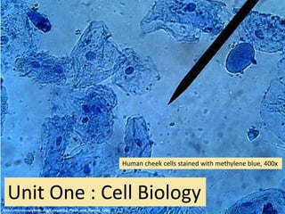
  • 1. Unit One : Cell Biology Human cheek cells stained with methylene blue, 400x http://microscopy4kids.org/Comparing_Plant_and_Animal_Cells
  • 2. 1.1 Introduction to Cells Below are some of the oldest fossilized cells on Earth, Prokaryotes from 3.0 to 3.5 billion years ago • Essential idea: The evolution of multicellular organisms allowed cell specialization and cell replacement.
  • 3. Cell theory states that: 1. All living things are composed of cells (or cell products) 2. The cell is the smallest unit of life 3. Cells only arise from pre-existing cells 1.1 U.1 According to the cell theory, living organisms are composed of cells.
  • 4. 1. All living things are composed of cells (or cell products) Living organisms are composed of cells or cell products Volvox, a type of green algae. It forms spherical colonies of up to 50,000 cells
  • 5. 2. The cell is the smallest unit of life Specialized structures within cells (organelles) carry out different functions. Organelles cannot survive alone.
  • 6. 3. Cells only arise from pre-existing cells: • Cells multiply through division • All life evolved from simpler ancestors • Mitosis results in genetically identical diploid daughter cells • Meiosis generates haploid gametes (sex cells) http://cx.aos.ask.com/question/aq/700px- 700px/why-do-cells-divide_462b38a6-0721-4835- 9cbd-e4b9e1a0fb59.jpg
  • 7. 1.1 A.1 Questioning the cell theory using atypical examples, including striated muscle, giant algae and aseptate fungal hyphae. Exceptions to the Cell Theory Example 1: striated muscle • Challenges the idea that a cell has one nucleus are multi-nucleated. This does not conform to the standard view of a small single nuclei within a cell • Muscle cells have more than one nucleus per cell • Muscle Cells called fibers can be very long (300mm)
  • 8. Exceptions to the Cell Theory Example 2: Acetabularia • Acetabularia is a giant algae is a single-celled organism that challenges both the idea that cells must be simple in structure and small in size. • As a comparison in size a human blood cell (the most abundant cell in a human is about 100 micrometers. cells average between 5000 – 100,000 micrometers. 1.1 A.1 Questioning the cell theory using atypical examples, including striated muscle, giant algae and aseptate fungal hyphae.
  • 9. Exceptions to the Cell Theory Example 3: fungal hyphae • challenges the idea that a cell is a single unit. • Fungal hyphae are again very large with many nuclei and a continuous cytoplasm • The tubular system of hyphae form dense networks called mycelium 1.1 A.1 Questioning the cell theory using atypical examples, including striated muscle, giant algae and aseptate fungal hyphae.
  • 10. Exceptions to the Cell Theory Example 4: Bone: •There are some cells that secrete material outside of the cell membrane •The secretions solidify and dominate the structure 1.1 A.1 Questioning the cell theory using atypical examples, including striated muscle, giant algae and aseptate fungal hyphae.
  • 11. Exceptions to the Cell Theory Example 5: Red Blood Cells • Mammalian erythrocytes are unique among the vertebrates as they are non-nucleated cells in their mature form. • The lack of a nucleus provide more space for hemoglobin. • In mammals, erythrocytes also lose all other cellular organelles such as their mitochondria, Golgi apparatus and endoplasmic reticulum. 1.1 A.1 Questioning the cell theory using atypical examples, including striated muscle, giant algae and aseptate fungal hyphae.
  • 12. 1.1 U.2 Organisms consisting of only one cell carry out all functions of life in that cell. You probably know: • Movement • Reproduction • Homeostasis • Growth • Respiration • Excretion • Nutrition • Transport • Repair In this course the functions are refined: • Metabolism - the web of all the enzyme- catalyzed reactions in a cell or organism, e.g. respiration • Response - Living things can respond to and interact with the environment • Homeostasis - The maintenance and regulation of internal cell conditions, e.g. water and pH • Growth - Living things can grow or change size / shape • Reproduction - Living things produce offspring, either sexually or asexually • Excretion – the removal of metabolic waste • Nutrition – feeding by either the synthesis of organic molecules (e.g. photosynthesis) or the absorption of organic matter An easy way to remember Metabolism, Response, Homeostasis, Growth, Reproduction, Excretion and Nutrition is: MR H GREN
  • 13. How does this paramecium show the functions of life? 1.1 U.2 Organisms consisting of only one cell carry out all functions of life in that cell.
  • 14. http://umanitoba.ca/Biology/BIOL1030/Lab1/biolab1_3.html#Ciliophora Homeostasis – contractile vacuole fill up with water and expel it through the plasma membrane to manage the water content Reproduction – The nucleus can divide to support cell division by mitosis, reproduction is often asexual Metabolism – most metabolic pathways happen in the cytoplasm Growth – after consuming and assimilating biomass from food the paramecium will get larger until it divides. Response – the wave action of the cilia moves the paramecium in response to changes in the environment, e.g. towards food. Excretion – the plasma membrane control the entry and exit of substances including expulsion of metabolic waste Nutrition – food vacuoles contain organisms the paramecium has consumed
  • 15. How does this photosynthetic algae show the functions of life? 1.1 U.2 Organisms consisting of only one cell carry out all functions of life in that cell.
  • 16. Homeostasis – contractile vacuole fill up with water and expel it through the plasma membrane to manage the water content Reproduction – The nucleus can divide to support cell division, by mitosis (these cells are undergoing cytokinesis) Metabolism – most metabolic pathways happen in the cytoplasm Growth – after consuming and assimilating biomass from food the algae will get larger until it divides. Response – the wave action of the cilia moves the algae in response to changes in the environment, e.g. towards light. Excretion – the plasma membrane control the entry and exit of substances including the diffusion out of waste oxygen Nutrition – photosynthesis happens inside the chloroplasts to provide the algae with food
  • 17. • All organisms need to exchange substances such as food, waste, gases and heat with their surroundings. These substances must be exchanged between the organism and its surroundings. • As the size of a structure increases the surface area to volume ratio decreases. Therefore the rate of exchange (diffusion/radiation) decreases. This is true for organelles, cells, tissues, organs and organisms. • As cells become larger they may not be able to take in essential nutrients need for survival. • The rate of exchange of substances therefore depends on the organism's surface area that is in contact with the surroundings. • The exchange depends on the volume of the organism, so the ability to meet the requirements depends on , which is known as the surface area to volume ratio. All organisms need to have a large surface to volume ration. • As organisms get bigger their volume and surface area both get bigger, but not by the same amount. This can be seen by performing some simple calculations concerning different-sized organisms. 1.1 U.3 Surface area to volume ratio is important in the limitation of cell size.
  • 18. 1 2 3 1 cm 10 cm 100 cm Assume we have 3 cubes: With sizes: What will happen to ratio between V and S.A. as their size increases? 1.1 U.3 Surface area to volume ratio is important in the limitation of cell size.
  • 19. Ratio of Surface Area to Volume ration of the 3 cells Cube Side Length Volume (x3) S.A. (6x2) Ratio (S.A./V) 1 1 cm 2 10 cm 3 100 cm 1 cm3 1 000 cm3 1 000 000 cm3 6 cm2 600 cm2 60 000 cm2 6 0.6 0.06 1.1 U.3 Surface area to volume ratio is important in the limitation of cell size.
  • 20. 1.1 U.5 Specialized tissues can develop by cell differentiation in multicellular organisms. • In humans 220 distinct highly specialized cell types have been recognized • All specialized cells and the organs constructed from them have developed as a result of differentiation
  • 21. Emergent properties arise from the interaction of component parts. The whole is greater than the sum of its parts. Multicellular organisms are capable of completing functions that individual cells could not undertake - this is due to the interaction between cells producing new functions. 1.1 U.4 Multicellular organisms have properties that emerge from the interaction of their cellular components. The advantage is that when these functions work together they can benefit the organism as a whole
  • 22. 1.1 U.4 Multicellular organisms have properties that emerge from the interaction of their cellular components. When studying the individual parts of a living thing, there are many times when the part alone do not allow the prediction of the properties of the whole living thing. As an example: the toaster parts to the right. Only when we combine them to form the toaster can these properties be determined. There is nothing supernatural about the emergent properties rather it is simply the combination of the parts that results in new properties emerging. As a model consider a toaster. The toaster is the system and is composed of 400 different parts. We can study the parts individually how they function and the properties they posses. Some of these would be the properties of : • Steele • Mica • Plastic Toaster Project
  • 23.
  • 24. 1.1 U.6 Differentiation involves the expression of some genes and not others in a cell’s genome. • All (diploid) cells of an individual organisms share an identical genome - each cell contains the entire set of genetic instructions for that organism • BUT not all genes are expressed (activated) in all cells • In (totipotent) embryonic stem cells the entire genome is active • Newly formed cells receive signals which deactivate (or more rarely activate) genes, e.g. a skin cell does not need to be able to produce hemoglobin (the pigment in red blood cells that carries oxygen)
  • 25. 1.1 U.7 The capacity of stem cells to divide and differentiate along different pathways is necessary in embryonic development and also makes stem cells suitable for therapeutic uses. Stem cells are unspecialized cells that can: • Can continuously divide and replicate • Have the capacity to differentiate into specialized cell types Totipotent Can differentiate into any type of cell. Pluripotent Can differentiate into many types of cell. Multipotent Can differentiate into a few closely-related types of cell. Unipotent Can regenerate but can only differentiate into their associated cell type (e.g. liver stem cells can only make liver cells). Potency
  • 26. Learn about stem cells using the tutorials 1.1 U.7 The capacity of stem cells to divide and differentiate along different pathways is necessary in embryonic development and also makes stem cells suitable for therapeutic uses.
  • 27. 1.1 A.4 Ethics of the therapeutic use of stem cells from specially created embryos, from the umbilical cord blood of a new-born baby and from an adult’s own tissues. Comparison of stem cell sources Embryo Cord blood Adult Differentiation Can differentiate into any cell type Limited capacity to differentiate (without inducement only naturally divide into blood cells) Limited capacity to differentiate (dependent on the source tissue) Genetic damage Less chance of genetic damage than adult cells Due to accumulation of mutations through the life of the adult genetic damage can occur Compatibility Stem cells are not genetically identical to the patient Fully compatible with the patient as the stem cells are genetically identical
  • 28. Comparison of stem cell sources Embryo Cord blood Adult Ease of extraction Can be obtained from excess embryos generated by IVF programs. Easily obtained and stored. Though limited quantities available Difficult to obtain as there are very few and are buried deep in tissues Ethics of the extraction Can only be obtained by destruction of an embryo Umbilical cord is removed at birth and discarded whether or not stem cells are harvested Adult patient can give permission for cells to be extracted Growth potential Almost unlimited Reduced potential (compared to embryonic cells) Tumor risk Higher risk of development Lower risk of development 1.1 A.4 Ethics of the therapeutic use of stem cells from specially created embryos, from the umbilical cord blood of a new-born baby and from an adult’s own tissues.
  • 29. 1.1 A.4 Ethics of the therapeutic use of stem cells from specially created embryos, from the umbilical cord blood of a new-born baby and from an adult’s own tissues.
  • 30. 1.1 U.7 Use of stem cells to treat Stargardt’s disease and one other named condition.
  • 31. Photo has been altered to show how someone with Macular Degeneration might see the Mona Lisa. 1.1 U.7 Use of stem cells to treat Stargardt’s disease and one other named condition.
  • 32. 1.1 U.7 Use of stem cells to treat Stargardt’s disease and one other named condition.
  • 33. The problem • Affects around one in 10,000 children • Recessive genetic (inherited) condition • The mutation causes an active transport protein on photoreceptor cells to malfunction • The photoreceptor cells degenerate • the production of a dysfunctional protein that cannot perform energy transport • that causes progressive, and eventually total, loss of central vision The treatment • Embryonic stem cells are treated to divide and differentiate to become retinal cells • The retinal cells are injected into the retina • The retinal cells attach to the retina and become functional • Central vision improves as a result of more functional retinal cells The future • This treatment is still in at the stage of limited clinical trials, but will likely be in usage in the future 1.1 U.7 Use of stem cells to treat Stargardt’s disease and one other named condition.
  • 34. 1.1 U.7 Use of stem cells to treat Stargardt’s disease and one other named condition. Leukemia
  • 35. 1.1 U.7 Use of stem cells to treat Stargardt’s disease and one other named condition. Leukemia The problem • Cancer of the blood or bone marrow, resulting in abnormally high levels of poorly-functioning white blood cells. The treatment • Hematopoetic Stem Cells (HSCs) are harvested from bone marrow, peripheral blood or umbilical cord blood • Chemotherapy and radiotherapy used to destroy the diseased white blood cells • New white blood cells need to be replaced with healthy cells. • HSCs are transplanted back into the bone marrow • HSCs differentiate to form new healthy white blood cells The benefit • The use of a patient’s own HSCs means there is far less risk of immune rejection than with a traditional bone marrow transplant.
  • 36. 1.1 A.4 Ethics of the therapeutic use of stem cells from specially created embryos, from the umbilical cord blood of a new-born baby and from an adult’s own tissues. Arguments for Therapeutic Cloning • Stem cell research may pave the way for future discoveries. • May be used to cure serious diseases or disabilities. • Stem cells can be taken from embryos that have stopped developing and would have died anyway (e.g. abortions) • Cells are taken at a stage when the embryo has no nervous system and can arguably feel no pain Arguments Against Therapeutic Cloning • Involves the creation and destruction of human embryos (at what point do we afford the right to life?) • Embryonic stem cells are capable of continued division and may develop into cancerous cells and cause tumors • With additional cost and effort, alternative technologies may fulfill similar roles (e.g. nuclear reprogramming of differentiated cell lines) • Religious or moral objections due to the ‘playing God’ argument.
  • 37. 1.2 Ultrastructure of cells • Essential idea: Eukaryotes have a much more complex cell structure than prokaryotes. The background image to the right is an electron micrograph of a plant cell. It clearly shows the complex structures present in eukaryote cells. http://botit.botany.wisc.edu/Resources/Botany/Plant%20Cell/Electron%20Micrographs/General%20Cell/Young-Cell.jpg
  • 38. Microscopes are needed to study cell (Due to there small size) with high magnification and resolution (clarity) are needed to observe cells and their components. LARGEST to smallest  Cells (up to 100um)  Organelles (up to 10 um)  Bacteria (1um)  Viruses(100nm)  Membranes (10nm)  Molecules (1nm)  Atoms 1.2 U.3 Electron microscopes have a much higher resolution than light microscopes.
  • 39. 1.2 U.3 Electron microscopes have a much higher resolution than light microscopes. Resolution is defined as the shortest distance between two points that can be distinguished Magnification is defined as magnifying or enlarged image
  • 40. 1.2 U.3 Electron microscopes have a much higher resolution than light microscopes. • Light microscopes are limited in resolution by the wavelengths of visible light (400–700 nm). • Electrons have a much shorter wavelength (2 – 12 pm) therefore electron microscopes have a much higher resolution • Conversions from millimeters to micro and nano meter above in green. Comparison of Resolution Millimetres (mm) Micrometres (μm) Nanometres (nm) Human eye 0.1 100 100,000 Light microscopes 0.0002 0.2 200 Electron microscopes 0.000001 0.001 1 Electron Microscope
  • 41. 1.2 U.3 Electron microscopes have a much higher resolution than light microscopes. • Light microscopes allow us to see the structure of cells • Electron microscopes allow us to see the ultrastructure of cells, such as these pancreatic exocrine cells • Electron microscopes can see viruses (0.1μm diameter) , but light microscopes cannot Ultrastructure is all the structures of a biological specimen that are at least 0.1nm in their smallest dimension http://medcell.med.yale.edu/systems_cell_biology_old/liver_and_pancreas/images/exocrine_pancreas_em.jpg
  • 42.
  • 43.
  • 44.
  • 45.
  • 46. 1.2 U.1 Prokaryotes have a simple cell structure without compartmentalization.
  • 47. 1.2 S.1 Drawing of the ultrastructure of prokaryotic cells based on electron micrographs.
  • 48.
  • 49. 1.2 U.1 Prokaryotes have a simple cell structure without compartmentalization. Ultrastructure of E. coli as an example of a prokaryote • E. Coli is a model organism used in research and teaching. Some strains are toxic to humans and can cause food poisoning. • We refer to the cell parts/ultrastructure of prokaryotes rather than use the term organelle as very few structures in prokaryotes are regarded as organelles.
  • 50. Prokaryotes reproduce asexually using the process of binary fission • The two DNA loops attach to the membrane • The membrane elongates and pinches off (cytokinesis) forming two separate cells • The two daughter cells are genetically identically (clones) • They have no proteins on there DNA. Making it impossible to make a chromosome and go through the process of mitosis. 1.2 A.2 Prokaryotes divide by binary fission. http://cronodon.com/images/Bacteria_dividing_3b.jpg http://youtu.be/vTzH1P3aQjg
  • 51. Eukaryotes “those having a true nucleus,” • Eukaryotic cells are forms of life composed of mutually independent parts the maintain various vital process’ • They area form of life consisting of animals, plants, fungus, protest, and moneran. • These cells contain membrane bound organelles, which dramatically increase the efficiency of all metabolic processes. • Any organized body system is composed of these cells 1.2 U.2 Eukaryotes have a compartmentalized cell
  • 52. 1.2 U.2 Eukaryotes have a compartmentalized cell There are several advantages in being compartmentalized: • Efficiency of metabolism - enzymes and substrates can localized and much more concentrated • Localized conditions - pH and other such factors can be kept at optimal levels. The optimal pH level for one process in one part of the cell • Toxic / damaging substances can be isolated, e.g. digestive enzymes (that could digest the cell itself) are stored in lysosomes • Numbers and locations of organelles can be changed dependent on the cell’s requirement.
  • 53. Free ribosomes • 80S Ribosomes (approx. 20nm diameter) - larger than the ribosomes found in prokaryotes • No membrane • These appear as dark granules in the cytoplasm • Synthesizes proteins to function in the cytoplasm, for use within the cell, e.g. enzymes
  • 54. 1.2 A.1 Structure and function of organelles within exocrine gland cells of the pancreas and within palisade mesophyll cells of the leaf. Nucleus • Generally spherical with a double membrane • Pores (holes) are present in the membrane • Contains genetic information in the form of chromosomes (DNA and associated histone proteins) • Uncoiled chromosomes are referred to as chromatin – they stain a dark colour and are concentrated at the edges of the nucleus • mRNA is transcribed in the nucleus (prior to use in protein synthesis in the cytoplasm) • mRNA leaves the nucleus via the pores (DNA is too large to move through the pores)
  • 55. Mitochondria • Has a double membrane • A smooth outer membrane and a folded inner membrane • The folds are referred to as cristae • Variable in shape • Site of ATP production by aerobic respiration (if fat is used as a source of energy it is digested here) 1.2 U.2 Eukaryotes have a compartmentalized cell
  • 56. Rough Endoplasmic Reticulum (rER) • The consists of flattened membrane sacs, called cisternae • Often located near to the nucleus • 80S Ribosomes are attached to the outside of the cisternae are ribosomes • rER synthesizes proteins which are transported, by vesicles, to the golgi apparatus for modification before secretion outside the cell Smooth Endoplasmic Reticulum • no ribosomes present • we are not studying this structure it this course
  • 57. Golgi apparatus • This organelle also consists of flattened membrane sacs called cisternae, like rER. • Different to rER:  No attached ribosomes  Often sited close to the plasma membrane  The cisternae are shorter and more curved that those of the rER • The Golgi apparatus processes (modifies) proteins from from the rER. The proteins are then repackaged in vesicles for secretion outside the cell.
  • 58. Vesicles • A single membrane with fluid inside • Very small in size • Used to transport materials inside of a cell http://commons.wikimedia.org/wiki/File:C_Golgi.jpg 1.2 U.2 Eukaryotes have a compartmentalized cell
  • 59. Lysosomes • Generally spherical with a single membrane • Formed from Golgi vesicles. • They contain digestive enzymes for breakdown of: o ingested food in vesicles o unwanted/damaged organelles o The cell itself • High concentration of enzymes (a type of protein) cause this organelle to stain heavily and hence appear dark on micrographs 1.2 U.2 Eukaryotes have a compartmentalized cell
  • 60. Vacuoles • Single membrane with fluid inside • In Plant cells vacuoles are large and permanent, often occupying the majority of the cell volume • In animals vacuoles are smaller and temporary and used for various reasons, e.g. to absorb food and digest it. 1.2 U.2 Eukaryotes have a compartmentalized cell
  • 61. Flagellum (Flagella pl.) • Thin projection (usually singular) from the cell surface. • Contain microtubules • Used to move the cell 1.2 U.2 Eukaryotes have a compartmentalized cell
  • 62. Cilia • Thin projections from the cell surface. • Contain microtubules • Used to either move the cell or to move the fluids next to the cell 1.2 U.2 Eukaryotes have a compartmentalized cell
  • 63. Microtubules • Small cylindrical fibres called microtubules • Have a variety of functions, e.g. part of the structure of flagella and they play a role in cell division Centrioles • Consist of two groups of nine triple microtubules • Are mainly found in animal cells, not present in vascular plants or fungi. 1.2 U.2 Eukaryotes have a compartmentalized cell
  • 64. Chloroplast • Many, but not all, plant cells contain chloroplasts • A double membrane surrounds the chloroplast • Inside are stacks of thylakoids • Each thylakoid is a disc composed of a flattened membrane • The shape of chloroplasts is variable but is usually ovoid • The site of photosynthesis and hence where glucose is produced. • Starch grains maybe present if photosynthesis is happening quickly 1.2 A.1 Structure and function of organelles within exocrine gland cells of the pancreas and within palisade mesophyll cells of the leaf.
  • 65. Cell wall • an extracellular component not an organelle. • secreted by all plant cells (fungi and some protists also secrete cell walls). • Plant cell walls consist mainly of cellulose which is: o Permeable - does not affect transport in and out of the cell o Strong – gives support to the cell and prevent the plasma membrane bursting when under pressure o Hard to digest –resistant to being broken down, therefore lasts along time without the need for replacement/maintenance 1.2 U.2 Eukaryotes have a compartmentalized cell
  • 66. Two Types of Eukaryotic Cells: Plant Cells • Cell wall is present. • Chloroplasts containing the green pigment, chlorophyll – are present. • Autotrophs – can make their own food. • Cells are angular shaped. • Large central vacuole is present. • Centrioles absent. • Flagellum are uncommon. Animal Cells • Cell wall is absent. • Chloroplasts are absent. • Heterotrophs – depend on other organisms for food. • Cells are rounded and not very regular. • Vacuoles are small and scattered. • Centrioles are present. • Flagellum may be present.
  • 67. This is a good drawing, but the nucleus should show a double membrane and small pores (gaps) in the membrane.
  • 69. 1.2 S.2 Drawing of the ultrastructure of eukaryotic cells based on electron micrographs. Draw a single palisade mesophyll cell from the image below. You should be able to label the following structures: • cell wall • plasma membrane • chloroplasts • vacuole • nucleus • cytoplasm • mitochondria http://www.lifesci.sussex.ac.uk/home/Julian_Thorpe/TEM19.htm
  • 70. 1.2 S.2 Drawing of the ultrastructure of eukaryotic cells based on electron micrographs. Draw a pancreas exocrine cell from the image to the right. You should be able to label the following structures: • plasma membrane • mitochondria • rER • Nucleus • Secretory granules (dark spheres) http://medcell.med.yale.edu/systems_cell_biology_old/liver_and_pancreas/images/exocrine_pancreas_em.jpg
  • 71. What organelles can you identify? Think about the role of the organelles that occur most common and deduce the function of the cell.
  • 72. Evidence & conclusions: • Nucleus present • No cell wall – this is an animal cell • ER is present and dominates the cell • Mitochondria • Lots of secretory granules/vesicles near the inside border
  • 73. http://www.vcbio.science.ru.nl/images/tem-plant-cell.jpg What organelles can you identify? Think about the role of the organelles that occur most common and deduce the function of the cell.
  • 74. http://www.vcbio.science.ru.nl/images/tem-plant-cell.jpg Evidence & conclusions: • Cell wall present – must be a plant cell • No chloroplasts – must be found inside the stem or in the roots • Mitochondria • Vacuoles relatively small – the cell does not have a storage, transport or support function • Nucleus relatively large / cell size small – likely to be a new cell recently undergone mitosis • Possibly recently divided cell tissue from a plant root This is: a plant cell found at the root-tip
  • 75. Essential idea: The structure of biological membranes makes them fluid and dynamic. 1.3 Membrane Structure
  • 76. The Oil droplet stays together and makes a perfect circular shape. The oil molecules are Hydrophobic Oil Molecules are non- polar and water molecules are polar. 1.3 U.1 Phospholipids form bilayers in water due to the amphipathic properties of phospholipid molecules.
  • 77. Remember the properites of fatty acids (non polar) When put into water, an emergent property is that phospholipids will self-organise to keep their heads ‘wet’ and their tails ‘dry’. These phospholipids are associated with spontaneously into stable micelles because of their amphipathic properties. micelle liposome 1.3 U.1 Phospholipids form bilayers in water due to the amphipathic properties of phospholipid molecules.
  • 78. Singer-Nicholson fluid mosaic model http://commons.wikimedia.org/wiki/File:Cell_membrane_detailed_diagram_en.svg?uselang=en-gb 1.3 S.3 Analysis of the falsification of the Davson-Danielli model that led to the Singer-Nicolson model. Key features of the current model: • Phospholipid molecules form a bilayer - phospholipids are fluid and move laterally • Peripheral proteins are bound to either the inner or outer surface of the membrane • Integral proteins - permeate the surface of the membrane • The membrane is a fluid mosaic of phospholipids and proteins • Proteins can move laterally along membrane
  • 79. Membrane fluidity 1.3 A.1 Cholesterol in mammalian membranes reduces membrane fluidity and permeability to some solutes. Though it is difficult to determine whether the membrane is truly either a solid or liquid it can definitely be said to be fluid. It is important to regulate the degree of fluidity: • Membranes need to be fluid enough that the cell can move • Membranes need to be fluid enough that the required substances can move across the membrane • If too fluid however the membrane could not effectively restrict the movement of substances across itself The hydrophobic hydrocarbon tails usually behave as a liquid. Hydrophilic phosphate heads act more like a solid.
  • 80. Phospholipid molecules have a polar (charged) phosphate head and long non-polar lipid tails • The head is hydrophilic (attracted to water) • The tails are hydrophobic (repelled by water) 1.3 U.1 Phospholipids form bilayers in water due to the amphipathic properties of phospholipid molecules. When drawing a diagram of a phospholipid this is a good example which shows all the key features
  • 81. Cholesterol in the membrane : 1. Makes the phospholipids pack more tightly and regulates the fluidity and flexibility of the membrane. 2. The presence of cholesterol disrupts the regular packing of the of the hydrocarbon tails of phospholipid molecules - this is increases the flexibility as it prevents the tails from crystallising and hence behaving like a solid. 3. Cholesterol also reduces the permeability to hydrophilic/water soluble molecules and ions such as sodium and hydrogen. 1.3 U.3 Cholesterol is a component of animal cell membranes.
  • 82. Cholesterol http://www.cholesterol-and-health.com/images/Cholesterol_Structure.jpg http://www.uic.edu/classes/bios/bios100/lectf03am/cholesterol.jpg Hydroxyl group makes the head polar and hydrophilic - attracted to the phosphate heads on the periphery of the membrane. Carbon rings – it’s not classed as a fat or an oil, cholesterol is a steroid Non-polar (hydrophobic) tail –attracted to the hydrophobic tails of phospholipids in the centre of the membrane
  • 83. Proteins: Three major types 1. Integral Proteins are permanently embedded, many go all the way through from the inside of the cell to the outside of the cell. 2. Peripheral Proteins usually have a temporary association with the membrane, they can be attach to the surface of the membrane 1.3 U.2 Membrane proteins are diverse in terms of structure, position in the membrane and function.
  • 84. 1.3 U.2 Membrane proteins are diverse in terms of structure, position in the membrane and function. 3. Glycoproteins: Are proteins with an oligosaccaride (oligo = few, saccharide = sugar) chain attached. They are important for cell recognition by the immune system and as hormone receptors
  • 85. 1.3 U.2 Membrane proteins are diverse in terms of structure, position in the membrane and function. Proteins associated with membranes have many functions. We are going to look at six function for membrane proteins. Try and remember the acronym TRACIE Transport Receptor Anchor Cell to Cell Communication Intercellular joints Enzymes
  • 86. Transport: Protein channels (facilitated) and protein pumps (active) Receptors: Hormones binding sites (insulin, glucagon, etc.) Anchorage: Cytoskeleton attachments and extracellular matrix Cell to cell communication: Example: Macrophages and Helper T Cells Intercellular joining's: Tight junctions and plasmodesmata Enzymatic activity: Metabolic pathways (e.g. electron transport chain) 1.3 U.2 Membrane proteins are diverse in terms of structure, position in the membrane and function.
  • 87. Our current model of the cell membrane is called the Singer-Nicholson fluid mosaic model This model was first proposed in by Singer-Nicolson in 1972 Before then Davson-Danielli model was widely accepted …
  • 88. 1.3 S.2 Analysis of evidence from electron microscopy that led to the proposal of the Davson-Danielli model. The Davson-Danielli model: • A protein-lipid sandwich • Lipid bilayer composed of phospholipids (hydrophobic tails inside, hydrophilic heads outside) • Proteins coat outer surface • Proteins do not permeate the lipid bilayer Old model for the cell membrane
  • 89. 1.3 S.3 Analysis of the falsification of the Davson-Danielli model that led to the Singer- Nicolson model. Interpreting the image: • The fracture occurs along lines of weakness, including the centre of membranes. • The fracture reveals an irregular rough surface inside the phospholipid bilayer • The globular structures were interpreted as trans- membrane proteins. Falsification of the Davson-Danielli model– freeze fracturing Conclusion: This is contrary to the Davson-Danielli model which only involves proteins coating the surface of the membrane. A new model is needed to explain the presence of as trans-membrane proteins.
  • 90. Singer-Nicholson fluid mosaic model There is strong evidence for this model: Biochemical techniques • Membrane proteins were found to be very varied in size and globular in shape • Such proteins would be unable to form continuous layers on the periphery of the membrane. • The membrane proteins had hydrophobic regions and therefore would embed in the membrane not layer the outside
  • 91. Singer-Nicholson fluid mosaic model There is strong evidence for this model: Fluorescent antibody tagging • Within 40 minutes the red and green markers were mixed throughout the membrane of the fused cell. • This showed that membrane proteins are free to move within the membrane rather than being fixed in a peripheral layer. • red or green fluorescent markers attached to antibodies which would bind to membrane proteins • The membrane proteins of some cells were tagged with red markers and other cells with green markers. • The cells were fused together.
  • 93. 1.4 Membrane transport • Essential idea: Membranes control the composition of cells by active and passive transport.
  • 94. Polar Head: Attracted to polar molecules (Like water) Non-polar fatty acid tails Will repel charged molecules (like ions) 1.4 U.1 Particles move across membranes by simple diffusion, facilitated diffusion, osmosis and active transport. Due to the polar head and non polar fatty acid chain, some molecules pass through the membrane easily (diffusion), or go through proteins (facilitated diffusion), others need energy to get them through (active transport). Very large molecules come in and out using their own membrane (endo and exo- cytosis)
  • 95. 1.4 U.1 Particles move across membranes by simple diffusion, facilitated diffusion, osmosis and active transport. Movement of materials is controlled in 5 ways: 1. Simple diffusion 2. Osmosis 3. Facilitated diffusion 4. Active Transport 5. Membrane aided The Phospholipid bilayer is selectively permeable controlled entry/exit of molecules
  • 96. 1.4 U.1 Particles move across membranes by simple diffusion, facilitated diffusion, osmosis and active transport. 1. Simple Diffusion is a form of passive transport, a net movement of particles from an area of high concentration to an area of low concentration. This is often through a partially permeable membrane. PASSIVE: DOES NOT REQUIRE ENERGY Concentration gradient: the difference in concentration of substances between two locations
  • 97. 1. Simple Diffusion: movement from an area of high concentration to an area of low concentration 1.4 U.1 Particles move across membranes by simple diffusion, facilitated diffusion, osmosis and active transport.
  • 98. 2. Osmosis Most cell are partially permeable membrane, water flows with the concentration gradient. When a cell is submerged in water, the water molecules pass through the cell membrane from an area of low solute concentration (outside the cell) to one of high solute concentration (inside the cell) a. Hypertonic concentration of solute (material other then water) is higher outside of the cell. Water moves out of the cell. Cell get smaller b. Isotonic concentration of solute is the same inside the cell as outside of the cell. Equal movement of water into and out of the cell. Cells maintain there size. c. Hypotonic concentration of solute is lower inside of the cell. Water moves into the cell. Cell expands 1.4 U.1 Particles move across membranes by simple diffusion, facilitated diffusion, osmosis and active transport.
  • 99. Turgor (Plant Cells) • When the outside water enters the plant cell the vacuole becomes bigger and the cytoplasm swells • This causes the membrane to be pushed out towards the cell wall • When cells are fully “swelled” like this with the membranes pushing against the cell wall they are described as Turgid. This turgor pressure gives plants their strength. If plants did not have this they would be wilted 1.4 U.1 Particles move across membranes by simple diffusion, facilitated diffusion, osmosis and active transport.
  • 100. Plasmolysis (Animal Cells) • When this happens the cell wall stays intact but the membrane shrivels up away from it • This is called Plasmolysis • Cells in this condition are called plasmolysis cells we can look at this easily by placing a layer of red onion cells in salt water 1.4 U.1 Particles move across membranes by simple diffusion, facilitated diffusion, osmosis and active transport.
  • 101. OSMOlARITY REGULATION regulation of the osmotic pressure of an organism's body fluids to maintain the homeostasis of the organism's water content; that is, it maintains the fluid balance and the concentration of electrolytes (salts in solution) to keep the fluids from becoming too diluted or too concentrated. Saline solution used for eye drops, flushing out wound is 0.9% NaCl. Ringers Lactate Solution (Show in the right) Ringer's lactate solution used to replace lost fluids due to trama or dehydration, it is isotonic with normal blood plasma 1.4 A.2 Tissues or organs to be used in medical procedures must be bathed in a solution with the same osmolarity as the cytoplasm to prevent osmosis.
  • 102. 3. Facilitated Diffusion: Large and polar molecules can’t get across the membrane via simple diffusion. Protein Channels or Carrier Proteins imbedded in the membrane, recognize a particular molecule and help it to move across the membrane. 1.4 U.1 Particles move across membranes by simple diffusion, facilitated diffusion, osmosis and active transport.
  • 103. 1.4 A.1 Structure and function of sodium–potassium pumps for active transport and potassium channels for facilitated diffusion in axons. 1. At one stage during a nerve impulse there are relatively more positive charges inside. 2. This voltage change causes potassium channels to open, allowing potassium ions to diffuse out of the axon. 3. Once the voltage conditions change the channel rapidly closes again. Example: Potassium channels in axons are voltage gated. They enable the facilitated diffusion of potassium out of the axon
  • 104. 4. Active transport uses energy from the hydrolysis of ATP (see below) to pump molecules against the concentration gradient 1.4 U.1 Particles move across membranes by simple diffusion, facilitated diffusion, osmosis and active transport.
  • 105. Primary active transport requires ATP. Integral protein pumps use the energy from the hydrolysis of ATP to move ions or large molecules across the cell membrane. Molecules are moved against their concentration gradient 1.4 U.1 Particles move across membranes by simple diffusion, facilitated diffusion, osmosis and active transport. Remember: 3 Sodium (Na+) out making the neuron -70 mv 2 Potassium (K+) in. With the use of ATP
  • 106. 5. Membrane aided • Endocytosis: The taking in of external substances by an inward pouching of the plasma membrane, forming a vesicle • Exocytosis: The release of substances from a cell (secretion) when a vesicle joins with the cell plasma membrane. 1.4 U.2 The fluidity of membranes allows materials to be taken into cells by endocytosis or released by exocytosis.
  • 107. http://commons.wikimedia.org/wiki/File:Endocytosis_types.svg “Cell eating” “Cell drinking” 1.4 U.2 The fluidity of membranes allows materials to be taken into cells by endocytosis or released by exocytosis.
  • 108. 1.5 Origin of cells Essential idea: There is an unbroken chain of life from the first cells on Earth to all cells in organisms alive today. Two billion year old prokaryotic fossils of cells.
  • 109. Spontaneous Generation • For much of history, people believed that animals could come from non-living sources. They thought: – Frogs developed from falling drops of rain – mice arose from sweaty underwear – and flies arose from decaying meat. • This is called abiogenesis, also known as spontaneous generation 1.5 A.1 Evidence from Pasteur’s experiments that spontaneous generation of cells and organisms does not now occur on Earth. Watch as a rock and a leaf (picture on the left) turn into a frog and a fish, amazing!
  • 110. • A 17th century recipe for the spontaneous generation of mice: – Place sweaty underwear and husks of wheat in an open mouthed jar – Wait 21 days while sweat from the underwear penetrates the husks of wheat and changes them into mice – This recipe was widely accepted until the late 1860’s 1.5 A.1 Evidence from Pasteur’s experiments that spontaneous generation of cells and organisms does not now occur on Earth.
  • 111. 1.1 U.1 According to the cell theory, living organisms are composed of cells. Cell theory states that: • All living things are composed of cells (or cell products) • The cell is the smallest unit of life • Cells only arise from pre-existing cells Most of these debates involved the nature of cellular regeneration, and the idea of cells as a fundamental unit of life. Cell theory was eventually formulated in 1839. The ideas in the cell theory contradict the theory of spontaneous generation
  • 112. 1.5 U.1 Cells can only be formed by division of pre-existing cells. Cells can only be formed by division of pre-existing cells: • Cells multiply through division • Mitosis results in genetically identical diploid daughter cells • Meiosis generates haploid gametes (sex cells) What evidence do we have to support this theory?
  • 113. 1.5 U.1 Cells can only be formed by division of pre-existing cells. Some of the ideas of biology held back by the idea of spontaneous generation: 1. Cells are highly complex structures and no mechanism has been found for producing cells from simpler subunits.
  • 114. 1.5 U.1 Cells can only be formed by division of pre-existing cells. 2. All known examples of growth from cell to tissue, tissue to organ, organ to multicellular organism, are all a result of cell division.
  • 115. 1.5 U.1 Cells can only be formed by division of pre-existing cells. 3. Viruses are produced from simpler subunits, but they do not consist of cells, and they can only be produced inside the host cells that they have infected.
  • 116. 1.5 U.1 Cells can only be formed by division of pre-existing cells. 4. Genetic code is universal each of the 64 codons (a codon is a combination of 3 DNA bases) produces the same amino acid in translation, regardless of the organism. The logical deduction is that all cells have arisen as the result of cell division from a single common ancestor.
  • 117. Pasteur’s experiment: Supported the ideas of the Cell theory and put to rest the idea that cells appear spontaneously. 1.5 A.1 Evidence from Pasteur’s experiments that spontaneous generation of cells and organisms does not now occur on Earth.
  • 118. 1.5 A.1 Evidence from Pasteur’s experiments that spontaneous generation of cells and organisms does not now occur on Earth. Method: • Two experiments were setup • In both, Pasteur added nutrient broth to flasks and bent the necks of the flasks into S shapes • Each flask was then heated to boil the broth in order than all existing microbes were killed. • After the broth had been sterilized, Pasteur broke off the swan necks from the flasks in Experiment 1, exposing the nutrient broth within them to air from above. • The flasks in Experiment 2 were left alone. Results: • The broth in experiment 1 turned cloudy whilst the broth in experiment 2 remained clear. • This indicates that microbe growth only occurred in experiment 1. Conclusion: Pasteur rejected the hypothesis of spontaneous generation as for growth of microbes to occur a source of contamination was needed.
  • 119. There were times in the history of the Earth when cells did not exist. The first cells must have arisen from non-living material or life was transported here from elsewhere in the universe. As illustrated to the right, an asteroid may have carried the first cells. This theory is called Panspermia. 1.5 U.2 The first cells must have arisen from non-living material.
  • 120. Some of the key problems are: 1. Non-living synthesis of simple organic molecules, e.g. sugars and amino acids 2. Assembly of these organic molecules into polymers 3. Formation of membranes to package the organic molecules 4. Compartmentalization by formation of polymers that can self-replicate (enabling inheritance) 1.5 U.2 The first cells must have arisen from non-living material. We will focus on the organic evolution from materials on Earth.
  • 121. 1. Non-living synthesis of simple organic molecules, e.g. sugars and amino acids What are the raw materials around during Prebiotic Earth? 1.5 U.2 The first cells must have arisen from non-living material. Little or no free oxygen Oxygen very reactive, would remove electrons needed for reactions Considerable energy Heat, light, electrical energy needed to make/break bonds Volcanism, sun provided much UV, lightning Chemical ‘building blocks’ to be built into more complex molecules The reactions required time ~ 0.8 billion years to cause life to come about. This is a long time, but not long compared to the life of the Universe (~10-20 billion years)
  • 122. • H2O vapor • NH3 • N2 • H2 • H2S • CH4 • Virtually no O2 1.5.U.2 The first cells must have arisen from non-living material. 1. Non-living synthesis of simple organic molecules Below are the materials available in the Prebiotic Earth. These material must have been used to build the first cells on Earth. We know that all living things have the same overall chemistry. This suggests a common origin Below is the early Earth atmospheric composition:
  • 123. 1.5 U.2 The first cells must have arisen from non-living material. 2. Assembly of these organic molecules into polymers Primitive ‘Soup’ (ocean surface) • Haldane and Oparin (~1920s) proposed that life arose from nonliving conditions by means of a series of changes (and increasing complexity) in molecular composition as a result of the reducing, high energy environment that existed on early Earth (between 4.6-3.8 bybp (billion years before present) • Proposed the Prebiotic Broth Hypothesis • Not tested until 1950s • Tested by Stanley Miller and Harold Urey
  • 124. 2. Assembly of these organic molecules into polymers (Miller / Urey Experiment) • Created an ancient earth atmosphere in globes – H2O vapor – H2 – NH3 – CH4 • Electrical discharge • Heated via mantle in lower chamber • Paper chromatography showed that 13 of the 20 amino acids had formed • Similar experiments have shown that all 20 amino acids, lipids, nucleotide bases, and ATP, if phosphate present. 1.5.U.2 The first cells must have arisen from non-living material.
  • 125. 2. Assembly of these organic molecules into polymers • In 1977 hydrothermal vents were discovered, this discovery added evidence to the theory that life may have formed close to the oceans surface. • Today, hydrothermal vents provide basic materials to support living organisms in environments where there is no light, very little energy, and little other perturbation • Ammonia is produced in abundance at such locations, suggesting that it could possibly be built into nucleic acid bases and the possibly into amino acids 1.5 U.2 The first cells must have arisen from non-living material. Hyperlink
  • 126. 1.5 U.2 The first cells must have arisen from non-living material. • DNA though very stable and effective at storing information is not able to self- replicate – enzymes are required • However RNA can both store information and self-replicate - it can catalyze the formation of copies of itself. • In ribosomes RNA is found in the catalytic site and plays a role in peptide bond formation 3. Formation of polymers that can self-replicate (enabling inheritance)
  • 127. •The current “most accepted“ theory of life evolving hypothesizes an RNA world •RNA in the early world would have functioned as a self replicating molecule, eventually developing a number of minimal catalytic properties self-replicates Proteins take over catalysis DNA becomes long term storage and major coding molecule Packaging evolves - RNA codes and catalyses RNA Molecular Reproduction 1.5 U.2 The first cells must have arisen from non-living material.
  • 128. The RNA World Hypothesis • Proposed by Walter Gilbert, mid 1980s • Concept of RNA catalyzing critical pre-biotic and early biological reactions • RNA would give rise to RNA as an informational molecule • RNA would give rise to protein • RNA might bind amino acids proteins • RNA might give rise to DNA • Proteins might take over some functions • DNA would take over informational functionality • Eventually giving rise to the DNA RNA protein scheme found today 1.5 U.2 The first cells must have arisen from non-living material.
  • 129. 1.5 U.2 The first cells must have arisen from non-living material. 4. Compartmentalization The formation of membranes to package the organic molecules. Experiments have shown that phospholipids natural assemble into bilayers, if conditions are correct. These techniques are often used by drug companies as a delivery method for medications. Formation of the bilayer creates an isolated internal environment. The formation of an internal environment means that optimal conditions, e.g. for replication or catalysis can be maintained. Two ideas A. Microsphere B. Ocean froth
  • 130. 1.5 U.3 The origin of eukaryotic cells can be explained by the endosymbiotic theory. The endomembrane system A series of compartments that work together to package, label, and ship proteins and molecules. In your cells, the endomembrane system is made up of both the endoplasmic reticulum and the Golgi apparatus. These compartments are folds of membranes that form tubes and sacs in your cells. Two organelles are not part of this network, Mitochondria and chloroplasts
  • 131. http://youtu.be/q71DWYJD-dI 1.5 U.3 The origin of eukaryotic cells can be explained by the endosymbiotic theory. Endosymbiotic theory explains the existence of several organelles of eukaryotes. The theory states that the organelles (e.g. mitochondria and chloroplasts) originated as symbioses between separate single-celled organisms, http://highered.mcgraw-hill.com/sites/9834092339/student_view0/chapter26/animation_-
  • 132. First Eukaryotes (about 2 billion years ago) • Development of internal membranes – create internal micro-environments – advantage: specialization = increase efficiency • natural selection! infolding of the plasma membrane DNA cell wall plasma membrane Prokaryotic cell Prokaryotic ancestor of eukaryotic cells Eukaryotic cell endoplasmic reticulum (ER) nuclear envelope nucleus plasma membrane 1.5 U.3 The origin of eukaryotic cells can be explained by the endosymbiotic theory.
  • 133. Ancestral eukaryotic cell Eukaryotic cell with mitochondrion internal membrane system aerobic bacterium mitochondrion Endosymbiosis Endosymbiosis • Evolution of eukaryotes – origin of mitochondria – engulfed aerobic bacteria, but did not digest them – mutually beneficial relationship • natural selection! 1.5 U.3 The origin of eukaryotic cells can be explained by the endosymbiotic theory.
  • 134. mitochondrion chloroplast Eukaryotic cell with chloroplast & mitochondrion Endosymbiosis photosynthetic bacterium • Evolution of eukaryotes – origin of chloroplasts – engulfed photosynthetic bacteria, but did not digest them – mutually beneficial relationship • natural selection! Eukaryotic cell with mitochondrion 1.5 U.3 The origin of eukaryotic cells can be explained by the endosymbiotic theory.
  • 135. Theory of Endosymbiosis • Evidence – Structural • mitochondria & chloroplasts resemble bacterial structure – Genetic • mitochondria & chloroplasts have their own circular DNA, like bacteria – Functional • mitochondria & chloroplasts move freely within the cell • mitochondria & chloroplasts reproduce independently from the cell Lynn Margulis • Divide by binary fission independently of host cell • Have 70S ribosomes • Have circular DNA • Have 2 membranes, one resembles the host. Example: 1.5 U.3 The origin of eukaryotic cells can be explained by the endosymbiotic theory.