
Mais conteúdo relacionado
Semelhante a Venous Thrombosis Emboli disease and how to manage it
Semelhante a Venous Thrombosis Emboli disease and how to manage it (20)
Último
Último (20)
Venous Thrombosis Emboli disease and how to manage it
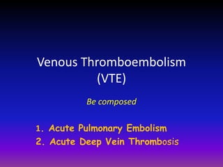
- 1. Venous Thromboembolism (VTE) Be composed 1. Acute Pulmonary Embolism 2. Acute Deep Vein Thrombosis
- 2. VTE Pathophysiology Virchow’s Triad Circulatory Stasis Hyper- cogulability Endothelial Injury Rudolf Virchow, 1856
- 3. 3 Virchow’s Triad Hypercoagulability Malignancy Nonmalignant thrombophilia Pregnancy Postpartum status (<4wk) Estrogen/ OCP’s Genetic mutations (Factor V Leiden) Protein C & S deficiency, AT III, anti ds DNA ,ACA IgG ,ACA IgM FactorVIII, Prothrombin mutations, Venous Statis Bedrest > 24 hr Recent cast or external fixator Long-distance travel or prolong automobile travel more 10 hours Venous Injury Recent surgery requiring endotracheal intubation Recent trauma (especially the lower extremities and pelvis) Recent venous access chateter jugular-subclavian, femoralis
- 4. How to Diagnose Acute Pulmonary Embolism ( PE )
- 5. Clinical Presentation of acute PE • Clinical Signs and Symptoms are frequently vague , Less specific and rarely “classic” • Untreated mortality rate of 20% - 30%, reduce to 5% with timely intervention Eur Heart J 2000; 21: 130-6
- 6. ESC Guidelines, 2008 Clinical Presentation Suspected Acute PE • The Classic Triad to Suspected acute PE : • Dyspnea • Pleuritic pain or chest pain • Hemoptysis Occurs in less than 20% of patients with documented acute PE
- 7. How to increase clinical validity if clinical acute PE less specific ? Should be used Well’s Score • Low probability category : 10% of acute PE • Intermediate probability category: 30% of acute PE • High probability category : 65% of acute PE Just be applicated in Normotensive PE Ann Intem Med, 2006;
- 8. ECG and Thorax Photo More Specific but rare expose in PE
- 9. SI QIII TIII Specific but Rare
- 10. Hampton’s Hump
- 11. Westermark ‘ s Sign
- 12. Diagnosis Suspected of PE Clinical Presentations ECG X photo Thorax + Hemodynamic State : • Normotensive Suspected non-high risk acute PE • Hypotensive Suspected high risk acute PE
- 13. Clinical Suspected high-risk PE i.e with shock or hypotension CT immediately available Echocardiography PE-specific Fibrinolytic or embolectomy Search for the other causes Fibrinolytic /embolectomy not justified Normal Abnormal No other tests available or patient unstable CT available and patient stabilized RV overload Search for the other causes No Yes No Yes CTA ESC Guidelines, 2008
- 14. Clinical Suspected non-high-risk PE i.e Normotensive Well’s Score No PE No treatment or Investigate further PE Anticoagulant Treatment 500 mg/L PE Anticoagulant Treatment < 500 mg/L No Treatment D-dimer Low / intermediate probability High probability No PE No Treatment CTA ESC Guidelines, 2008 CTA
- 15. PE Well’s Score
- 16. Pretest Probability of Wells Score ESC Guidelines, 2008 Variables Points Previous acute DVT or PE Recent surgery or immobilization Cancer +1.5 +1.5 +1 Haemoptysis +1 Heart Rate >100 beats/min Clinical signs of DVT +1.5 +3 Alternative diagnosis less likely than PE - 3 Low Intermediate High 0 -1 2 -6 7 PE unlikely PE likely 0 - 4 > 4
- 17. D- dimer
- 18. FIBRINOGEN THROMBUS FIBRIN POLIMER SOLUBLE ( UNSTABLE ) THROMBUS FIBRIN POLIMER INSOLUBLE ( STABLE ) D DIMER TROMBIN XIII a PLASMIN
- 19. Quantitative D-dimer Brand Name sensitivity specificity ELISA 90 – 100 % 30 – 40 % Latex agglutination 70 - 90 % 70 – 90 % Whole blood (the simpli – RED) 85 - 95 % 65 – 68 %
- 20. D-dimer Test Fibrin split product Circulating half-life of 4-6 hours Positive Value : ≥ 500 mg/dl(PE), ≥ 900 mg/dl(DVT) has many False Positives . Negative Value : < 500 mg/dl(PE),<900mg/dl(DVT) has a high and 93-100% negative predictive value ( 93 – 100 % ) to rule out acute PE False Positives: VTE Pregnant Patients Post-partum < 1 week Malignancy Surgery within 1 week Advanced age > 80 years Sepsis Hemmorrhage CVA AMI Collagen Vascular Diseases Hepatic Impairment
- 21. CTA 21
- 22. CTA Scan of Acute PE (Short Axis ) Normal PE
- 23. CTA of Acute PE (Short Axis) 23 Normal PE normal
- 24. CTA Scan of Acute PE Normal PE
- 25. ECHCARDIOGRAPHY
- 28. Echocardiography of PE RV overload RV LV LV RV Crescent Shape (Normal) D – Shape (PE)
- 29. Echocardiography of PE Normal kinetic in Apex Hypo kinetic basal McConnell’s Sign RV LV
- 30. The aim of Blood Test 1. Blood Gas Analysis – Follow Up Clinical Progress in Hospitalized 2. Thrombophilia testing - How long anticoagulant to be given ? Indication to screening test : History of unexplained or rcurrent thrombosis and /or pregnancy complication. Test Include : ACAIgG ,ACAIgM, Protein S & C, AT III, APC, Fibrinogen, Homo- cystein,CRP, Factor VIII. Thrombophilia proven Long life anticoagulant maintenance Thrombophilia unproven 3- 12 bulanantcoagulant maintenance
- 32. Anticoagulants for Acute PE TYPES THERAPEUTIC DOSE RECOMMENDATION Grade of Evidence Unfractionated Heparin - Bolus 5000 Unit per IV - maintain 1000 U/hr per drip - aPTT target 2-3 times of unl - given for 5 days Non-high risk PE 1 A Enoxaparin -1 mg/kg every 12 hours - given for 5 days Non-high risk PE LMWH over UFH 1 A Fondaparinux - 5 mg / 24 hr for <50 kg weight - 7.5 mg/24 hr for 50-100 kg wght - 10 mg / 24 hr for >100 kg weight - given for 5 days Non-high risk PE Indefinite Warfarin / Coumadin - 2-10 mg/day - INR target 2.5-3 - given overlapping in 3rd day during UHF/Enoxa/Fonda Usual maintenance 1 A Am J Med, 2007; 120: 18-25
- 33. Fibrinolytic for Acute PE TYPES THERAPEUTIC DOSE RECOMMENDATION Grade of Evidence Streptokinase - Bolus 250.000 U/IV for 30 minutes - Maintain 100.000 U/hr for 24 hours High risk PE 1 C Urokinase - Bolus 4400 U/kg for 10 mnt - Maintain 2000 U/kg/hr for 12 hours High risk PE 1 C tPA 100 mg IV over 2 hours High risk PE 1 C Note: No evidence catheter-directed method (local) has greater benefit than systemic intravenous Am J Med, 2007; 120: 18-25
- 34. Embolectomy for Acute PE TYPES RECOMMENDATION Grade of Evidence Mechanical percutaneous embolectomy (MPE) High risk PE who are unable to receive fibrinolytic therapy 2 C Note: - Grade 1: Strong recommendation - Grade 2: Weaker recommendation - Level of evidence A: RCTs with consistent result - Level of evidence B: RCTs with inconsistent result - Level of evidence C: Observational studies Am J Med, 2007; 120: 18-25
- 35. CASE 1
- 36. Mrs. AN, 37 y.o • Dyspnea, 1 bed pillow, couging up blood since 3 days ago. • Left leg swelling then massaged about 6 days earlier and diagnosed DVT by doctor • Denied Asthma, TBC and heart disease • C M, JVP stable,TD 120/80 mmHg.HR 105 x /mnt Lung : Wheezing & Rales (-) Left Leg: Edema & Warm . Os ke RS - ECG : Sinus Tachycardi - Thorax Photo : WNL , WD/ Suspect Low Risk PE What Next ? Well’s Score
- 37. Pretest Probability of Wells Score Variables Points Previous acute DVT or PE Recent surgery or immobilization Cancer +1.5 +1.5 +1 Haemoptysis +1 Heart Rate >100 beats/min Clinical signs of DVT +1.5 +3 Alternative diagnosis less likely than PE - 3 Low Intermediate High 0 -1 2 -6 7 PE unlikely PE likely 0 - 4 > 4 Points +1.5 - - +1 +1.5 +3 - 7 High PE Likely
- 38. Suspect Low Risk PE Well Score , High Probability What Next ? CTA
- 39. What Next ? - Unfractionated Heparin - Low Molecule Weight Heparin - Fondaparinux Injectie Anticoagulant 5 days: Oral anticoagulant - Maintenance
- 40. Deep Vein Thrombosis ( DVT )
- 41. Background DVT is common but elusive illness In 1644, Schenk In 1846,Virchow The worldwide incidence 100 cases per 100,000 person, annually The incidence in Asians is 2.2 - 62.5 %
- 42. Complication of DVT 1. Pulmonary Embolism is the cause of death in 0.9 % 2. Post Thrombotic Syndrome 60 - 85 % ( Venous Hypertension Venous Reflux - CVI ) 3. Phlegmasia Cerulea Dolens 1 – 6 %
- 43. The Surgical risk of Deep Vein Thrombosis • 16 - 30 % in general surgical • 45 - 70 % in orthopedic surgery • 7 - 45 % in gynecologic surgery • 25 % in urologic surgery
- 44. Clinical Diagnosis of Suspected DVT . Conditions Sensitivity Specificity Calf pain Calf tenderness Ankle edema Leg swelling Redness Superficial venous distention Homans’sign 66 – 91 % 56 – 82 % 40 – 76 % 35 – 97 % 15 – 26 % 27 – 33 % 13 – 43 % 3 – 19 % 26 – 74 % 23 – 52 % 8 – 88 % 90 – 91 % 30 – 91 % 39 – 84 % Source Modified from LeClere JR 1954,
- 46. Lower Extremity of DVT 1. Distal / Calf DVT : Small thrombus , Asymptomatic Tibial and Peroneal Vein Complete Spontaneous lysis Incidence >> to be PE 20 %- 30 % 2. Proximal / Thigh DVT : Large thrombus , Symptomatic Iliac ,Femoral, Popliteal, vein. Uncomplete spontaneus lysis Incidence << to be PE 60 %
- 49. VENOGRAPHY / CTA/ MRA DVT D - DIMER Clinical COMPRESSION ULTRASOUND (cus) Modality Diagnostic Test Well‘s Score no yes
- 50. DVT Exclude Clinical Suspected DVT Well’s Score Low Probability or Intermediate High Probability D - dimer CUS < 900 ≥900 Uncollaps Collaps Exclude DVT Ultrasound D -dimer ≥ 900 < 900 Exclude DVT CUS 3-7 days / Venography Uncollaps Collaps DVT CUS Collaps Uncollaps Exclude DVT DVT
- 51. DVT Wells’Score
- 52. Wells Score Clincal Features Score - Active cancer 1 - Paralysis, recent plaster cast 1 - Recent immobilisation or surgery 1 - Tenderness along entire deep vein system 1 - Swelling of entire leg 1 - > 3 cm difference in calf circumference compared with other leg 1 - Pitting oedema 1 - Collateral superficial veins (non Varicose) 1 - Alternative diagnosis as likely as DVT -2 High probability > 3 Moderate probability 1-2 Low Probability < 1 Interobserver reliability kappa 0.85 The prevalence of disease in each pre-test category was 85,33 and 5 % for the high, moderate,and low probability groups, respectively. Wells PS et al. Lancet 1997 ; 350 : 1795-8
- 53. Ultrasound
- 54. NORMAL COMPRESSION ULTRASOUND ( C U S ) Proximal DVT Uncollaps DVT By Pressure Collaps normal V V V
- 55. CUS SQUEEZE DEEP VEIN THROMBOSIS NORMAL Squeeze Distal Distal DVT
- 56. Anticoagulants for Acute DVT TYPES THERAPEUTIC DOSE RECOMMENDATION Grade of Evidence Unfractionated Heparin - Bolus 5000 Unit per IV - maintain 1000 U/hr per drip - aPTT target 2-3 times of unl - given for 5 days - Proximal - Distal with severe risK 1 A Enoxaparin -1 mg/kg every 12 hours - given for 5 days - Proximal - Distal with severe risK 1 A Fondaparinux - 5 mg / 24 hr for <50 kg weight - 7.5 mg/24 hr for 50-100 kg wght - 10 mg / 24 hr for >100 kg weight - given for 5 days -Proximal - Distal with severe risK 1 A Warfarin / - 2-10 mg/day Usual 1 A
- 57. Fibrinolytic for Acute DVT TYPES THERAPEUTIC DOSE RECOMMENDATION Grade of Evidence Streptokinase - Bolus 250.000 U/IV for 30 minutes - Maintain 100.000 U/hr for 24 hours Ilio Femoral 1 C Urokinase - Bolus 4400 U/kg for 10 mnt - Maintain 2000 U/kg/hr for 12 hours Iliofemoral 1 C tPA 100 mg IV over 2 hours iliofemoral 1 C Note: No evidence catheter-directed method (local) has greater benefit than systemic intravenous Am J Med, 2007; 120: 18-25
- 58. Thrombolytic Treatment Illiofemoral DVT < 14 days Percutaneus intervention is Performed in Catheter laboratory. Technique : - Popliteal vein is accessed by ultrasound guidance - 5 Fr multisidehole catheter and wire is placed Thrombus obstruc. - urokinase 80,000 IU / hr ( side arm ) + 80,000 /hr ( multisidehole) - Infusion heparin following bolus 5,000 IU. Evaluation by Contras Catheter after overnight . .
- 59. Temporary Vena Cava Filter (VCF) Indication : - DVT + Recurrent Pulmonary Embolism despite adequate anticoagulation - Contraindication for anticoagulant in Proximal DVT progress to illiac vein - After pulmonary embolectomy - Floating Thrombus in inferior vena cava VCF Performed in Catheter laboratory
- 60. Case 1
- 61. Mr. An 60 Yo - Right leg swelling, pain and warm continuously causing difficult to walk since 5 days ago. - 6 days ago he’s just returned from America by plane for 24 hours and sleep soundly due to drinking tranquilizers. - Denied limb trauma history
- 62. Mr.An 60 Yo - Sweeling on right thigh to calf, pitting edema with diameter 4,5 cm greater thanleft leg, felt warm and reddish. - Homans’ Sign positive Wells’ Score What next??? WD/Suspect DVT
- 64. Wells Score Clincal Features Score Score - Active cancer 1 - Paralysis, recent plaster cast 1 - Recent immobilisation or surgery 1 - Tenderness along entire deep vein system 1 - Swelling of entire leg 1 - > 3 cm difference in calf circumference compared with other leg 1 - Pitting oedema 1 - Collateral superficial veins 1 - Alternative diagnosis as likely as DVT -2 High probability > 3 Moderate probability 1-2 Low Probability < 0 Interobserver reliability kappa 0.85 The prevalence of disease in each pre-test category was 85,33 and 5 % for the high, moderate,and low probability groups, respectively. Wells PS et al. Lancet 1997 ; 350 : 1795-8 - - 1 - 1 1 1 - 4 -
- 65. Mr.An 60 yo High probability Wells’ Score What next WD/Suspect DVT What next Ultrasound
- 66. Uncollaps
- 68. Mr.An 60 yo High probability Wells’ Score WD/Suspect DVT Ultrasound Acute DVT Heparin What Next
- 70. Sebelum terapi Sesudah terapi