(March 13, 2024) Overview of Preclinical Small Animal and Multimodal Imaging

In this webinar, we reviewed some of the most commonly used preclinical imaging modalities, including magnetic resonance imaging (MRI), positron emission tomography (PET), computer tomography (CT), ultrasound, photoacoustic, bioluminescence, fluorescence, dual-energy x-ray absorptiometry (DEXA/DXA), and intravital microscopy. For each modality, we spent time reviewing the basics of how each worked, the strengths and considerations of each, and some key application areas and example images. Finally, we discussed the benefits of multimodal imaging and reviewed a few papers utilizing a variety of imaging modalities to help support their research outcomes. We ended with a very brief introduction to Scintica Instrumentation and our philosophy behind the various products we represented. However, the main focus of the webinar was on education, and not our diverse product portfolio.

Recomendados
Recomendados
Mais conteúdo relacionado
Semelhante a (March 13, 2024) Overview of Preclinical Small Animal and Multimodal Imaging
Semelhante a (March 13, 2024) Overview of Preclinical Small Animal and Multimodal Imaging (20)
Mais de Scintica Instrumentation
Mais de Scintica Instrumentation (20)
Último
Último (20)
(March 13, 2024) Overview of Preclinical Small Animal and Multimodal Imaging
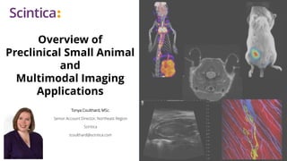
- 1. Overview of Preclinical Small Animal and Multimodal Imaging Applications Tonya Coulthard, MSc. Senior Account Director, Northeast Region Scintica tcoulthard@scintica.com
- 2. Preclinical Imaging • In most cases, adaptation of clinical imaging to preclinical research • Magnetic Resonance Imaging (MRI) • Positron Emission Tomography (PET) • Computer Tomography (CT) • Ultrasound • Some modalities have gone from preclinical research to the clinic • Photoacoustic • While some modalities designed specifically for preclinical research • Bioluminescence (BLI) • Intravital Microscopy (IVM) 2
- 3. Benefits vs. in vitro, ex vivo, in situ • Intact subject involvement of all organs and immune system • Longitudinal studies Elucidation of disease mechanism over time Reduced variability - imaging subject used as own control Supports 3R’s (Replace, Reduce, Refine) • Multimodal imaging Possible Preclinical Imaging 3 Day 0 Baseline Image Day 1 Induce Model Day 2 Imaging Timepoint, Disease Progression Day 3 Imaging Timepoint, Disease Progression Day 4 End Point Imaging, Tissue Collection
- 4. Preclinical Imaging 4 • Size of imaging subject and imaging target increased spatial resolution • Physiological differences, i.e. heart rate increased temporal resolution • Cost of novel target/contrast agents increased sensitivity • Animal welfare integrated anesthesia, heating, physiological monitoring, etc. Considerations – “From Mouse to Man”
- 5. Modality Overview Overview • How each modality works • Strengths vs. weaknesses • Application areas and example images Focus • MRI, PET, CT, Ultrasound, Photoacoustic, Bioluminescence/Fluorescence, DEXA, Intravital Microscopy • Other preclinical imaging modalities - SPECT, Magnetic Particle Imaging, Raman Imaging, etc. 5 James ML, Gambhir SS. Physiological reviews. 2012
- 6. Magnetic Resonance Imaging (MRI) 6 James ML, Gambhir SS. Physiological reviews. 2012
- 7. Magnetic Resonance Imaging (MRI) 7 Images acquired using Aspect Imaging’s M-Series. T2 weighted FSE on Mouse Abdomen T1 weighted SE on Mouse Abdomen 400µm resolution 4:54m:s 7 excitations 420µm resolution 6:12m:s 11 excitations
- 8. Magnetic Resonance Imaging (MRI) 8 Key Strengths • No limit to depth of penetration • High spatial resolution • Excellent soft tissue contrast • No ionizing radiation • Quantitative data • Clinically translatable Key Limitations/Considerations • Lower sensitivity to contrast agents • Can be expensive • Permanent magnet options at lower cost, and complexity to install • Relatively low temporal resolution
- 9. Magnetic Resonance Imaging (MRI) • Anatomical and morphological • Neurology • Cancer Biology • Cardiovascular Biology • Cell Tracking • Ex vivo Imaging 9 Images acquired using Aspect Imaging’s M-Series.
- 10. Positron Emission Tomography (PET) 10 James ML, Gambhir SS. Physiological reviews. 2012 Lancelot S, Zimmer L. Trends in Pharmacological Sciences. 2010
- 11. Positron Emission Tomography (PET) Isotope Half-Life • 15O (Oxygen) • 2 minutes • 13N (Nitrogen) • 10 minutes • 11C (Carbon) • 20 Minutes • 18F (Fluorine) • 110 Minutes 11 Lancelot S, Zimmer L. Trends in Pharmacological Sciences. 2010
- 12. Positron Emission Tomography (PET) 12 Key Strengths • No limit to depth of penetration • Excellent sensitivity • Quantitative data • Clinically translatable Key Limitations/Considerations • Requires cyclotron/generator • Relatively expensive • Ionizing radiation from isotopes • Limited spatial resolution • Should be combined with another modality (i.e. MRI or CT) for anatomical co-registration
- 13. Positron Emission Tomography (PET) • Neurology • Cancer Biology • Cardiovascular Biology • Bone & Disease Imaging • Biodistribution 13 Images acquired using Sedecal’s SuperArgus PET.
- 14. Computed Tomography (CT) 14 James ML, Gambhir SS. Physiological reviews. 2012 Images acquired using Sedecal’s SuperArgus CT.
- 15. Computed Tomography 15 Key Strengths • No limit to depth of penetration • High spatial resolution • Good temporal resolution • Bone and lung imaging is very strong • Clinically translatable Key Limitations/Considerations • Poor sensitivity • Primarily anatomical information – bone and air-filled structures • Limited soft tissue resolution • Ionizing radiation Images acquired using Sedecal’s SuperArgus CT.
- 16. Ultrasound 16 James ML, Gambhir SS. Physiological reviews. 2012
- 17. Ultrasound • High-frequency (i.e. shorter wavelength) soundwaves are needed when imaging small animal models - High-resolution - Shallow depth of penetration • Select highest frequency possible to visualize structure of interest 17 Images acquired using S-Sharp’s Prospect T1.
- 18. Ultrasound 18 Key Strengths • Relatively inexpensive • Quantitative data • No ionizing radiation • Good soft tissue contrast • High-temporal resolution • Clinically translatable Key Limitations/Considerations • Limited depth of penetration • Primarily anatomical information • Expanded with the use of contrast agents • Limited to imaging soft-tissue only (no bone or air structures)
- 19. Ultrasound • Cancer Biology • Cardiovascular Biology • Developmental Biology • Image Guided Injection • Gene/Drug Delivery 19 Images acquired using S-Sharp’s Prospect T1.
- 20. Photoacoustic 20 James ML, Gambhir SS. Physiological reviews. 2012 Images acquired using Photosound’s TriTom.
- 21. Photoacoustic 21 Key Strengths • Superior depth of penetration compared to other optical techniques • Good temporal resolution • Clinically translatable Key Limitations/Considerations • Limited to imaging soft tissue only (no bone or air structures) • Coupling of instrument to subject required • Limited anatomical/structural information Images acquired using Photosound’s TriTom.
- 22. Bioluminescence/Fluorescence 22 James ML, Gambhir SS. Physiological reviews. 2012 Bioluminescence (BLI) Fluorescence (FLI)
- 23. Bioluminescence/Fluorescence BLI • Imaging target must express a luciferase enzyme • Appropriate substrate must be present FLI • Appropriate fluorophore must be selected • Imaging target must either express, or contain, the fluorophore 23
- 24. Bioluminescence/Fluorescence 24 BIOLUMINESCENCE • Key Strengths • Relatively inexpensive • Excellent sensitivity • Multiplexing capabilities • No ionizing radiation • High throughput • Key Limitations/Considerations • Limited depth of penetration • Poor spatial resolution • Substrate and co-factors required • Not clinically translatable FLUORESCENCE • Key Strengths • Relatively inexpensive • Multiplexing capabilities • No ionizing radiation • High throughput • Clinically translatable • Key Limitations/Considerations • Limited depth of penetration • Poor spatial resolution • Surface weighted images • Autofluorescence
- 25. Bioluminescence/Fluorescence • Cancer Biology • Neurology • Biodistribution Studies • Cell Tracking 25 Images acquired using Vilber’s Newton FT500.
- 26. Dual Energy X-Ray Absorptiometry (DEXA) 26 Figure from Luo, Yunhua. 2017. Chapter 3 Parameter Unit of Measure Description (available on whole animal, or from each ROI) BMC g Bone Mineral Contents (Bone Mass) BMC = bone density x bone area Fat g Fat mass Fat Ratio % Fat Ratio = Fat/Total Mass Lean g Fat free mass Lean Ratio % Lean Ratio = Lean/Total Mass Total Mass g Total Mass = Fat + Lean + Bone BMD g/cm2 Bone Mineral Density Bone Area cm2 Bone Area in Image Tissue Area cm2 Tissue Area in Image
- 27. Dual Energy X-Ray Absorptiometry (DEXA) 27 Key Strengths • Rapid scan times • Whole body imaging is possible • Very low ionizing rations levels per scan – allows for longitudinal imaging • Clinically translatable Key Limitations/Considerations • Provide only 2D images • Low soft tissue contrast Images acquired using Osteosys’s iNSiGHT.
- 28. Intravital Microscopy 28 James ML, Gambhir SS. Physiological reviews. 2012 Images acquired using IVIM Technology’s IVIM.
- 29. Intravital Microscopy 29 Key Strengths • Excellent spatial resolution • Multiplex capabilities • Dynamic/real-time information about microscopic cellular events • Yields quantitative measures Key Limitations/Considerations • Poor depth of penetration - Improved with two photon over confocal • Small field of view • Can require multiple laser excitations • Surgical preparation of models Images acquired using IVIM Technology’s IVIM.
- 30. When to use Which Imaging Modality Primary Considerations • Feasibility – cost to purchase equipment + infrastructure costs + running/maintenance costs • Type of Information – anatomical, functional, molecular • Imaging Target, Resolution, and Depth Required Then ask yourself the following • Do you need to image over time? • Do you have the ability to engineer cells, what contrast agents do you have access to? • Would multimodal imaging be beneficial? 30
- 31. Multimodal/Multiplex Imaging • Defined • Use of two or more complimentary imaging modalities, or contrast/reporting agents used within a single experiment – may be simultaneously or sequentially • Goal • Acquire as much data as possible to tackle a biological question more holistically then would be possible with a single modality, as well as to approach the biological question from multiple scale levels • Purpose • Elucidate the various biological mechanisms of disease, as well as to fully understand the response to therapeutic interventions 31
- 33. Audience Poll
- 34. Our Mission Providing instrumentation to scientists and the preclinical research community to advance research, science and medicine. 34
- 36. WWW.SCINTICA.COM 36 Model Development In Vitro Studies General Laboratory Equipment Product Portfolio – General Products Anesthesia & Ventilators Surgical Monitoring Impact Device Stereotaxic Device Image Guided Injection Hypoxia Chambers – In Vivo Hypoxia Workstation Colony Counter Bioprinter Hypoxia Incubator Gel Documentation Chemiluminescent Blot Imaging Microtome & Cryostat
- 37. MRI 37 M-Series Compact, Cryogen-free, Self-shield M7+ SimPET for simultaneous PET-MRI
- 38. Eyes Real-time, in vivo, screening of PET, SPECT, or optical tracers 38 Cardiac Imaging β-eye |PET γ-eye |SPECT φ-eye |SPECT PET & SPECT SuperArgus State-of-the-art PET/CT
- 39. Optical 39 Cardiac Imaging Newton Bioluminescence, 3D tomography, fluorescence (NIR-I & NIR-II) IVM All-in-one intravital confocal/two-photon microscopy system
- 40. TriTom 3D Tomographic Photoacoustic and Fluorescence DXA & PIA 40 Cardiac Imaging iNSiGHT Shielded dual energy x- ray absorptiometry
- 41. Ultrasound 41 Cardiac Imaging Doppler Flow Velocity System Pulsed Doppler Ultrasound for Cardiovascular Research Prospect T1 High Frequency Ultrasound
- 42. Audience Poll
- 43. Multimodal Imaging – Journal Review 43
- 44. Multimodal Imaging – Journal Review 44
- 45. Multimodal Imaging – Journal Review 45
- 46. Multimodal Imaging – Journal Review 46
- 47. Summary - Preclinical Imaging 47 • Introduction to Preclinical Imaging • Considerations of adaptation from clinical imaging • Preclinical only imaging modalities • Modality Overview • MRI, PET, CT, Ultrasound, Photoacoustic, Bioluminescence, Fluorescence, DEXA, Intravital Microscopy • Importance of Multimodal Imaging
- 48. Q&A Session WWW.SCINTICA.COM INFO@SCINTICA.COM Please enter your questions in the Q&A section. Thank You!