The maxillary sinuses were first illustrated and described by Leonardo Da Vinci in 1489 and later documented by the English anatomist Nathaniel Highmore in 1651.
The maxillary sinus, or antrum of Highmore, lies within the body of the maxillary bone and is the largest and first to develop of the paranasal sinuses.
Shape- a pyramid-shaped cavity; base- adjacent to the nasal wall; apex- pointing to zygoma.
Size- insignificant until eruption of permanent dentition; average dimensions of adult sinus- 2.5–3.5 cm wide, 3.6–4.5 cm tall, and 3.8–4.5 cm deep; estimated volume of approximately 12–15 cm.
Extent- Anteriorly, extends to canine and premolar area. sinus floor usually has its most inferior point near the first molar region.
Surgical aspect of implants and recent advancespulakmishra1988
This document discusses dental implants, including their history, uses, techniques, planning and surgical procedures. It provides details on implant types, the two-stage surgery process involving initial fixture installation and a later procedure to attach the abutment. Techniques for implant planning, drilling, tapping and fixture installation are described. Considerations for soft and hard tissue management are also covered.
Screw vs cement retained implant prosthesisApurva Thampi
This is a journal club presentation featuring a recent article regarding a screw and cement retained implant prosthesis.
the presentation and all its related material is available on request. Mail me at apurvathampi@gmail.com
This document discusses terminology and techniques for dental implant impressions. It defines terms like cover screws, healing caps, transfer copings, and implant analogues. It explains that impressions are needed to capture the implant position, depth, axis, and soft tissue contour. The document outlines two main impression techniques - open tray (using pick-up copings) and closed tray (using transfer copings). It notes the advantages and disadvantages of each technique. Abutment level impressions are also discussed for customization and laboratory abutment selection. Gingival simulation is described as a technique to simulate the soft tissue around implants.
This document provides an overview of implant supported overdentures. It defines overdentures and discusses the advantages and disadvantages compared to fixed prostheses. It describes different prosthetic options and classifications of prosthesis movement. Treatment options for mandibular and maxillary overdentures using various numbers and positions of implants are outlined. The focus is on removable prostheses that are partially retained by and supported by dental implants.
This document provides an outline and overview of surgical techniques for maxillary sinus elevation. It begins with an introduction describing how maxillary sinus pneumatization can compromise implant placement in the maxilla. It then describes the anatomy of the maxillary sinus and surgical armamentarium. The remainder of the document details different surgical approaches to maxillary sinus elevation, including the lateral window technique with and without grafting materials, and discusses considerations for graft materials and membrane barriers.
This is a power point presentation on sinus floor elevation, describing the various techniques, biological aspects and clinical outcomes from a periodontist point of view. It also includes a brief review on the anatomy of maxillary sinus and management of complications.
The document discusses the indirect sinus lift technique for implant placement in the posterior maxilla with limited bone height. It describes how the technique involves using osteotomes to elevate the sinus floor by at least 5-6 mm without membrane perforation. This creates space for grafting material and implant placement with high survival rates of 93.5-100%. The technique offers a minimally invasive alternative to the lateral window approach with advantages of avoiding membrane visualization and permitting treatment in a single stage.
This document discusses considerations for implant placement and restoration in the esthetic zone. It covers:
1. Factors to consider pre-surgery like bone quality and site evaluation using the Garber classification system.
2. Implant positioning factors such as buccolingual and mesiodistal position, angulation, depth, and their influence on esthetics and function.
3. Techniques to develop the emergence profile like using healing abutments, provisional restorations, and custom abutments.
4. The multidisciplinary approach involving prosthodontists and consideration of soft tissues, abutment materials, and impression techniques.
Sinus lift surgery is used to augment the posterior maxilla when there is insufficient bone height for dental implants. There are direct and indirect sinus lift procedures, with the direct approach involving raising the sinus membrane through a lateral window created in the maxillary sinus wall. Grafting material such as autologous bone is placed to increase bone volume, allowing implant placement after 6 months. Indirect sinus lift is less invasive and has a shorter healing time, using osteotomes to lift the sinus membrane from the alveolar crest when 4-7mm of bone is present. Contraindications include sinus infections or tumors, allergies, steroid use, radiation, smoking, and mental impairment.
This document provides an overview of dental implant sinus lift procedures. It begins with brief anatomy of the maxillary sinus and defines a dental implant. It then discusses patient evaluation, including radiographic assessment and anatomical limitations for implantation. Classification systems for the posterior maxilla are presented. The document reviews indications, contraindications, and surgical techniques for sinus lift procedures, including direct and indirect methods. It also discusses graft materials, post-operative instructions, and potential complications.
The content covers majority of the aspect of immediate implant placement - why immediate implants?, case selection, decision making, classifications, surgical technique, healing following immediate implant placement, immediate implants in infected sockets/periapical infections, literature reviews and recommendations for clinical practice.
Classification and impression techniques of implants/ dentistry dental implantsIndian dental academy
Indian Dental Academy: will be one of the most relevant and exciting
training center with best faculty and flexible training programs
for dental professionals who wish to advance in their dental
practice,Offers certified courses in Dental
implants,Orthodontics,Endodontics,Cosmetic Dentistry, Prosthetic
Dentistry, Periodontics and General Dentistry.
Anterior Single Tooth implants in the Esthetic ZoneDoreen Bello
This document discusses considerations for placing single-tooth implants in the anterior esthetic zone. It outlines 7 levels of difficulty for implant success based on factors like bone quality and quantity, papilla health, and smile line visibility. Guidelines are provided for pre-treatment evaluation of soft tissue, bone dimensions, and root morphology. Surgical techniques like conservative flap design and use of osteotomes are recommended. Prosthetic factors like implant positioning and emergence profile are also discussed. The document describes traditional and immediate loading approaches for restoration.
Various studies have examined immediate implants versus delayed implants. Immediate implants have the advantages of preserving alveolar bone and providing esthetics by maintaining tooth alignment, but risks include unfavorable implant angulation if the extracted tooth was misaligned or inadequate bone beyond the apex. Delayed implants allow primary soft tissue healing for 6-10 weeks before placement and have benefits like adequate soft tissue and minimized microorganisms, as well as highly osteogenic bone filling in of defects. Studies found high survival rates for both immediate and delayed implants, though immediate implants showed slightly deeper pockets and more membrane exposure on average.
The document discusses various impression techniques used for dental implants. It describes the key components used, such as implant analogues and impression copings. The most common impression materials are vinyl polysiloxanes and polyether rubbers due to their dimensional stability and detail reproduction. Direct open tray techniques involve exposing the impression coping screws and incorporating the copings into the impression tray. Indirect closed tray techniques retain the copings in the mouth and reattach them to analogues in the lab. Factors like implant angulation, number of implants, and interarch space determine whether open or closed tray methods are preferred. Accurate transfer of the implant positions is crucial for passive fitting of the final prosthesis.
The socket shield technique at molar sitesNaveed AnJum
The socket-shield technique for avoiding postextraction tissue alteration was first described in 2010. The technique was developed for hopeless teeth in anterior esthetic sites but has not yet been described for molar sites. Managing postextractive ridge changes in the posterior region by prevention or regeneration remains a challenge. The socket shield aims to offset these ridge changes wherever possible, preserving the patient’s residual tissues at immediate implants.
This document discusses cement retention versus screw retention for dental implants. Both methods can be used if done properly. Cement retention is simpler but risks residual cement being left under gums, which can lead to peri-implantitis. Screw retention allows easy removal but requires access holes. Residual subgingival cement is the major problem, as it is difficult to fully remove and can cause inflammation and bone loss over time.
loading protocols in dental implants about indications and contraindications of conventional , immediate,progressive and delayed loading of dental implants
Dental implants are artificial tooth roots inserted into the jaw to hold replacement teeth. There are several types but all rely on a process called osseointegration where the implant fuses with the jaw bone. Implants can replace single teeth, multiple teeth, or a full arch. They have advantages over other options like preserving bone, improved function and aesthetics. However, they also have longer treatment times and costs compared to other options. Placement involves surgery to insert the implant which then fuses with the bone before an abutment and crown are added to restore bite and appearance.
Ridge preparation for implant placement - part 1Hesham El-Hawary
- criteria of ideal ridge
- implants timing protocol
- implants planning and case selection
- clinical types of bone
- preventive methods to preserve the alveolar ridge
This document provides information about maxillary sinus augmentation. It begins with an introduction discussing the anatomy of the maxillary sinus and the need for sinus augmentation when there is inadequate bone height for dental implant placement. It then describes the two main techniques for sinus augmentation - direct sinus lift using a lateral window approach and indirect sinus lift using a crestal approach. The document provides details of the surgical procedures, instrumentation, and grafting materials used for both techniques. It emphasizes the importance of thorough preoperative evaluation and planning to ensure successful outcomes.
Soft tissue considerations for implant placementGanesh Nair
pre and post soft tissue considerations prior and post implant placement including various surgical technique for simple and advanced soft tissue augmentation
Sinus lift procedures. final copy of presentation pptxNAMITHA ANAND
This document discusses maxillary sinus lift procedures. It begins with the anatomy of the maxillary sinus, including its bony walls, blood supply, and Schneiderian membrane. It then covers clinical assessment of the sinus and various factors that can affect sinus health. The document discusses contraindications for sinus lift procedures and techniques for reducing complications. It also covers classifications of sinus lifts, different surgical techniques, potential intraoperative and postoperative complications, and instrumentation used. In summary, the document provides an overview of maxillary sinus anatomy and considerations, techniques, and risks associated with sinus lift procedures.
Sinus Lift with implant placement-surgical approachspsangeetaporiya
This document provides an overview of sinus grafting procedures for dental implants. It defines maxillary sinus grafting as a procedure to increase bone thickness in the posterior maxilla when there is insufficient bone for dental implants. The document describes the surgical anatomy of the maxillary sinus and the conventional lateral window approach for sinus grafting. This involves raising a bone window to access the sinus membrane, elevating the membrane to increase bone height, then grafting and often simultaneously placing dental implants. The document outlines indications, contraindications and postoperative care for sinus grafting.
This document discusses keys to success for dental implant placement in the posterior maxilla. The posterior maxilla presents several challenges for implants due to poor bone quality and limited bone height from sinus pneumatization. A case report describes using the crestal approach for sinus elevation and placing an implant bicortically stabilized between the crest and sinus floor. This provides high initial stability and allows placement of longer implants. The author found this technique resulted in higher success rates compared to conventional protocols. Bicortical stabilization of implants is an effective approach but requires expertise to avoid membrane perforation.
Implant placement in posterior maxilla by Dr. Ajay SinghAjay Singh
Implant placement in posterior maxilla. Dental implant therapy into the posterior
maxilla has always been and continues to be a
challenge due to various limitations in this
region such as poor bone density, sinus
pneumatization, lateral and vertical bone
resorption, high occlusal forces and area of
limited access. Further, if the implant is
placed into poor density posterior maxilla,
the bone which forms around the osseointegrated
implants does not show very high
bone to implant surface contact (BIC) percentage,
thus in several cases the implant even
after successful osseointegration may fail
once it is restored in function.
Vertical ridge augmentation is sometimes required for dental implant placement. The presentation looks at various conventional and newer techniques for ridge augmentation in the oral cavity.
Journal Club On Pre-extractive Interradicular Implant Bed Preparation: Case P...Shilpa Shiv
Journal Club On Pre-extractive Interradicular Implant Bed Preparation: Case Presentations of a Novel Approach to Immediate Implant Placement at Multirooted Molar Sites, IJPRD 2013.
This document discusses implant site preparation and assessment. It covers evaluating bone quality and quantity, classifying alveolar ridge defects, and assessing risk factors. Both hard and soft tissue augmentation procedures are described, including guided bone regeneration using grafts and membranes. Autogenous bone grafts from sites like the mandibular symphysis are discussed, along with harvesting techniques and factors for success. The goal of implant site preparation is to develop adequate bone volume and quality for implant placement and long term function.
Dental implants are artificial fixtures placed surgically into the jawbone to replace missing teeth. There are different types of implants including subperiosteal, transosseous, and endosteal implants, with endosteal implants being the most common today. The process of osseointegration, where bone bonds to the implant surface without soft tissue interference, was discovered in 1952 and refined for successful dental implant treatment. The surgical procedure for placing implants involves two stages - the initial surgery to place the implant fixture followed by a second surgery once osseointegration is complete to place the abutment and crown. Factors like patient health, bone quality and quantity, surgical technique, and loading conditions can
Sinus lift with dental implants Placement.(with Clinical Photographs) Dr. ...All Good Things
Hi. This is Dr. Amit T. Suryawanshi. Oral & Maxillofacial surgeon from Pune, India. I am here on slideshare.com to share some of my own presentations presented at various levels in the field of OMFS. Hope this would somehow be helpful to you making your presentations. All the best & your replies are welcomed!
This document provides an overview of dental implants including:
1. Definitions of dental implants and their purpose in retaining prosthetics.
2. The basic parts of a modern dental implant including the implant, abutment screw, and healing cap.
3. Popular implant systems from manufacturers like Nobel Biocare and 3i.
4. The concept of osseointegration and theories of implant-bone integration.
5. The surgical technique for placing implants including drilling protocols and healing periods.
6. Advanced techniques like sinus lifts and bone grafting to support implants.
This document discusses dental implants, including their uses, types, placement locations, and procedures. Key points include:
- Dental implants provide support for fixed or removable prostheses by being implanted in the oral tissues or bone.
- Implant placement depends on the patient's condition and dental needs, as well as the quality and location of available bone.
- Common types include endosteal implants placed within the bone, and various metallic and non-metallic implant materials.
- Placement procedures involve initial surgery to insert the implant fixture, a healing period, attachment of an abutment, and final prosthetic restoration. Success rates above 90% are achievable when properly placed.
This document discusses soft tissue grafting procedures around dental implants. It begins by explaining the importance of soft tissue integration for implant success. It then describes the anatomy of periodontal and peri-implant soft tissues. Various grafting techniques are discussed including modified palatal roll, epithelialized palatal graft, alloderm grafting, and subepithelial connective tissue grafting. Indications and surgical principles/techniques for each method are provided. The goal of these grafts is to establish an adequate zone of keratinized attached tissue around implants for long term health of the peri-implant tissues.
This document provides an overview of immediate implant placement. It defines key terminology, discusses the advantages of immediate placement which include preserving bone and soft tissue and reducing treatment time. Guidelines for extractions that allow for immediate placement are described, such as atraumatic extractions and osteotomy preparation techniques. Factors such as implant selection, the use of grafts, and loading protocols are summarized. Potential disadvantages including lack of control of implant position and difficulty obtaining primary stability are also mentioned. The document concludes with a review of literature on case reports evaluating immediate placement.
JOURNAL CLUB ON THE OUTCOME OF ORAL IMPLANTS PLACED IN BONE WITH LIMITED BU...Shilpa Shiv
This document summarizes a study that evaluated the outcomes of oral implants placed in bone with limited bucco-oral dimensions over a 3-year period. 100 implants were placed in 28 patients. The study found that the implants had a 100% survival rate over 3 years and that the marginal bone levels around the implants remained stable. The results indicate that implants can successfully be placed in sites with up to 4.5mm of bucco-oral bone width without the need for bone grafting, providing patients maintain good oral hygiene.
Split ridge and expansion techniques are effective for the correction of moderately resorbed edentulous ridges in selected cases.
Transverse expansion is based on osseous plasticity obtained by corticotomy. It progressively allows for an adequate transversal intercortical diameter large enough to insert one or several dental implants.
The gap created by sagittal osteotomy expansion undergoes spontaneous ossification, following a mechanism similar to that occurring in fractures.
Dental implants are prosthetic devices implanted into the jawbone to support dental prostheses like dentures or bridges. This document discusses dental implant terminology, the science of osseointegration where bone directly attaches to the implant, rationales for implants over other options, classification of implants, components, surgical procedures, and success criteria. Implants have advantages over traditional bridges and dentures by avoiding abutment tooth preparation, reducing bone loss, and improving function. Careful patient evaluation and treatment planning is required for optimal implant placement and long term success.
Dental Implants Procedures and ComplicationsBALAKRISHNA341
This document discusses dental implants, including the stages of implant placement and factors considered during treatment planning and surgery. It describes the preoperative examination, implant placement procedures such as flap design and bone drilling, and factors such as healing time and abutment selection. Key stages include preoperative examination and planning, implant placement surgery, and maintenance of implants after restoration. Success relies on maintaining the health of the implant environment through regular recalls and cleaning.
This document discusses various pre-prosthetic hard tissue procedures including: recontouring alveolar ridges through alveoloplasty and Dean's alveoloplasty; reducing maxillary tuberosities, palatal exostoses, mylohyoid ridges, and genial tubercles; removing tori and bone augmentation of atrophic maxillary and mandibular ridges through onlay grafts, sinus lifts, and hydroxyapatite grafts. The goal is to modify oral anatomy and eliminate undercuts/protuberances to facilitate dental prosthesis placement through reshaping bony areas.
Basic Surgical Techniques for Endosseous Implant Placement discusses the history and process of dental implants. It describes how Branemark discovered that titanium bonds directly to living bone, called osseointegration. The document outlines the 4 steps of a typical surgical procedure: 1) initial surgery, 2) osseointegration period, 3) abutment connection, and 4) final prosthetic restoration. It also discusses factors that influence osseointegration like biocompatible materials and atraumatic surgery.
Dental implants can be used to support crowns, bridges, or dentures for patients who are missing one or more teeth. There are several types of implants based on placement location and material. Implant surgery involves placing the implant fixture into the jawbone, with some procedures allowing the implant to heal below gum tissue or protruding above gum tissue. Regular dental visits are needed after implant placement to monitor bone and soft tissue health around the implants.
This document describes a technique using an autologous fibular strut graft as a "biological intramedullary nail" to treat complex nonunions of long bones. 22 patients with nonunions of the humerus, femur or tibia were treated with this technique, with a mean time to union of 17 weeks. Complications included 3 cases of nerve palsy and 3 superficial infections, all of which resolved. The technique provides biological fixation and osteogenesis without the need for reaming or additional hardware. It is presented as a simple option for difficult nonunions, especially in osteoporotic bone.
Osteoporosis - Definition , Evaluation and Management .pdfJim Jacob Roy
Osteoporosis is an increasing cause of morbidity among the elderly.
In this document , a brief outline of osteoporosis is given , including the risk factors of osteoporosis fractures , the indications for testing bone mineral density and the management of osteoporosis
Travel Clinic Cardiff: Health Advice for International TravelersNX Healthcare
Travel Clinic Cardiff offers comprehensive travel health services, including vaccinations, travel advice, and preventive care for international travelers. Our expert team ensures you are well-prepared and protected for your journey, providing personalized consultations tailored to your destination. Conveniently located in Cardiff, we help you travel with confidence and peace of mind. Visit us: www.nxhealthcare.co.uk
These lecture slides, by Dr Sidra Arshad, offer a simplified look into the mechanisms involved in the regulation of respiration:
Learning objectives:
1. Describe the organisation of respiratory center
2. Describe the nervous control of inspiration and respiratory rhythm
3. Describe the functions of the dorsal and respiratory groups of neurons
4. Describe the influences of the Pneumotaxic and Apneustic centers
5. Explain the role of Hering-Breur inflation reflex in regulation of inspiration
6. Explain the role of central chemoreceptors in regulation of respiration
7. Explain the role of peripheral chemoreceptors in regulation of respiration
8. Explain the regulation of respiration during exercise
9. Integrate the respiratory regulatory mechanisms
10. Describe the Cheyne-Stokes breathing
Study Resources:
1. Chapter 42, Guyton and Hall Textbook of Medical Physiology, 14th edition
2. Chapter 36, Ganong’s Review of Medical Physiology, 26th edition
3. Chapter 13, Human Physiology by Lauralee Sherwood, 9th edition
Clinic ^%[+27633867063*Abortion Pills For Sale In Tembisa Central19various
Clinic ^%[+27633867063*Abortion Pills For Sale In Tembisa Central Clinic ^%[+27633867063*Abortion Pills For Sale In Tembisa CentralClinic ^%[+27633867063*Abortion Pills For Sale In Tembisa CentralClinic ^%[+27633867063*Abortion Pills For Sale In Tembisa CentralClinic ^%[+27633867063*Abortion Pills For Sale In Tembisa Central
Adhd Medication Shortage Uk - trinexpharmacy.comreignlana06
The UK is currently facing a Adhd Medication Shortage Uk, which has left many patients and their families grappling with uncertainty and frustration. ADHD, or Attention Deficit Hyperactivity Disorder, is a chronic condition that requires consistent medication to manage effectively. This shortage has highlighted the critical role these medications play in the daily lives of those affected by ADHD. Contact : +1 (747) 209 – 3649 E-mail : sales@trinexpharmacy.com
Histololgy of Female Reproductive System.pptxAyeshaZaid1
Dive into an in-depth exploration of the histological structure of female reproductive system with this comprehensive lecture. Presented by Dr. Ayesha Irfan, Assistant Professor of Anatomy, this presentation covers the Gross anatomy and functional histology of the female reproductive organs. Ideal for students, educators, and anyone interested in medical science, this lecture provides clear explanations, detailed diagrams, and valuable insights into female reproductive system. Enhance your knowledge and understanding of this essential aspect of human biology.
Travel vaccination in Manchester offers comprehensive immunization services for individuals planning international trips. Expert healthcare providers administer vaccines tailored to your destination, ensuring you stay protected against various diseases. Conveniently located clinics and flexible appointment options make it easy to get the necessary shots before your journey. Stay healthy and travel with confidence by getting vaccinated in Manchester. Visit us: www.nxhealthcare.co.uk
Cell Therapy Expansion and Challenges in Autoimmune DiseaseHealth Advances
There is increasing confidence that cell therapies will soon play a role in the treatment of autoimmune disorders, but the extent of this impact remains to be seen. Early readouts on autologous CAR-Ts in lupus are encouraging, but manufacturing and cost limitations are likely to restrict access to highly refractory patients. Allogeneic CAR-Ts have the potential to broaden access to earlier lines of treatment due to their inherent cost benefits, however they will need to demonstrate comparable or improved efficacy to established modalities.
In addition to infrastructure and capacity constraints, CAR-Ts face a very different risk-benefit dynamic in autoimmune compared to oncology, highlighting the need for tolerable therapies with low adverse event risk. CAR-NK and Treg-based therapies are also being developed in certain autoimmune disorders and may demonstrate favorable safety profiles. Several novel non-cell therapies such as bispecific antibodies, nanobodies, and RNAi drugs, may also offer future alternative competitive solutions with variable value propositions.
Widespread adoption of cell therapies will not only require strong efficacy and safety data, but also adapted pricing and access strategies. At oncology-based price points, CAR-Ts are unlikely to achieve broad market access in autoimmune disorders, with eligible patient populations that are potentially orders of magnitude greater than the number of currently addressable cancer patients. Developers have made strides towards reducing cell therapy COGS while improving manufacturing efficiency, but payors will inevitably restrict access until more sustainable pricing is achieved.
Despite these headwinds, industry leaders and investors remain confident that cell therapies are poised to address significant unmet need in patients suffering from autoimmune disorders. However, the extent of this impact on the treatment landscape remains to be seen, as the industry rapidly approaches an inflection point.
Cell Therapy Expansion and Challenges in Autoimmune Disease
Closed Sinus Lift Surgery
1.
2. Boyne (1960)
Boyne & James (1980) : Bladed Implant
• two‐stage :
1. Autogenous particulate iliac bone
2. Blade implants were placed (after 3month)
Tatum (1986)
• TransAlveolar Tech (A) :
Socket Former -- Green Stick F -- Root for Implant
Summer (1994)
• TransAlveolar Tech (B) :
Using tapered Osteotomes : conservative Tech
Drilling was not performed
3. Two‐stage technique using the lateral
window approach
One‐stage technique using a lateral or a
transalveolar approach
How to decision?
Amount of residual bone available
Possibility of achieving primary stability
4. the most important of which is the
availability of bone
removable prostheses, short implants,
cantilevered restorations (Carranza)
5. Placement of short implants :
• most canservative : at least 6 mm of residual
bone
Tilted Implant
Extra‐long zygomatic implants
Minor augmentation – TransAlveolar Tech
Lateral approach (one or two satge):
• Most Invasive
Shortened Dental Arch
6. distal aspect of the implant is more apical than
at the time of implant placement
8. Implant success rates are equal to or
better than that of implants placed in
nongrafted maxillary bone
Interocclusal dimension is normal or only
moderately increased
9.
10. Advantage : more conservative and less
invasive approach
Disadvantage : possibility of perforation
Elevated by up to 5 mm without perforating
the membrane
14. Chemotherapy or radiotherapy of the head and neck
area
Immunocompromised patients
Medical conditions affecting Bone metabolism
Uncontrolled diabetes
Drug or alcohol abuse
Patient non‐compliance
Psychiatric conditions
21. The goal of sinus elevation and bone
augmentation is to lift the schneiderian
membrane from the floor of the sinus
originally used as a preprosthetic surgical
procedure
22. 5 mm of existing native bone
1 mm of remaining bone
Ability to achieve implant stability
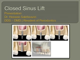
23. 1. Presurgical patient preparation
2. Local anesthesia (buccal and palatal)
3. Mid‐crestal incision with or without
releasing
4. Implant positions are marked
5. Crestal floor to the floor of the maxillary
sinus, measured
24. Exact position of the implant site
is first marked with a small round bur
25. 6. Pilot drills
7. The first Tapered
Osteotome:
• Greenstick fracture
an osteotome is chosen to minimize the
force needed to fracture the compact bone.
26. 8. The seccond Tapered Osteotome:
• Increase the fracture area
diameter slightly larger then the first one
27. 9. The third Osteotome used is straight
diameter about 1–1.5 mm smaller than that
of the implant to be placed
28.
29. Advantage:
• Reduce the risk of membrane perforation
• Reduce the risk of benign paroxysmal positional
vertigo
Disadvantage :
• More time consuming
30. 1. the straight osteotome
penetrates the sinus floor
2. Form and diameter that are
suitable for the implant
3. Final step before placing the
implant is to check that the
preparation is patent to the
planned insertion depth
31. 1. osteotomes are not supposed to enter the
sinus cavity
2. sinus membrane must by tested for any
perforations
34. Antiseptic rinsing 0.1–0.2% CHX:
• Twice daily - 3 weeks
Antibiotic prophylaxis:
• for 1 week in cases where bone substitutes are
used
35. There is still controversy with regards to
the necessity of using grafting material to
maintain the space for new bone formation
after elevating the sinus
• Rosen 1999
• Nedir 2010
• Schmidlin 2008
• Petursson 2009
• Bragger 2004
36. Radiograph taken at the 5‐year follow‐up visit of an implant placed in
the first quadrant utilizing the osteotome technique without grafting
material. A new cortical bony plate at the inferior border of the
maxillary sinus is clearly visible, but no bony structure can be
detected apical to the implant
37. Radiograph (same patient) taken after 5 years in function of an
implant placed in the second quadrant utilizing the osteotome
technique with xenograft grafting material. A dome‐shaped structure
is clearly visible, documenting a definite increase in bone volume
compared to the initial situation. The “dome” is surrounded by a new
cortical bony plate.
38. (a) Radiograph taken immediately after implant insertion with
the osteotome technique and grafting material, showing a
cloudy dome‐shaped structure extending 2–3 mm apical to
the implant. (b) Radiograph of the same implant taken 1 year
later showing significant reduction of the size of the
“dome”, but the new bony structure is clearly visible apical to
the implant
39. Autogenous bone is often referred to as
the gold standard
Anorganic bovine bone-derived mineral
(ABBM)
demineralized freeze-dried bone allograft
(shrinkage)
40.
41.
42. Increased bone height from 2 to 7 mm
(average, 3.8 mm)
If more vertical bone height is needed, the
lateral wall osteotomy approach may be
more advantageous.
43. the implant survival rate was 96% when
residual bone height was 5 mm or more,
but dropped to 85.7% when residual bone
height was 4 mm or less
44. Transalveolar sinus floor elevation
technique was most predictable when the
residual alveolar bone height was ≥5 mm
and with implants of ≥8 mm.
45. Indications:
Filling material for the sinus:
(either alone or in combination with a bone substitute)
Membrane to seal the lateral window
for protection of the schneiderian membrane after
detachment from the underlying bone
Close a membrane rupture.
46. L-PRF can be used successfully as the sole filling
material during sinus augmentation, but only if it is used
simultaneously with the placement of implants
47. when L-PRF as the sole filler material,
natural bone regeneration occurred around
the implants
• 10 mm vertical bone gain with the Window technique
• 3.5 to 4 mm with the Transalveolar approach
All gain in radio opaque areas in the sinus,
observed on (CBCT), is “Vital” bone, with
0% substitute remnants.
48. When implants have to be placed in a second
stage L-PRF can also be used, but it should
be mixed with a bone substitute to delay its
resorption.
Lambert:
• use of pure autologous bone resulted in greater loss of
the augmented volume after 5 weeks
A mixture (DBBM) and L-PRF resulted in
more new bone formation than when DBBM
was used exclusively.
49. Fibrin glue : newly formed epithelium
L-PRF for the treatment of perforations of the schneiderian membrane
1. L-PRF membranes are resilient
2. Pliable
3. easy to manipulate
4. have an adhesive nature
They have the capacity to seal the connection to
the sinus in minutes
56. Tapping can be bother some patients with
dense cortical bone and for those with
loose trabecular bone
benign paroxysmal positional vertigo
(BPPV)
patients must be able to open wide enough
58. the most common intraoperative
complication is perforation of the sinus
membrane.
• Smaller perforations : using Tissue Fibrin Glue
• Larger perforations : achieved through a Lateral
Window
• Perforations before any grafting material
procedure should be aborted or
shorter implants used
59. Postoperative infections after transalveolar
sinus lift are rare complications
Postoperative hemorrhage
Nasal bleeding
Hematomas
Loosening of cover screws resulting in:
• Suppuration
• (BPPV)
60. In the posterior maxilla:
• High initial stability
• Rough surface geometry.
Implants with
• Slightly conical morphometry or
• implants with a Wider implant neck
tend to give better primary stability
62. Residual bone height of ≥8 mm and an
oblique sinus floor:
• standard implant placement using a short implant
• elevation of the maxillary sinus floor using the
osteotome technique without grafting material
63. Residual bone height of 5–7 mm and a
relatively flat sinus floor:
• elevation of the maxillary sinus floor using the
osteotome technique with grafting material that is
resistant to resorption
64. Residual bone height of 5–7 mm and an
oblique sinus floor:
• elevation of the maxillary sinus floor using the
lateral approach with grafting material, and
simultaneous implant placement (one‐stage)
65. Residual bone height of 3–4 mm and a flat
or oblique sinus floor:
• elevation of the maxillary sinus floor using the
lateral approach with grafting material, and
simultaneous implant placement (one‐stage)
66. Residual bone height of 1–2 mm and a flat
or oblique sinus floor:
• elevation of the maxillary sinus floor using the
lateral approach with grafting material and
delayed implant placement
4–8 months later (two‐stage)