
Recomendados
Mais conteúdo relacionado
Mais procurados
Mais procurados (20)
Semelhante a Digestive system full
Semelhante a Digestive system full (20)
Mais de simranjeet kaur
Último
Último (20)
Digestive system full
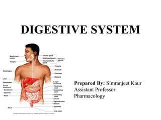
- 1. DIGESTIVE SYSTEM Prepared By: Simranjeet Kaur Assistant Professor Pharmacology
- 2. Essential Terms Digestion • process of mechanically or chemically breaking down food Absorption • passage of small molecules into blood and lymph Digestive system • organs which carry out process of digestion and absorption Metabolism • all the chemical reactions of the body
- 3. Digestive System 1. Composed of GI tract and accessory organs 2. Breaks down ingested food for use by the body 3. Digestion occurs by mechanical and chemical mechanisms 4. Excretes waste products or feces through process of defecation
- 4. GI Tract / Alimentary Canal • Continuous tube from mouth to anus • Mouth • Pharynx • Esophagus • Stomach • Small intestine • Large intestine
- 5. Accessory Digestive Organs • Provide mechanical and chemical mechanisms to aid digestion • Teeth • Tongue • Salivary glands • Liver • Gallbladder • Pancreas
- 6. Figure 23.1
- 7. Functions of Digestive System 1. Ingestion 2. Secretion 3. Mixing and propulsion • Motility 1. Digestion • Mechanical and chemical 1. Absorption 2. Defecation
- 8. Layers of GI Tract •From deep to superficial: •Mucosa •Submucosa •Muscularis •Serosa
- 9. Figure 23.2
- 10. Layers of GI Tract • Mucosa – Epithelium • Type varies – Lamina propria – areolar connective tissue • MALT – mucus-associated lymphatic tissue – Muscularis mucosae – smooth muscle • Submucosa – Areolar connective tissue – Blood and lymphatic vessels – Neurons – submucosal plexus
- 11. Layers of GI Tract • Muscularis – Skeletal and smooth muscle – Neurons – myenteric plexus • Serosa – Areolar and simple squamous epithelium – Visceral peritoneum
- 12. Peritoneum • Mesothelium • Parietal peritoneum • Visceral peritoneum • Peritoneal cavity • Retroperitoneal
- 13. Figure 23.3a
- 14. Figure 23.3b
- 15. Figure 23.3c
- 16. Figure 23.3d
- 17. Folds of Peritoneum • Greater omentum – Adipose tissue • Falciform ligament – Liver to anterior abdominal wall • Lesser omentum • Mesentery – Small intestine to posterior abdominal wall • Mesocolon
- 18. Neural Innervation of GI Tract • Regulated by autonomic nervous system • Enteric division – Myenteric plexus / plexus of Auerbach – Submucosal plexus / plexus of Meissner • Able to function independently from rest of nervous system • Linked to CNS by extrinsic sympathetic and parasympathetic nerves • Sympathetic nerves decrease GI secretions & motility • Parasympathetic nerves increase GI secretion and motility
- 19. Mouth Parts of Digestive System • Mouth formed by several parts: • Cheeks • Lips / labia • Labial frenulum • Orbicularis • Vestibule • Oral cavity proper • Fauces • Hard and soft palate • Uvula • Palatoglossal and palatopharyngeal arch
- 20. Figure 23.4
- 21. Tongue • Skeletal muscle and mucous membrane • Helps form floor of oral cavity • Extrinsic muscles • Intrinsic muscles • Lingual frenulum • Papillae – Fungiform – Filiform – Circumvallate – Foliate • Lingual glands – Lingual lipase
- 22. Salivary Glands • Release saliva to oral cavity • 3 pairs of salivary glands • Parotid • Submandibular • Sublingual
- 23. Composition of Saliva • 99.5 % water • 0.5% other solutes – Ions – Mucus – Immunoglobulin A – Enzymes • Salivation controlled by autonomic nervous system • Stimulated by various mechanisms
- 24. Figure 23.5
- 25. Teeth • External regions 1. Crown 2. Root 3. Neck • Internal components 1. Enamel 2. Dentin • Cementum 3. Pulp cavity • PulpRoot canals • Apical foramen
- 26. Figure 23.6
- 27. Teeth • Dentitions • Deciduous teeth – first set • Permanent teeth – secondary • Carry out mechanical digestion by mastication • Creates bolus • Salivary amylase • Breakdown starch • Lingual lipase • Breakdown triglycerides
- 28. Figure 23.7
- 29. Table 23.1
- 31. Pharynx • Composed of skeletal muscle • Lined by mucous membrane • Nasopharynx • Oropharynx • Laryngopharynx
- 32. Esophagus • Collapsible muscular tube through esophageal hiatus of diaphragm • Mucosa – Submucosa contains areolar connective tissue • Muscularis – Skeletal muscle – Upper and lower esophageal sphincter • Adventitia – Attaches esophagus to nearby structures • Secrets mucus and transports food
- 33. Figure 23.8
- 34. Deglutition • Stages of swallowing • Voluntary – Mouth to oropharynx • Pharyngeal – Deglutition center in medulla oblongata and pons – Closing of epiglottis – Involuntary • Esophageal – Involuntary – Peristaltic contractions
- 35. Figure 23.9a,b
- 36. Figure 23.9c
- 37. Table 23.2
- 38. Stomach • Serves as mixing chamber and storage area for ingested food • Rugae allow for increased volume • 4 main regions 1. Cardia 2. Fundus 3. Body 4. Pylorus – Pyloric antrum and canal – Pyloric sphincter – Lesser and greater curvatures
- 39. Figure 23.10a
- 40. Stomach Histology 1. Mucosa – Surface mucous cells – Lamina propria – Muscularis mucosae – Gastric glands and pits – Parietal cells – Chief cells – G cells 2. Submucosa – areolar connective tissue 3. Muscularis – 3 layers of smooth muscle 4. Serosa
- 41. Figure 23.11a
- 42. Figure 23.11b
- 43. Figure 23.11c
- 44. Mechanical and Chemical Digestion • Mixing waves caused by peristaltic movement • Chyme released in process of gastric emptying • Proton pumps bring H+ into the lumen • Carbonic anhydrase forms carbonic acid to provide H+ and bicarbonate ions (HCO3 - )
- 45. Figure 23.12
- 46. Mechanical and Chemical Digestion • Chemical digestion stimulated by nervous system • Parasympathetic neurons release acetylcholine – Works with gastrin – HCl released in presence of histamine • Pepsin begins digestion of proteins – Stomach protected by alkaline mucus secretion • Gastric lipase digests triglycerides • Few molecules absorbed by stomach – Water, ions, short-chain fatty acids, alcohol
- 47. Table 23.3 pt 1
- 48. Table 23.3 pt 2
- 49. Liver • Superficially has four lobes – right, left, caudate, and quadrate • The largest gland in the body
- 50. Liver: Associated Structures The falciform ligament: – Separates the right and left lobes anteriorly – Suspends the liver from the diaphragm and anterior abdominal wall The ligamentum teres: – Is a remnant of the fetal umbilical vein – Runs along the free edge of the falciform ligament
- 51. Liver: Associated Structures • The Lesser omentum anchors the liver to the stomach • The hepatic blood vessels enter the liver at the Porta hepatis • Gallbladder - rests in a recess on the inferior surface of the right lobe; stores bile for digestion of fats
- 52. Figure 23.14a
- 53. Figure 23.14b
- 54. Liver: Microscopic Anatomy Lobules are hexagonal shaped and the structural and functional units of the liver – Composed of hepatocyte (liver cell) plates radiating outward from a central vein – Portal triads are found at each of the six corners of each liver lobule Portal triads consist of a bile duct and – Hepatic artery – supplies oxygen-rich blood to the liver – Hepatic portal vein – carries venous blood with nutrients from digestive viscera
- 55. Microscopic Anatomy of the Liver Figure 23.24c, d
- 56. • Liver sinusoids – enlarged, leaky capillaries located between hepatic plates • Kupffer cells – hepatic macrophages found in liver sinusoids
- 57. Bile Duct System • Bile secreted by hepatocytes • Bile canaliculi • Bile ducts • Right and left hepatic ducts • Common hepatic duct • Common bile duct • Gallbladder for temporary storage of bile • Cystic duct
- 58. Blood Supply of Liver • Hepatic artery provides oxygenated blood • Hepatic portal vein provides deoxygenated blood – Nutrients, drugs, toxins, microbes • Hepatic artery and vein carry blood to sinusoids – Substances exchanged by hepatocytes – Blood drains to central vein and eventually hepatic vein • Portal triad – Hepatic portal vein – Hepatic artery – Bile duct
- 59. Figure 23.15
- 60. Bile • 800-1000 mL/day • pH 7.6 – 8.6 • Water • Bile acids • Bile salts – Emulsification • Cholesterol • Lecithin • Bile pigments – Bilirubin • Stercobilin
- 61. Liver Functions • Metabolism of: – Carbohydrates – Lipids – Proteins • Process drugs and hormones • Excrete bilirubin • Synthesize bile salts • Storage – Glycogen – Vtamins – Minerals • Phagocytosis • Activate Vitamin D
- 62. Small Intestine • Adapted for digestion and absorption • 3 m (10 ft) living • 6.5 m (21 ft) without muscle tone • Duodenum • Jejunum • Ileum • Ileocecal sphincter – Connection to large intestine
- 63. Figure 23.16a
- 64. Figure 23.16b
- 65. Histology of Small Intestine • Mucosa • Cell types – Absorptive – Goblet – Endocrine – Paneth • Lysozyme • Intestinal glands (crypts of Lieberkühn) – S cells • Hormone secretin – CCK cells • Hormone – cholecystokinin (CCK)
- 66. Figure 23.17a
- 67. Small Intestine Structural modifications of the small intestine wall increase surface area: – Villi – fingerlike extensions of the mucosa – Microvilli – tiny projections of absorptive mucosal cells’ plasma membranes
- 69. Small Intestine: Histology The epithelium of the mucosa is made up of: – Absorptive cells and goblet cells • Cells of intestinal crypts secrete intestinal juice • Peyer’s patches are found in the submucosa and are collections of lymphatic/wbc tissue • Brunner’s glands in the duodenum secrete alkaline mucus
- 70. Pancreas Posterior to great curvature of the stomach 1. head - enlarged portion in C-curve of the duodenum 2. body - tapers off beneath the stomach 3. tail - terminal part near the end 4. pancreatic duct - merges with bile duct to duodenum a. hepatopancreatic ampulla (merging of both) 5. accessory duct - empties into duodenum, smaller
- 72. Pancreas - histology • Made of glandular epithelial cells • Pancreatic islets (of Langerhans) (1% of all cells) a. hormones: glucagon, insulin, somatostatin • Acini - (99% of the cells in pancreas) a. mixture of enzymes called "pancreatic juice"
- 73. Figure 23.13a
- 74. Figure 23.13c
- 75. Histology of Pancreas • Glandular epithelial cells – 99% exocrine clusters – Secrete pancreatic juice • Fluid and enzymes • Pancreatic islets (islets of Langerhans) – 1% endocrine cells – Hormones • Glucagon • Insulin • Somatostatin – Pancreatic polypeptide
- 76. Pancreatic Juice • 1200-1500 mL/day • pH 7.1-8.2 • Water • Salts • Sodium bicarbonate • Enzymes – Pancreatic amylase – Trypsin • Entereokinase – Chymotrypsin – Carboxypeptidase – Elastase – Pancreatic lipase – Ribonuclease and deoxyribonuclease
- 77. Bile Bile leaves the liver via: – Bile ducts, which fuse into the common hepatic duct – The common hepatic duct, which fuses with the cystic duct • These two ducts form the bile duct
- 78. Gallbladder and Associated Ducts
- 79. Hepatocytes Hepatocyte functions include: – Production of bile – Processing bloodborne nutrients – Storage of fat-soluble vitamins – Detoxification
- 80. The Gallbladder • Thin-walled, green muscular sac on the ventral surface of the liver • Stores and concentrates bile by absorbing its water and ions • Releases bile via the cystic duct, which flows into the bile duct
- 81. Large Intestine • Is subdivided into the cecum, appendix, colon, rectum, and anal canal • The saclike cecum: – Lies below the ileocecal valve in the right iliac fossa – Contains a wormlike vermiform appendix
- 82. Colon Has distinct regions: • ascending colon • hepatic flexure • transverse colon • splenic flexure • descending colon • sigmoid colon
- 83. Large Intestine
- 84. Colon • The transverse and sigmoid portions are anchored via mesenteries called mesocolons • The sigmoid colon joins the rectum • The anal canal, the last segment of the large intestine, opens to the exterior at the anus
- 85. Anus • Internal sphincter - smooth muscle (involuntary) • External sphincter - skeletal muscle (voluntary)
- 86. Mesenteries of Digestive Organs
- 87. Mesenteries of Digestive Organs
- 88. Mesenteries of Digestive Organs Figure 23.30d