
Recomendados
Mais conteúdo relacionado
Mais procurados
Mais procurados (20)
Semelhante a SDS- Polyacrylamide Gel Electrophoresis
Semelhante a SDS- Polyacrylamide Gel Electrophoresis (20)
Mais de Shylesh M
Mais de Shylesh M (14)
Último
Último (20)
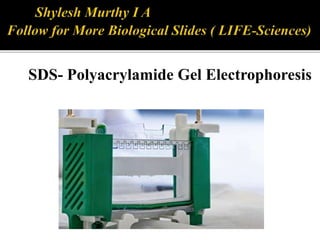
SDS- Polyacrylamide Gel Electrophoresis
- 1. SDS- Polyacrylamide Gel Electrophoresis
- 2. Standard test that used to determine the charged molecules, mainly proteins and nucleic acids. widely used in biochemistry ,forensics, genetics and molecular biology to separate and identify proteins according to their molecular weight. This method separates proteins based primarily on their molecular weights. Laemmli system of SDS-PAGE was first introduced in 1970s.
- 3. Separates protein in an electric field. Migrate through a liquid or semisolid medium when subjected to an electric field from anode to cathode terminal. Molecules flow at different rates depend on the molecular size of proteins.
- 4. -the proteins samples are having uniformed structure and charge the separation will depend on their molecular weight only. -SDS-treated proteins have very similar charge-to-mass ratios, and similar shapes. During PAGE, the rate of migration of SDS-treated proteins is effectively determined by molecular weight. -Small proteins migrate faster through the gel under the influence of the applied electric field, whereas large proteins are successively retarded, due to the sieving effect of the gels.
- 5. SDS –page coated large proteins migrate slowly through the gel matrix and small proteins migrate quickly through the matrix. The nearer the band to the well.The large the molecular size of protein.
- 6. Negatively charged detergent sodium dodecyl sulfate. Used to denature and linearize the proteins Coated the proteins with negatively charged.
- 7. SDS-PAGE is differentiated into two systems. *continuous sds-page *discontinuous sds-page. • Polyacrylamide is used to form a gel, a matrix of a pores which allow the molecules migrate at different rates.
- 8. The size of pores is determined by the concentration of acrylamide. The higher the concentration, the smaller the size of pores. Discontinuos sds-page consist of two different gels. *stacking gel -4%of acrylamide *separating gel-range from 5-15% of acrylamide.
- 9. Preparation of gel Clean the plates and combs. Set up the plates on the rack. Pour the separating gel. Pour the stacking gel. Gel storage.
- 11. Visualizes the band under UV light. Types of stains; 1. Coomassie Blue; * Coomassie Brilliant Blue staining The Coomassie dyes R-250 and G-250 bind to proteins stoichiometrically through their sulfonic acid groups. * . The interactions between dye and protein are Van der Waals and ionic. The sulfonic acid groups interact with positive amine groups. Therefore coomassie dye binds to wide range of proteins. * Limited to ~100ng of protein. 1. Silver stain; *most sensitive test *detection limit 0.1-1.0ng of protein
- 13. Determine purity of protein samples Determine molecular weight of protein Identifying disulfide bonds between protein Quantifying proteins Blotting applications
- 14. SDS-page is a technique that used to separate proteins according to their molecular size through the gel. Proteins are unfolded and migrate from cathode to anode terminal at different rates. Molecular weight is determined by compare the result with a standard curve of relative motility of standard proteins.
- 15. • P.K .Gupta molecular biology second edition. • https://www.google.co.in/search?q=sds+page +principle.
- 16. Thank you