
Recomendados
Mais conteúdo relacionado
Mais procurados
Mais procurados (20)
Semelhante a HYPERBILIRUBINEMIA FINAL.pptx
Semelhante a HYPERBILIRUBINEMIA FINAL.pptx (20)
Último
Último (20)
HYPERBILIRUBINEMIA FINAL.pptx
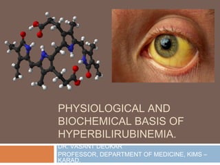
- 1. PHYSIOLOGICAL AND BIOCHEMICAL BASIS OF HYPERBILIRUBINEMIA. DR. VASANT DEOKAR PROFESSOR, DEPARTMENT OF MEDICINE, KIMS – KARAD.
- 2. COMPETENCIES ◻ To understand the biochemical and physiological basis of hyperbilirubinemia. ◻ To come to a accurate diagnosis behind the reason for the hyperbilirubinemia based on physical and biochemical findings.
- 3. LEARNING OBJECTIVES ◻ By the end of the lecture the student should know: ◻ The etiology and epidemiology of hyperbilirubinemia. ◻ To be able to enumerate the different signs and symptoms of hyperbilirubinemia. ◻ To distinguish between different types of hyperbilirubinemia.
- 4. INTRODUCTION ◻ Jaundice, also known as hyperbilirubinemia, is defined as a yellow discoloration of the body tissue resulting from the accumulation of excess bilirubin. ◻ Deposition of bilirubin happens only when there is an excess of bilirubin, and this indicates increased production or impaired excretion. The normal serum levels of bilirubin are less than 1 milligram per deciliter (mg/dL). ◻ However, the clinical presentation of jaundice with peripheral yellowing of the eye sclera, also called scleral icterus, is best appreciated when serum bilirubin levels exceed 3 mg/dl. ◻ With further increase in serum bilirubin levels, the skin will progressively discolor ranging from lemon yellow to apple green, especially if the process is long-standing; the green color is due to biliverdin.
- 6. BILIRUBIN ◻ Bilirubin is an important metabolite of heme (ferroprotoporphyrin IX), a coordination complex that serves to coordinate iron in various proteins. ◻ It is a potentially toxic substance. However, the body has developed mechanisms for its safe detoxification and disposition. ◻ Bilirubin and its metabolites also provide the distinctive yellow color to bile and stool and a lesser degree, urine.
- 7. PATHOPHYSIOLOGY ◻ It includes 3 phases: Prehepatic, Hepatic and Posthepatic. ◻ PREHEPATIC ◻ Bilirubin is the end product of heme, which is released by senescent or defective RBCs. In the reticuloendothelial cells of spleen, liver and bone marrow, heme released from the RBC undergoes a series of reactions to form the final product bilirubin:
- 8. ◻ HEPATIC ◻ Hepatocellular uptake - The bilirubin released from the reticuloendothelial system is in an unconjugated form (i.e., non-soluble) and gets transported to the hepatocytes bound to albumin which accomplishes solubility in blood. ◻ The albumin-bilirubin bond is broken, and the bilirubin alone is then taken into the hepatocytes through a carrier-membrane transport and bound to proteins in the cytosol to decrease the efflux of bilirubin back into the plasma.
- 9. . ◻ Conjugation of bilirubin - This unconjugated bilirubin then proceeds to the endoplasmic reticulum, where it undergoes conjugation to glucuronic acid resulting in the formation of conjugated bilirubin, which is soluble in the bile. This is rendered by the action of UDP- glucuronosyl transferase
- 10. ◻ POSTHEPATIC ◻ Bile secretion from hepatocytes- Conjugated bilirubin is now released into the bile canaliculi into the bile ducts, stored in the gallbladder, reaching the small bowel through the ampulla of Vater and finally enters the colon.
- 11. ◻ Intestinal metabolism and Renal transport- The intestinal mucosa does not reabsorb conjugated bilirubin due to its hydrophilicity and large molecular size. The colonic bacteria deconjugate and metabolize bilirubin into urobilinogen’s, 80% of which gets excreted into the feces and stercobilin and the remaining (10 to 20%) undergoes enterohepatic circulation. Some of these urobilin’s are excreted through the kidneys imparting the yellow pigment of urine.
- 12. ◻ Dysfunction in prehepatic phase results in elevated serum levels of unconjugated bilirubin while insult in post hepatic phase marks elevated conjugated bilirubin. Hepatic phase impairment can elevate both unconjugated and conjugated bilirubin. ◻ Increased urinary excretion of urobilinogen can be due to increased production of bilirubin, increased reabsorption of urobilinogen from the colon, or decreased hepatic clearance of urobilinogen.
- 14. EPIDIMEOLOGY ◻ The prevalence of jaundice differs among patient populations; newborns and elderly more commonly present with the disease. ◻ The causes of jaundice also vary with age. Around 20 percent of term babies are found with jaundice in the first week of life, primarily due to immature hepatic conjugation process. ◻ Congenital disorders, overproduction from hemolysis, defective bilirubin uptake, and defects in conjugation are also responsible for jaundice in infancy or childhood. ◻ Hepatitis A was found to be the most afflicting cause of jaundice among children. Bile duct stones, drug-induced liver disease, and malignant biliary obstruction occur in the elderly population.
- 15. ◻ Men have an increased prevalence of alcoholic and non- alcoholic cirrhosis, chronic hepatitis B, malignancy of pancreas, or sclerosing cholangitis. In contrast, women demonstrate higher rates of gallbladder stones, primary biliary cirrhosis, and gallbladder cancer.
- 16. ETIOLOGY
- 20. HISTORY ◻ A thorough questioning regarding the use of drugs, alcohol or other toxic substances, risk factors for hepatitis (travel, unsafe sexual practices), HIV status, personal or family history of any inherited disorders or hemolytic disorders is vital. ◻ Other points such as duration of jaundice; and the presence of any coexisting signs and symptoms, like a joint ache, rash, myalgia, changes in urine and stool, presence of weight loss can help narrow down the etiology, further.
- 21. PHYSICAL EXAMINATION ◻ Physical examination begins by evaluation of body habitus and nutritional status. Temporal and proximal muscle wasting suggests malignancy or cirrhosis. ◻ Stigmata of chronic liver disease, which includes spider nevi, palmar erythema, gynecomastia, caput medusae, Dupuytren contractures, parotid gland enlargement, and testicular atrophy. ◻ Palpable lymph nodes can also direct the clinician towards malignancy (left supraclavicular & periumbilical). Increased volume status of the patient evidenced by jugular venous distension can be a sign of right-sided heart failure, suggesting hepatic congestion.
- 22. Clinical findings in a patient with hyperbilirubinemia
- 23. ◻ The abdominal examination should provide information on the presence of hepatosplenomegaly, or ascites. Jaundice with ascites indicates either cirrhosis or malignancy with peritoneal spread. ◻ Right upper quadrant tenderness with palpable gallbladder (Courvoisier sign) suggests obstruction of the cystic duct due to malignancy.
- 24. EVALUATION
- 25. ◻ Hepatocellular workup: viral serologies, autoimmune antibodies, serum ceruloplasmin, ferritin. ◻ Cholestatic workup: Additional tests include abdominal ultrasound, CT, magnetic resonance cholangiopancreatography (MRCP), endoscopic retrograde cholangiopancreatography (ERCP), percutaneous transhepatic cholangiography (PTC), endoscopic ultrasound (EUS).
- 26. TREATMENT/ MANAGEMENT ◻ Treatment of choice for jaundice is the correction of the underlying hepatobiliary or hematological disease, when possible. ◻ Pruritis associated with cholestasis can be managed based on the severity. For mild pruritis, warm baths or oatmeal baths can be relieving. Antihistamines can also help with pruritis. Patients with moderate to severe pruritis respond to bile acid sequestrants such as cholestyramine or colestipol.
- 27. ◻ Other less effective therapies include rifampin, naltrexone, sertraline, or phenobarbital. If medical treatments fail, liver transplantation may be the only effective therapy for pruritis. ◻ Jaundice is an indication for hepatic decompensation and may be an indication for liver transplant evaluation depending on the severity of the hepatic injury.