Dental tooth nomenclature
•Transferir como PPTX, PDF•
2 gostaram•1,113 visualizações

This PPT contains tooth nomenclature, Notation System, needs of operative dentistry, Caries nomenclature, Non carious lesion

Recomendados
Mais conteúdo relacionado
Mais procurados
Mais procurados (20)
Semelhante a Dental tooth nomenclature
Semelhante a Dental tooth nomenclature (20)
Mais de Shraddha Joshi
Mais de Shraddha Joshi (20)
Último
Último (20)
Dental tooth nomenclature
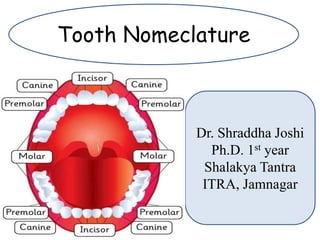
- 1. Tooth Nomeclature Dr. Shraddha Joshi Ph.D. 1st year Shalakya Tantra ITRA, Jamnagar
- 2. Definition 2 definitions: • Deciduous: Primary • Permanent: Secondary Elements of tooth: • Water • Organic materials • Inorganic materials Elements
- 7. Incisor- Function as cutting or shearing instruments for food. Canines- possess the longest roots of all teeth and are located at the corners of the dental arch. Premolars- acts like the canines in the tearing of food and are similar to molars in grinding of food. Molars- are located nearest the TMJ, which serves as the fulcrum during function.
- 9. Enamel (Hard tissue) Dentin (Hard tissue) Odontoblast layer Pulp chamber (Soft tissue) Gingiva (Soft tissue) PDL (Soft tissue) Cementum (Hard tiissue) Alveolar bone (Hard tissue) Pulp canals Apical foramen Dental tissues
- 10. 3 Parts: • Anatomical Crown • Anatomical Root • Pulp chamber
- 12. It includes a sequence of Arabic numbers (1-32) for permanent and the alphabet system (A-T) for Deciduous teeth, moving clockwise around the dentition. Universal Numbering System Universal Tooth Numbering System. Zsigmondy & Palmar Tooth Numbering System. FDI Tooth Numbering System
- 14. Deciduous teeth
- 15. Permanent teeth
- 16. In this system, the teeth that should be there are numbered. If the wisdom teeth is missing missing, then first number will be 2 instead of 1 acknowledging the missing tooth. If the teeth had removed or teeth are missing, the missing teeth will be numbered as well. Advantages: Separate number/ alphabet is given for individual tooth. Easy to visualize.
- 17. Disadvantages: Confusing when comparing with palmar notation system. Confusing and difficult to remember. It doesnot consider the jaw quadrant clearly.
- 18. Zsigmondy/ Palmar Numbering System
- 19. Advantages: System is simple to use. Easier for beginner due to less confusion as permanent teeth and deciduous teeth are indicated differently. Disadvantages: No differentiation between right upper, right lower, left upper, left lower. No provision to identify supernumerary tooth.
- 20. FDI tooth Numbering System Federation Dentaire Internationalae system. It is two digit system. The first digit indicates the quadrant (5 through 8) and the second digit indicates the tooth type (1 through 5). For Primary teeth: 5- Maxillary right 6- Maxillary left 7- Mandibular left 8- Mandibular right For Permanent teeth: 1- Maxillary right 2- Maxillary left 3- Mandibular left 4- Mandibular right
- 21. FDI Notation for primary teeth
- 22. FDI Notation for Permanent teeth
- 23. Advantages: Very simple, Accurate, easy to memorise. Prevents Errors in differentiating left and right, upper and Lower Arches, and tooth type. Simple to teach and easy to understand. Disadvantages: In case of deciduous teeth, there can be confusion and it is difficult to memorise.
- 24. Current minimally intervention philosophy follows three concepts of disease treatment: 1. Identify— Identify and assess risk factors early. 2. Prevent— Prevent disease by eliminating risk factors. 3. Restore— Restore the health of the oral environment.
- 25. Following are the indications of operative dentistry: 1. Caries. 2. Non carious loss of tooth structures. 3. Malformed, Traumatized or fractured tooth. 4. Esthetic Improvement. 5. Replacement or repair of restoration. 6. Developmental defects.
- 26. Caries: Caries is most common disease affecting the teeth. Dental caries is microbiological disease of the teeth which results in localized dissolution and destruction of the calcified tissue, caused by the action of microorganisms and fermentable carbohydrates. Based on anatomy of the surface involved, dental caries can be of the following types: a. Pit and fissures carious lesions. b. Smooth surface carious lesions. c. Root caries.
- 27. Non-carious Loss of the Tooth Structure: 1. Attrition Mechanical wear of opposing teeth commonly seen on contacting occlusal, incisal and proximal surfaces. 2. Abrasion Loss of tooth material by mechanical wear other than contacting surfaces. It commonly occurs due to improper brushing and use of abrasive tooth powder. 3. Erosion Loss of dental hard tissue as a result of a chemical process not involving bacteria.
- 28. Malformed, Traumatized, or Fractured Teeth: Traumatic injuries may involve the hard dental tissues and the pulp which require restoration. Esthetic Improvement: Discolored teeth because of staining or other reasons look unesthetic and require restoration. Replacement or Repair of Restoration: Repair or replacement of previous defective restoration is indicated for operative treatment.
- 29. Developmental Defects: Defects like enamel and dentin hypoplasia, hypomineralization, amelogenesis and dentinogenesis imperfecta, tetracycline stains, peg- shaped laterals need operative intervention.
- 30. Objectives of operative dentistry are as follows: Diagnosis: Diagnosis is determination of nature of disease, injury or other defect by examination, test and investigation. Prevention: To prevent any recurrence of the causative disease and their defects, it includes the procedures done for prevention before the manifestation of any sign and symptom of the disease.
- 31. Interception: It includes the procedures undertaken after signs and symptoms of disease have appeared, in order to prevent the disease from developing into a more serious or full extent. Preservation: Preservation of the vitality and periodontal support of remaining tooth structure is obtained by preventive and interceptive procedures.
- 32. Restoration: Includes restoring form, function, phonetics and esthetics. Maintenance: After restoration is done, it must be maintained for providing service for longer duration.
- 33. Anatomical crown: It is part of tooth that is covered with enamel. It extends from cementoenamel junction (CEJ)to occlusal or incisal surface. Clinical crown: It is part of tooth that is visible in oral cavity. In case of gingival recession, the clinical crown is longer than anatomical crown.
- 35. Nomenclature of Dental Caries Primary Caries: Denotes lesions on unrestored surfaces, in other words it is the first attackon a tooth. Secondary (Recurrent) Caries: Lesions developing adjacent or beneath existing restorations are referred to as ‘secondary’ or ‘recurrent’ caries.
- 36. Radiograph showing recurrent caries
- 37. Residual Caries: Demineralizedtissue left in place before a restoration is placed. It can occur as a result of the clinician’s neglect or intentionally. Active Carious Lesion: A lesion which is progressing is described as an active carious lesion. Inactive/Arrested Carious Lesion: A lesion that has formed and then not progressed is referred to as an ‘arrested’ or ‘inactive’ carious lesion. Arrested carious lesions are often characterized by a large open cavities which no longer retain food and become self- cleansing.
- 38. Pit and Fissure Caries: Pit and fissure caries describes carieswhich occurs on occlusal surface of posterior teeth, buccal and lingual surfaces of molars and on lingual surfaces of maxillary incisors Smooth Surface Caries: Smooth surface caries occurs on the gingival third of buccal and lingual surfaces and on approximal surfaces. Root Caries: Root caries occur on exposed root cementum and dentin,usually following gingival recession.
- 39. Pits and Fissures caries
- 41. Root caries/ Radicular caries
- 42. Acute Dental Caries: Acute caries travels towards the pulp at a very fast speed. Rampant Caries: The name given to multiple active carious lesions occurring in the same patient, frequently involving surfaces of teeth that are usuallycaries-free. Rampant caries can be of following three types: 1. Early childhood caries is a term used to describe dental caries presenting in the primary dentition of young children. 2. Bottle caries or nursing caries is seen in the primary dentition of infants and young children as a consequence of sucking on a bottle or dummy containing cariogenic liquids. The clinical pattern is characteristic, with the four maxillary deciduous incisors most severely affected.
- 43. 3. Xerostomia induced rampant caries is commonly seen after radiotherapy of malignant lesions of the jaws, as the salivary glands may be damaged by the radiation resulting in reduced salivary flow. Chronic Dental Caries: Chronic caries progresses very slowly towards the pulp. Such lesions appear dark incolour and hard in consistency.
- 44. Non carious defect of teeth Attrition It is defined as a physiological, continuous, process resulting in loss of tooth structure from direct frictional forces between contacting teeth. It occurs both on occlusal and approximal surfaces. Attrition is accelerated by parafunctional mandibular movements, especially bruxism. Abrasion It refers to the loss of tooth substance induced by mechanical wear other than that of mastication. Abrasion results in saucer-shaped or wedge-shaped indentations with a smooth, shiny surface.
- 45. Abrasion
- 46. Attrition
- 47. Erosion: It can be defined as a loss of tooth substance by a chemical process that does not involve known bacterial action. The eroded area appears smooth, hard and polished. Abfraction: Abfractionsare the microfractures which appear in the enamel and possibly the dentine caused by flexion of the cervical area of the tooth under heavy loads. These lesions usually appear as wedge-shape defects with sharp line angles. Resorption: ‘Resorption’ is defined as “a condition associate with either a physiologic or a pathologic process resulting in the loss of dentin, cementum or bone”.
- 48. Erosion of teeth resulting in its shiny surface
- 49. Abfraction
- 50. Resorption
- 51. Localized Nonhereditary Enamel Hypoplasia Refers to the localized defects in the crown of the portion tooth caused by the injury to ameloblasts during the enamel matrix formation stage. These lesions may appear as isolated pits or widespread linear defects, depressions or loss of a part of the enamel. Injury to ameloblasts may be caused by: • Traumatic intrusion of deciduous teeth • Fluorosis • Exanthematous diseases • Deficiency of vitamins A, C or D • Hypocalcemia.
- 52. Localized Nonhereditary Enamel Hypocalcification Refers to localized defects in the crown of a tooth due to injury to the ameloblasts during the mineralization stage. In these defects, the enamel is normal in structure but its mineralization is defective. The colour of lesion varies from chalky to yellow, brown, dark brown or grayish.