SlideShare uma empresa Scribd logo
1 de 133
1
SURGICAL PATHOLOGY OF
PANCREAS
4 hours
SURGICAL PATHOLOGY OF PANCREAS – 4 hours
• C 8
– Acute pancteatitis: etiology, phisiopathology, clinical findings,
evaluation, diagnosis, differential diagnosis,
complications(pancreatic pseudocyst), treatment.
– Chronic pancteatitis: etiology, phisiopathology, clinical findings,
evaluation, diagnosis, differential diagnosis, complications,
treatment.
• C 9
– Malignant tumors of the pancreas: etiology, classification,
pathology, clinical findings, evaluation, diagnosis, complications,
treatment.
– Tumors of the Vater's ampulla: etiology, classification, pathology,
clinical findings, evaluation, diagnosis, complications, treatment;
– Tumors of the endocrine pancreas.
2
3
SURGICAL PATHOLOGY OF PANCREAS
Penetrating Injuries
• Penetrating injuries to the pancreas are usually diagnosed in the operating room.
• The pancreas is located in the retro peritoneum, surrounded by other viscera and major vascular
structures.
• An isolated injury to the pancreas is unusual, major vascular injuries are seen in 40% to 50% of
patients with penetrating pancreatic injuries,
PANCREATIC TRAUMA Penetrating Injuries of Pancreas
Pancreatic injury involving the ductal system
Retroperitoneal location of pancreas
4
SURGICAL PATHOLOGY OF PANCREAS
• Patients with penetrating pancreatic trauma have obvious indications for abdominal exploration.
• Preoperative serum amylase concentrations are not helpful; (elevated in 30% of patients with
penetrating pancreatic injuries).
Abdominal exploration,
• Signs of pancreatic injury include:
– knife or a projectile path that passes in proximity to the pancreas,
– a central hematoma in the upper abdomen, and
– injuries to the duodenum, vena cava, suprarenal aorta, or mesenteric vessels.
• In all these instances, the pancreas should be thoroughly explored:
• The anterior surface of the pancreas is visualized by entry into the lesser sac of the peritoneal
cavity by division of the gastrocolic ligament in a relatively avascular area to the left of the midline.
• The tail of the pancreas can be more fully visualized, especially in its posterior aspect, by
mobilization of the spleen and the tail of the pancreas as a unit.
• The posterior aspect of the body of the pancreas is visualized by development of the avascular
area at the inferior margin of the body and tail of the pancreas with a combination of sharp and
blunt dissection.
• The posterior aspect of the head of the pancreas can be exposed by an extensive Kocher
maneuver. In combination with entry into the lesser sac, this also allows for bimanual palpation of
the pancreatic head, with one hand placed on the anterior surface of the pancreas through the
hole in the lesser sac and the other hand placed behind the pancreas in the plane developed by
the Kocher maneuver.
PANCREATIC TRAUMA Penetrating Injuries of Pancreas
5
SURGICAL PATHOLOGY OF PANCREAS
• In the evaluation of penetrating pancreatic injuries, the key to operative management is the
determination of whether a ductal injury is present.
– Transduodenal intraoperative pancreatography has been recommended, but the use of this
technique in the identification of ductal injuries is controversial or dangerous.
– In certain circumstances, the use of intraoperative ERCP (endoscopic retrograde
cholangiopancreatography) eliminates this problem.
• Penetrating pancreatic injuries can be classified according to both location and severity.
• With respect to location, injuries can be subdivided into those of the
– head,
– body, and
– tail of the pancreas.
• With respect to severity, classification systems must be satisfactory to research applications but
also in the determination of the best treatment.
Classification
– Class I injuries are simple contusions of the pancreas;
– Class II injuries are lacerations of the parenchyma in the body or tail of the pancreas;
– Class III injuries are those with severe disruption of the head or body;
– Class IV injuries are those in which there is an associated injury to the duodenum.
PANCREATIC TRAUMA Penetrating Injuries of Pancreas
6
SURGICAL PATHOLOGY OF PANCREAS
• Class I injuries should be usually observed or simply drained externally. The type of drain used after
pancreatic injury is probably not important as long as adequate drainage is effected. This can be
accomplished with passive, sump, or closed suction systems.
• If drains are used, they should be left in place for at least 5 to 7 days to ensure that a drain tract
develops. The timing of drain removal should be based on both the amount and character of the
pancreatic drainage:
– Drain outputs in excess of 150 to 200 mL/day are suggestive of pancreatic fistula.
• The morbidity rate for patients with undrained pancreatic secretions is much greater than that for those
with drained pancreatic secretions.
• Class II injuries The treatment depends on the presence or absence of a ductal injury,
a determination that can be difficult to make.
• The argument for intraoperative pancreatography is made for class II injuries.
• The presence of a ductal injury in the head of the pancreas does not usually make a difference with
respect to treatment, because most of these injuries should be drained regardless.
• If a ductal injury is present in the body or tail of the pancreas, the appropriate treatment is resection of
the distal pancreas, whereas if no ductal injury is present, simple drainage is adequate.
• the difficulty lies in determining whether injuries to the body or tail of the pancreas do in fact include
ductal injury.
• Intraoperative pancreatography is helpful, regardless of the technique employed.
• Class III injuries of the body or tail of the pancreas, should be treated with a distal pancreatectomy.
Distal resection can include up to 80% of the gland if necessary.
PANCREATIC TRAUMA Penetrating Injuries of Pancreas
7
SURGICAL PATHOLOGY OF PANCREAS
• Class III injuries of the head of the pancreas should be drained. Resection of these injuries requires
internal drainage, near-total pancreatectomy, or pancreaticoduodenectomy. If the patient develops a
pancreatic fistula, the fistula can be controlled by the drains. If the fistula does not resolve with time, the
pancreas can be drained internally at a later date.
• Class IV injuries of the pancreas involve injuries to the duodenum as well as the pancreas.
– If the injuries to the duodenum and pancreas are simple, the duodenum can be repaired primarily,
and the pancreas can be drained, or a distal resection can be carried out if the pancreatic injury is
in the body or tail.
– For more complicated, combined injuries, pyloric exclusion can be done to minimize pancreatic
stimulation and protect the duodenal repair (see Duodenum).
– For massive injuries to the duodenum and head of the pancreas, pancreaticoduodenectomy with
reconstruction should be reserved for cases in which débridement of devitalized tissue results in a
de facto removal of the duodenum and head of the pancreas.
– Penetrating injuries to the ampulla of Vater may also require formal pancreaticoduodenectomy.
• Internal drainage of the pancreas has been suggested as a means of treating ductal injuries without the
need for resection of viable and functional pancreatic tissue.
• Distal pancreatectomy for traumatic injuries should be performed only after the pancreas has been
thoroughly mobilized and exposed.
• It is possible to perform a distal pancreatectomy without a concomitant splenectomy. This adds to
operative time and can increase the risk of bleeding, particularly if there is an associated injury to the
spleen treated with splenorrhaphy.
• Splenic salvage should be attempted, therefore, only in hemodynamically stable patients with minimal
or no associated intraabdominal or extraabdominal injuries.
PANCREATIC TRAUMA Penetrating Injuries of Pancreas
8
SURGICAL PATHOLOGY OF PANCREAS
PANCREATIC TRAUMA Penetrating Injuries of Pancreas
Internal drainage of a pancreatic injury involving the ductal system
with the creation of a Roux-en-Y jejunoileal limb and construction
of a pancreaticojejunal anastomosis.
9
SURGICAL PATHOLOGY OF PANCREAS
Complications of pancreatic injuries:
• Pancreatic fistulas, after trauma are characterized by persistent drainage of pancreatic enzymes and
secretions from the pancreatic injury for a number of weeks after injury. Most of these fistulas close
spontaneously, especially if there is no proximal obstruction of the pancreatic ductal system. In the rare
instances in which fistulas fail to close spontaneously, surgical intervention may be required.
• Pseudocysts, that develop after pancreatic trauma often resolve on their own or with percutaneous
aspiration. If, after 4 to 6 weeks of observation with serial ultrasound or CT scanning, the pseudocyst
does not show signs of resolution, it should be drained internally with a defunctionalized limb of
jejunum.
• Bleeding in the area of the pancreatic bed, is usually an early complication that is caused by
inadequate drainage with resultant autodigestion of the pancreas and surrounding tissue. Bleeding can
be avoided by identifying all injuries at the time of exploration and ensuring adequate drainage. If
massive bleeding does occur, it should be dealt with through operative intervention.
• Pancreatitis, another complication of pancreatic injury, is also related to inadequate drainage of
pancreatic secretions. Treatment consists of provision of adequate drainage and supportive care. If the
pancreatitis is localized to the distal pancreas and a trial of conservative management fails, distal
pancreatectomy should be considered.
PANCREATIC TRAUMA Penetrating Injuries of Pancreas
10
SURGICAL PATHOLOGY OF PANCREAS
Blunt Injuries
• The major difference between penetrating and blunt injuries of the pancreas concerns the diagnosis.
• Penetrating injuries are usually discovered on abdominal exploration for associated injuries, but
blunt injuries may occur in isolation, and the preoperative diagnosis can be difficult.
• Blunt pancreatic injuries are relatively rare, which increases the difficulty of diagnosis. So delays in
diagnosis of blunt injuries ranged up to several days, which induce an increased morbidity.
• The body of the pancreas lies directly anterior to the vertebral column and is vulnerable to crush injuries
when the anterior abdominal wall is forceably compressed, as can occur from a seat belt or a sharp blow
to the epigastrium. In such instances, the pancreas may be the only intraabdominal organ injured.
• A number of different means are available to make the diagnosis of blunt pancreatic injury:
– Physical examination of the abdomen is useful, but because of the retroperitoneal location of the
pancreas, the results can be misleadingly benign until a number of hours after injury.
– This emphasizes the importance of serial examinations.
– In most cases, the abdomen becomes progressively more tender to palpation during the first 24 to
48 hours after injury, and the need for abdominal exploration becomes more obvious.
– The physical examination of the abdomen is much less reliable in young children and in patients
with head injuries.
• The serum amylase concentration is elevated on admission in about 70% of patients with blunt
pancreatic injury.
• However, elevated serum amylase has a poor positive predictive value and also occurs in many patients
without pancreatic injury. The amylase concentration can be elevated because of trauma to other
organs, including the salivary glands and the ovaries.
• DPL(diagnostic peritoneal lavage) is of little help in the early diagnosis of pancreatic injury unless there
have been associated intraperitoneal injuries. The retroperitoneal location of the pancreas results in
minimal findings in the lavage fluid, and obtaining amylase concentrations in the lavage fluid is not
helpful.
PANCREATIC TRAUMA Blunt Injuries of Pancreas
11
SURGICAL PATHOLOGY OF PANCREAS
• CT scan of the abdomen allows for visualization of the retroperitoneum, including the pancreas.
In the case of isolated injury to the pancreas, the sensitivity of the CT scan is at its lowest shortly after
injury.
Although it may be a good test for the diagnosis of pancreatic injury after a number of hours have
passed, immediate CT of the abdomen will miss some pancreatic injuries, particularly if expert
interpretation is not available.
• Finally, ERCP is a means of diagnosing pancreatic injury.
ERCP is an attractive diagnostic method because it is less invasive than abdominal exploration and
also provides information about the status of the ductal system, but there are several practical
disadvantages of the technique.
– Most of the studies that have reported successful use of ERCP have involved stable patients
studied hours to days after injury and sent to a referral center specifically because of suspicion of
a pancreatic injury.
– These patients are a selected group, quite different from patients who are freshly injured.
– ERCP is not universally available and, even in large centers, is often unavailable at the odd hours
necessary for early diagnosis in acutely injured patients.
– Many endoscopists are fearful of inducing an exacerbation of pancreatitis in patients with mild
pancreatic injuries lacking ductal involvement.
• To summarize,
• the early diagnosis of blunt pancreatic injuries, particularly if they occur in isolation, can be
extremely difficult, and
• no single test allows for an easy and reliable diagnosis.
• A combination of serial abdominal examinations and serum amylase determinations, CT scan,
and ERCP in selected patients is the best diagnostic strategy available.
• These studies should be combined, with a low threshold for operative intervention if a pancreatic injury
is suspected.
PANCREATIC TRAUMA Blunt Injuries of Pancreas
12
SURGICAL PATHOLOGY OF PANCREAS
• Basic principles of exposure and operative management of blunt injuries of the pancreas are the
same as for penetrating injuries.
• In many instances of severe injury, the pancreas has already been transected by the trauma,
making the pancreatic resection somewhat simpler to carry out.
• Isolated injuries of the pancreas from blunt trauma also lend themselves to distal pancreatectomy with
splenic preservation.
• As in penetrating injury, splenic salvage should be attempted only in stable patients without
associated splenic rupture or severe associated intraabdominal or extraabdominal injuries.
• Complications of pancreatic injury are similar to those outlined for penetrating injuries.
PANCREATIC TRAUMA Blunt Injuries of Pancreas
13
SURGICAL PATHOLOGY OF PANCREAS
DEFINITION
• Acute pancreatitis is a complex disorder of the exocrine pancreas characterized by acute acinar cell
injury and both regional and systemic inflammatory responses.
• It is a common disease with a broad spectrum of clinical and pathologic findings that contribute to
considerable morbidity and mortality.
• Because pathogenic mechanisms are unclear, specific treatment is not available.
Empiric supportive care remains standard, and clinical outcomes have improved only to the
extent that critical care has evolved in recent years.
Most patients with acute pancreatitis have simple edematous pancreatitis, a self-limited and reversible
process.
In a small number of patients, fulminant or progressive disease develops, with pancreatic necrosis
that can lead to multiorgan system failure or death.
ACUTE PANCREATITIS
The problems related to acute pancreatitis
pose a formidable challenge to both
the clinical surgeon and the basic scientist.
14
SURGICAL PATHOLOGY OF PANCREAS
PATHOLOGY
ACUTE PANCREATITIS
Normal acinar cell ultrastructure. Cytoplasmic processing of the proenzymes is depicted, with apical
discharge into the acinar ductule by means of zymogen granule exocytosis.
15
SURGICAL PATHOLOGY OF PANCREAS
PATHOLOGY
• Acute pancreatitis is characterized by alterations in acinar cell structure and function as well as by
the development of acute regional and systemic inflammatory responses.
• The fundamental pathologic event is injury to the acinar cell.
• Acute pancreatitis is characterized by:
– Interstitial edema formation.
– Grossly, the gland becomes enlarged and edematous, with small areas of focal necrosis
involving either the pancreas or areas of adjacent retroperitoneal fat.
– Microscopically, edema is both interlobular and intralobular, and the acinar units appear
dispersed within the relatively sparse fibrous matrix.
ACUTE PANCREATITIS
The histologic features of acute edematous pancreatitis.
There is extensive edema formation, acinar cell cytoplasm
vacuolization, and polymorphonuclear cell (PMN)
infiltration.
16
SURGICAL PATHOLOGY OF PANCREAS
• Acute inflammation occurs rapidly, experimentally, the process begins within minutes.
• The initial cellular response involves the infiltration of polymorphonuclear leukocytes into the perivascular
regions of the pancreas.
• Within hours, mononuclear cells, including macrophages and lymphocytes, accumulate.
• Experimental evidence suggests that phagocyte-derived oxygen radicals and possibly other phagocytic
products are involved in a primary injury to pancreatic capillary endothelial cells.
• The resulting increase in microvascular permeability facilitates access to the acinar cell microenvironment for
circulating formed elements (additional neutrophils, monocytes, platelets) and humoral factors, such as
complement products and cytokines.
– The relative importance of this inflammatory injury to the pancreatic microvasculature is unclear, but it
provides one explanation for the process of local edema formation as well as for the systemic
microvascular sequelae of acute pancreatitis.
• Although clinical
– Edematous pancreatitis usually is a reversible disease characterized by acinar cell injury and edema
formation,
– frank pancreatic necrosis develops in 5% to 10% of patients, which can lead to irreversible regional
injury or multiorgan system failure.
Predictably, this group of patients is the primary source of morbidity and mortality associated with acute
pancreatitis.
• Histologic characteristics of advanced disease include
– extensive acinar cell necrosis,
– interstitial microabscess formation,
– extensive peripancreatic fat necrosis,
– microvascular thrombosis, and
– local hemorrhage.
• Pathologically, all these features appear to represent progression of processes already established with
acute edematous pancreatitis.
ACUTE PANCREATITIS
17
SURGICAL PATHOLOGY OF PANCREAS
PATHOPHISIOLOGY
• The cellular events that lead to acute pancreatitis may be initiated by a variety of different stimuli, and
the process has been considered a final common pathway. Current data offer considerably more
insight into the relevant pathogenic events than the simple historic concept that pancreatic autolysis
occurs.
• Acinar cell proteases (such as trypsin, chymotrypsin, carboxypeptidase, and elastase) and
phospholipases are normally synthesized in an inactive zymogen form.
• Peptide synthesis is accomplished and the proenzymes are packaged into cytoplasmic zymogen
granules.
• After apical exocytosis into the acinar ductal lumen, these precursors are transported with water and
bicarbonate through the pancreatic duct into the duodenum, where they are converted
enzymatically into active forms by enterokinase, a brush-border enzyme.
• A variety of endogenous protease inhibitors (α1-antitrypsin, β 2-macroglobulin, and pancreatic
secretory trypsin inhibitor) are normally found in pancreatic tissue and pancreatic secretions, and in
plasma in quantities sufficient to protect against premature or inappropriate activation of these
digestive enzymes.
• The specific mechanisms that initiate human disease are not known, the normal orderly secretory
sequence appears to be disrupted in acute pancreatitis.
• Inappropriate protease activation overcomes endogenous antiprotease defenses.
• In experimental acute pancreatitis, zymogen granules become localized with lysosomes and fuse to
form autophagic cytoplasmic vacuoles (zymogen lakes). These vacuoles move preferentially to the
basolateral acinar cell cytoplasm, rather than to the luminal apex.
ACUTE PANCREATITIS
18
SURGICAL PATHOLOGY OF PANCREAS
• Disordered discharge of the acinar cell contents through the basolateral cell membrane occurs.
• The mechanisms by which this cytoplasmic colocalization of zymogen granules and lysosomes occurs
are not known,
• Although evidence suggests that cytoskeletal alterations are associated with the loss of normal
acinar cell polarity and loss of the ability to achieve apical exocytosis.
• Trypsinogen is normally a major constituent of the zymogen granules, whereas cathepsin B is
usually abundant within the lysosomal fraction. Cathepsin B–induced trypsinogen cleavage is known
to generate activated trypsin, which, in turn, is capable of further cytoplasmic proenzyme conversion.
• The cytoplasmic liberation of activated proteases causes cell membrane injury, followed by the
disordered discharge of acinar cell contents through the basolateral cell membranes. For example,
intracellular trypsinogen cleavage has been demonstrated after either hypoxia- or acidosis-induced
acinar cell injury. Collectively, these findings suggest that the outcome of acinar cell injury involves
intracellular activation of endogenous proteases, leading to further injury and the local extracellular
discharge of acinar cell contents.
ACUTE PANCREATITIS
Diagram illustrating the loss of acinar cell polarity and
the process of cytoplasmic fusion of lysosomes and
zymogen granules. Disordered basolateral discharge of
activated proteases from the acinar cell follows.
19
SURGICAL PATHOLOGY OF PANCREAS
• Once initiated, this process of protease release perpetuates acinar cell injury and initiates a regional
acute inflammatory response, generating additional injury.
• The endogenous inflammatory system participates early in the development of acute pancreatitis.
• Both humoral and cellular factors / elements appear to be involved:
– Complement activation,
– Histamine release, and
– Bradykinin generation are demonstrable. Studies have suggested a role for
– Cytokines in the acute inflammatory process of pancreatitis.
– Serum levels of tumor necrosis factor (TNF) α, interleukin-6 (IL-6), and IL-1 are elevated in
animals with experimental pancreatitis and in patients with systemic complications of acute
pancreatitis. Administration of an IL-1 receptor antagonist, even after the onset of acute
pancreatitis, limited the degree of pancreatic inflammation. The administration of anti – TNF
antibody has both beneficial and deleterious effects on the development of local inflammation
and may reflect the different experimental models used.
– Neutrophil-mediated pancreatic capillary endothelial injury appears to occur early, when the
process is still reversible.
– NADPH oxidase–dependent oxygen radicals are directly implicated, whereas other phagocyte
products, such as elastase, collagenase, cathepsin G and D, phospholipases A2 and C, DNAase,
RNAase, glycosidases and other lysosomal hydrolases, platelet-activating factor, and
myeloperoxidase, are all potential participants in both the acinar and endothelial cell injury
processes.
– Chronic inflammatory cells, particularly macrophages, are recruited to the pancreas within hours
and share many of the proinflammatory products noted earlier. The microvascular injury
appears to amplify the inflammatory process
ACUTE PANCREATITIS
20
SURGICAL PATHOLOGY OF PANCREAS
ACUTE PANCREATITIS
Schematic diagram illustrating the inflammatory response in acute pancreatitis. Inflammatory effector
cells and plasma and tissue mediators are depicted. Increased microvascular permeability results
from capillary endothelial cell injury.
21
SURGICAL PATHOLOGY OF PANCREAS
• In addition to this localized pancreatic inflammation, evidence of a systemic response exists.
• Considerable data implicate the inflammatory process in the pathogenesis of pancreatitis-induced
multiorgan system failure.
• The systemic distribution of activated neutrophils, mononuclear cells and macrophages, complement
activation products, and other factors is clearly linked to remote organ dysfunction.
• A common event appears to be microvascular endothelial cell injury in diverse target organs.
• Pancreatitis-induced, polymorphonuclear leukocyte–dependent microvascular lung injury is one such
example This pathologic process is a likely explanation for the frequent pulmonary symptoms and the
occasional adult respiratory distress syndrome in patients with acute pancreatitis.
• Other target organs at risk for acute pancreatitis-induced injury are the liver, kidneys, and heart,
although the mechanisms involved are less clear.
• Complications of acute pancreatitis :
Early
– Shock
– Multiorgan failure
– Encephalopathy
– Coagulopathy
– Sepsis
– Hypocalcemia
Late
– Pseudocyst
– Diabetes
– Abscess
ACUTE PANCREATITIS
One consequence of the systemic
inflammatory response associated with
acute pancreatitis is neutrophil-
dependent, oxygen radical–mediated
alveolar capillary endothelial cell injury.
22
SURGICAL PATHOLOGY OF PANCREAS
• Common clinical associations with acute pancreatitis include:
• Ethanol ingestion acute pancreatitis
– Acetaldehyde, the hepatic product of ethanol metabolism, is known to induce acinar cell cytoskeletal
changes (microtubule disruption) as well as to increase acinar cell membrane permeability.
– Ethanol-induced elevations in plasma triglycerides provide a potential source of cytotoxic free fatty
– In addition, endogenous trypsin-inhibiting capacity in both the pancreas and in pancreatic duct fluid is
diminished after ethanol exposure acids.
– It appears that regional pancreatic blood flow is diminished after ethanol exposure, potentially adding
ischemia to other acinar cell insults
– The cytoskeletal disruption induced by acetaldehyde is consistent with the concept that the loss of acinar
cell polarity and associated proteinprocessing abnormalities lead directly to ethanol-induced acute
pancreatitis.
– Potentially important indirect ethanol effects include reduction in the level of protection afforded by the
endogenous antiprotease system and hypoxic stress induced by diminished pancreatic microvascular
blood flow.
• Pancreatic ductal obstruction
• Obstruction of the ampulla of Vater associated with pancreatic duct hypertension or increased
permeability may also lead to extravasation of acinar cell contents into the pancreatic interstitium.
– In this circumstance, the initial source of proenzyme cleavage is unknown because luminal small bowel
contents have no access to this compartment.
– Neutrophil and macrophage products do provide a potential source of proteases for proenzyme cleavage
and therefore represent a possible triggering mechanism.
– Experimentally, acute pancreatitis is reliably induced by bile duct or duodenal occlusion or by retrograde
injection of substances such as bile, duodenal aspirate, drugs (including ethanol), and other substances
into the pancreatic duct under pressure.
– Given the common clinical association of acute pancreatitis and gallstones or other occlusive lesions of
the pancreatic duct, it is likely that these are relevant experimental observations.
ACUTE PANCREATITIS
23
SURGICAL PATHOLOGY OF PANCREAS
• The role of the microcirculation in the development of acute pancreatitis is poorly understood.
– In addition to increased permeability, microvascular thrombosis or obstruction by leukoaggregates may
lead to local or regional tissue hypoxia.
– In nonacinar cell populations, it is well known that hypoxia-induced adenosine triphosphate depletion is
associated with cytoskeletal (microtubule and microfilament) disruption.
– A similar finding in acinar cells would be consistent with the pathogenic scheme summarized earlier.
Diminished microvascular blood flow occasionally initiates the process of acute pancreatitis.
– Clinically, hypoxic acinar cell injury is thought to be associated with events such as cardiopulmonary
bypass, thromboembolic disease, and myocardial infarction.
ACUTE PANCREATITIS
Ethanol- and acetaldehyde-induced changes in the pancreas include
acinar cell microtubule disruption, increased cell membrane permeability,
diminished trypsin-inhibiting capacity, and reduced regional blood flow.
Pancreatic duct hypertension or reflux through the ampulla
of Vater may lead to increased permeability of the pancreatic
duct with leakage of activated proteases into the pancreatic
interstitium. The result is self-perpetuating acinar cell injury.
24
SURGICAL PATHOLOGY OF PANCREAS
Incidence
• The precise incidence of AP is difficult to determine.
• Variations among populations are highly dependent on social factors such as ethanol use and on
environmental and hereditary determinants such as the incidence of gallstones.
Age
• AP can occur at any age but is most common in adults between 30 and 70 years of age.
• Patients with gallstone-induced pancreatitis are older (age 40 to 60 years), whereas those with
alcohol-associated pancreatitis are younger (age 30 to 40 years).
Sex ratio
• The sex distribution of acute pancreatitis depends on the clinical cause of the disease:
– women represents 68% of patients with gallstone-associated pancreatitis
– when alcohol is the primary association, most patients are men.
Mortality
• The mortality rates associated with AP range from 6% to 20.5%.
• Acute hemorrhagic or necrotizing pancreatitis is associated with mortality rates of 50% or more.
Necrotizing pancreatitis occurs in 5% to 10% of patients in most series of acute pancreatitis.
• More recent studies have reported lower mortality rates in AP, reflecting advances in critical care,
nutritional support, and antibiotic therapy.
ACUTE PANCREATITIS CLINICAL FEATURES
25
SURGICAL PATHOLOGY OF PANCREAS
ACUTE PANCREATITIS CLINICAL FEATURES
CLINICAL ASSOCIATIONS WITH ACUTE PANCREATITIS
 BILIARY TRACT STONE DISEASE
 ETHANOL
 OTHER:
• Trauma
• Postprocedural
• Postoperative
• Post-ERCP
• Direct
• Mechanical (nongallstone) obstruction
• Tumors of the pancreas, duodenum, or bile duct
• Duodenal obstruction
• Pancreas divisum
• Infection
• Hyperlipidemia
• Hyperparathyroidism
• Drugs
• Steroids
• Estrogen
• Glucocorticoids
• Diuretics
• Furosemide
• Thiazides
• Ethacrynic acid
• Diazoxide
• Calcium
• Coumadin
• Cimetidine
• Quinidine
• Phenformin
• Azothioprine
• Mercuric chloride
• Paracetamol
• Sulfonamides
• Tetracyclines
• L-Asparaginase
• Methyldopa
• Clonidine
• Pregnancy
• Idiopathic
26
SURGICAL PATHOLOGY OF PANCREAS
Etiology and Clinical Associations
• Clinical associations with AP can be divided into three broad categories:
– biliary stones,
– ethanol, and
– others
• Biliary tract stone disease and ethanol-induced pancreatitis account for most cases of acute
pancreatitis reported worldwide.
• The high incidence of idiopathic and other causes in India and Hong Kong may be related to endemic
infestation with Opisthorchis sinensis in these geographic areas.
Biliary Tract Stone Disease
• Because the intramural portion of the distal common bile duct is
shared with the pancreatic duct pancreatic ductal hypertension
may result when a gallstone obstructs the ampulla of Vater
during passage into the duodenum, the classic common channel
concept.
• If transient obstruction results in edema formation and ampullary
dysfunction, reflux of bile or duodenal contents into the pancreatic
duct may then initiate acute pancreatitis. If gallstone passage is
arrested within the ampulla, pancreatic ductal hypertension may additionally contribute.
• Choledocholithiasis is not always demonstrable in cases of suspected biliary pancreatitis.
ACUTE PANCREATITIS CLINICAL FEATURES
The common channel concept
27
SURGICAL PATHOLOGY OF PANCREAS
• In one review of 1450 patients with gallstone-associated pancreatitis shows the following aspects:
– gallstone impaction at the ampulla of Vater was identified in only 2%.
– Simple cholelithiasis was found in 72%,
– choledocholithiasis in 20%, and
– cholecystitis without apparent gallstones in 8%.
• Presumably, passage of a gallstone before diagnostic evaluation is routine. When stools are carefully screened
in patients with suspected biliary pancreatitis, gallstones can be demonstrated in 85% to 94% of the patients within 10 days of the onset of
disease.
Ethanol
• Ethanol use is the most common cause of AP in the United States.
• In addition to the pathophysiologic mechanisms proposed earlier, it is likely that genetic, dietary, and
environmental factors contribute.
• Pancreatic ductal hypertension is probable after
ethanol ingestion and this may occur by several
different mechanisms:
– The production and precipitation of protein
(stone protein) within the pancreatic duct
ACUTE PANCREATITIS CLINICAL FEATURES
Ethanol induced protein formation (stone protein)
within the pancreatic ductules.
28
SURGICAL PATHOLOGY OF PANCREAS
– Ethanol-induced increase in ampullary tension and therefore
resistance
– Ethanol-induced gastric acid secretion increases pancreatic
secretion indirectly through acid mediated secretin release.
• The combination of increased ampullary resistance and increased
ductal flow results in pancreatic ductal hypertension and appears
to enhance protease entry into the pancreatic interstitium.
Others
• A wide variety of other clinical conditions may be associated with AP
Postprocedural Pancreatitis
• Many surgical procedures in the upper abdomen are associated with
postoperative AP.
– After gastric resection the incidence of AP is between 0.6 -1.23%.
– After biliary tract surgery, particularly after common bile duct exploration, AP occurs with an
incidence of 0.5% to 3%.
– Direct manipulation or retraction of the pancreas or pancreatic duct appears to be the most
common cause.
– After endoscopic retrograde cholangiopancreatography (ERCP), AP develops in about 1% of
patients This is a predictable event, and the risk can be minimized by limiting the pressure used for contrast injection of the pancreatic duct.
– After coronary artery bypass surgery AP also occurs Although pancreatitis in this circumstance is
thought to result from ischemia.
Abdominal Trauma
• AP occurs in about 1% to 2% of patients with abdominal trauma, whether blunt or penetrating.
Contusion of the pancreatic parenchyma and pancreatic ductal injury both lead to extravasation of
activated enzymes to initiate the process.
ACUTE PANCREATITIS CLINICAL FEATURES
Ethanol-induced increases in
ampullary resistance
29
SURGICAL PATHOLOGY OF PANCREAS
Hyperlipoproteinemia
• Rare causes of AP are the hyperlipoproteinemias, types I and V. The initial acinar cell injury is thought
to result from the liberation of free fatty acids from circulating triglycerides by the local action of lipases
within the pancreatic microcirculation. Physical disruption of the microvascular endothelium by
cholesterol crystals may also occur.
Hyperparathyroidism
• AP is reported in 1% to 19% of patients with hyperparathyroidism. It is unclear whether increased
circulating plasma levels of calcium or parathyroid hormone are primarily responsible.
Hypercalcemia may be associated with the precipitation of calcium phosphate within the pancreatic
duct as well as with pancreatic hypersecretion.
Parathyroid hormone elevated levels may have a direct cytotoxic effect on acinar cells.
Drugs
• Drug-induced acute pancreatitis is relatively common and has been reported in association with many
pharmacologic agents. The mechanisms involved are largely unknown.
Infections
• The development of acute pancreatitis has been reported after a variety of bacterial, fungal, parasitic,
and viral infections. Postulated mechanisms include direct cytotoxic effects, coexisting
immunosuppression, and alterations in pancreatic blood flow.
Vascular Disease
• Impaired pancreatic blood flow arising from either anatomic lesions or functional events can induce
acute pancreatitis. IE: embolization of the pancreaticoduodenal artery after translumbar aortography,
celiac artery stenosis, ruptured abdominal aortic aneurysm, and myocardial infarction.
Immunologic Factors
• AP has been associated with systemic lupus erythematosus, rheumatoid arthritis, polyarteritis nodosa,
and Be87het syndrome. Systemic vasculitis, the associated glucocorticoid use, and the presence of
circulating antibodies to acinar cells are potentially related findings.
ACUTE PANCREATITIS CLINICAL FEATURES
30
SURGICAL PATHOLOGY OF PANCREAS
Obstruction of the Duodenum or the Pancreatic Duct
• Intrapancreatic or duodenal tumors, parasitic obstruction of the pancreatic duct, other obstructions of
the duodenum, and pancreas divisum are also associated with the development of pancreatic duct
hypertension and AP.
Pregnancy
• Acute pancreatitis has been linked to pregnancy with an incidence of 0.01% to 0.1%.
Because most reports include patients with gallstones, ethanol use, or other risk factors, it is unclear
whether pregnancy is an independent risk factor.
Presentation
• The cardinal clinical symptom for acute pancreatitis is epigastric pain that is visceral in character and
radiates into the back.
• The degree of discomfort is variable. The pain may be similar to that of acute peritonitis and may
mimic a perforated viscus. At the other extreme, pain may be minimal. Other common signs and
symptoms are nonspecific.
ACUTE PANCREATITIS CLINICAL FEATURES
COMMON SIGNS AND SYMPTOMS OF UNCOMPLICATED ACUTE EDEMATOUS PANCREATITIS
Sign or Symptom Frequency (%)
Abdominal pain 85–100
Nausea and vomiting 54–92
Anorexia 83
Fever 12–80
Abdominal mass 6–20
Ileus 50–80
31
SURGICAL PATHOLOGY OF PANCREAS
• Clinical signs of complex disease or necrotizing pancreatitis include
– jaundice
– hypotension
– retroperitoneal hemorrhage associated with pancreatic necrosis may become apparent as blood
dissects into the subcutaneous tissues, producing blue discoloration of the flanks (Grey Turner
sign), the umbilicus (Cullen sign), or the inguinal ligament (Fox sign). None of these signs is
common.
• The clinical course of patients with acute pancreatitis is exceedingly variable.
– The spectrum includes patients with minimal symptoms, few signs, and only mild
hyperamylasemia who require little or no treatment and have few, if any, complications.
– The opposite end of the spectrum, without apparent differentiating risk factors, is fulminant
pancreatic necrosis with hemorrhage and progressive, inexorable multisystem organ failure and
death.
• Several grading systems have been developed, to estimate risks and outcomes based on the
presenting clinical features, and the Ranson criteria are widely adopted.
• A patient with three or more of these findings has a mortality rate of about 30%.
• Pancreatitis-associated morbidity and mortality are directly correlated with the number of these criteria
present.
• The mortality rate is 28% if three criteria are met, 40% for five or six, and 100% for seven or eight.
• The APACHE II (Acute Physiology Score and Chronic Health Evaluation) scoring system for critically
ill patients has also been applied.
• Although not specific for acute pancreatitis, the APACHE score has some utility by predicting mortality
rates in critically ill patients.
ACUTE PANCREATITIS CLINICAL FEATURES
32
SURGICAL PATHOLOGY OF PANCREAS
On Admission
• Age above 55 years
• White blood cell count above 16,000/E6L
• Glucose level above 200 mg/dL
• Lactase dehydrogenase level above 350 IU/L
• Serum glutamic-oxaloacetic transaminase value above 250 IU/L
After 48 Hours
• Hematocrit decrease of 10%
• Blood urea nitrogen level increase of 5 mg/dL
• Ca2+ level below 8 mg/dL
• PaO2 level below 60 mmHg
• Base deficit value above 4 mEq/L
• Fluid sequestration greater than 6 L
ACUTE PANCREATITIS CLINICAL FEATURES
RANSON GRAVE PROGNOSTIC SIGNS ASSOCIATED WITH ACUTE
PANCREATITIS
33
SURGICAL PATHOLOGY OF PANCREAS
DIAGNOSIS
• The diagnosis of AP ultimately depends on a clinical judgment, based on the finding of epigastric
abdominal pain and tenderness with the laboratory finding of hyperamylasemia.
• No single laboratory or physical finding is pathognomonic.
Imaging
• Plain abdominal roentgenograms may show :
– A diffuse ileus and
– A solitary left upper abdominal sentinel loop are classic and are often seen on,
– The psoas muscle margins may be obscured by retroperitoneal edema;
– Pancreatic ascites may be apparent;
– Pancreatic calcifications imply preexisting chronic disease.
• Chest radiographs abnormalities are present at one third of patients with AP at the time of diagnosis.
– Segmental atelectasis,
– An elevated hemidiaphragm,
– Pleural effusions, or the presence of
– Early pulmonary parenchymal infiltrates.
• Barium studies of the gastrointestinal tract An upper gastrointestinal contrast study often demonstrates
– narrowing or spasm of the duodenum, with
– widening of the C loop secondary to pancreatic inflammation and edema formation.
• Ultrasound scanning provides a readily available, rapid, and noninvasive imaging of the pancreas.
– In the case of simple acute pancreatitis, an enlarged, echolucent gland is typically seen.
– Ultrasound examination yields information regarding cholelithiasis, choledocholithiasis, and the
status of the intrahepatic and extrahepatic biliary ducts.
– The technique is also valuable for assessing and sequentially evaluating peripancreatic fluid
collections or pancreatic pseudocysts.
ACUTE PANCREATITIS CLINICAL FEATURES
34
SURGICAL PATHOLOGY OF PANCREAS
• Computed tomography (CT) has similar capabilities to ultrasound, although sensitivity for detecting
cholelithiasis is lower. Intraluminal contrast in the duodenum and small bowel is necessary to optimize
CT imaging. Dynamic CT scanning with simultaneous intravenous contrast enhancement can give
valuable information in evaluating regional pancreatic perfusion and may provide an estimate of the
extent of pancreatic necrosis. CT may show gas bubbles in the pancreatic necrosis or peri pancreatic
fluid collections assessing the infection though the indication for surgical approach
• Needle aspiration either CT- or ultrasound-guided, allows access to peri pancreatic or pancreatic fluid
collections for diagnostic sampling and/or therapeutic drainage. Cultures may be obtained by this
route. Contrast injections can assess the relation of a pseudo cyst or abscess cavity to the pancreatic
duct. This latter information is important for any preoperative planning. Placement of therapeutic
drainage catheters is routinely combined with CT- and ultrasound-directed imaging procedures.
• ERCP The role in the diagnostic evaluation of the patient with AP is severely limited because of the
high risk of exacerbating existing inflammation. ERCP is generally reserved for the evaluation of a
patient with a suspected obstructive lesion and is timed to follow resolution of the acute phase of the
illness. In addition, it is appropriate to obtain ERCP in a patient with idiopathic acute pancreatitis after
a first recurrence of the disease. This strategy is designed to identify promptly anatomically
correctable causes of acute pancreatitis. ERCP is also useful to delineate the pancreatic duct after
injury, pseudo cyst drainage, or the development of pancreatic ascites. Lastly, ERCP combined with
urgent therapeutic sphincterotomy and gallstone extraction has been used for patients with impacted
ampullary gallstones.
ACUTE PANCREATITIS CLINICAL FEATURES
35
SURGICAL PATHOLOGY OF PANCREAS
Biochemical Markers
• Amylase. Amylase is released from the acinar cell into the pancreatic microcirculation in conjunction
with the pathophysiologic events described earlier.
• The laboratory finding of hyperamylasemia in a patient with clinical signs and symptoms of AP is the
usual means of confirming the diagnosis of AP.
• Efforts to correlate the degree of hyperamylasemia with disease severity or prognosis have been
consistently unsuccessful, and Ranson criteria are notable for the absence of serum amylase levels.
An important reason for this relates to:
– the relatively rapid clearance of amylase from plasma,
– the half-life being about 130 minutes.
– Pancreatitis resulting from a discrete event such as transient pancreatic duct obstruction with
gallstone passage is characterized by a single serum amylase peak with a rapid rise and prompt
clearance, both measured in terms of hours.
• A normal or minimally elevated serum amylase level may also be found in a patient with necrotizing
pancreatitis or with CP; in these instances, complete or nearly complete destruction of the acinar cell
population may have occurred, reducing the plasma amylase level.
• Additionally, a number of nonpancreatic sources of amylase exist, so that hyperamylasemia may
result from other pathology:
– Salivary glands,
– fallopian tubes, and
– the small bowel are important alternative amylase sources.
ACUTE PANCREATITIS CLINICAL FEATURES
36
SURGICAL PATHOLOGY OF PANCREAS
Clinical conditions associated with hyperamylasemia include the following:
ACUTE PANCREATITIS CLINICAL FEATURES
• Salivary gland injury
• Burns
• Cerebral trauma
• Multiple trauma
• Diabetic ketoacidosis
• Macroamylasemia
• Renal transplantation
• Renal dysfunction
• Pneumonia
• Pregnancy
• Fallopian tube pathology
• Drugs
• Afferent loop syndrome
• Acute appendicitis
• Dissecting aortic aneurysm
• Small bowel injury
• Perforated ulcer
• Small bowel obstruction
• Mesenteric infarction
37
SURGICAL PATHOLOGY OF PANCREAS
• In the case of salivary gland disease, plasma amylase isoenzyme determinations differentiate the
source. An accurate and more rapid amylase assay using a monoclonal antibody specific to salivary
isoamylase has been described.
• Gastrointestinal tract pathology other than pancreatitis may lead to increased amylase absorption
through the intestine or peritoneum and relatively mild elevations of pancreatic amylase in the
circulation.
• Lipase. Lipase is derived primarily from pancreatic acinar cells, and its elevation is also taken as
evidence of acinar cell injury in acute pancreatitis. Lipase is also nonspecific and has not proved more
useful than serum amylase determinations in clinical use.
• Other Serum Enzymes. Other acinar cell products, such as immunoreactive trypsin, chymotrypsin,
elastase, ribonuclease, and phospholipase A2, may be detectable in plasma after the onset of acute
pancreatitis. Measurement techniques are not in wide clinical use, but institutional enthusiasm for one
particular assay or another has led to an abundant literature.
• Ribonuclease and phospholipase A2 plasma elevations may correlate with more complex disease.
• Methemalbumin. Methemalbumin results from the proteolytic conversion of hemoglobin into oxidized
hematin that is conjugated with plasma albumin.
Acinar cell protease release into the circulation increases red blood cell exposure to proteases and
accelerates this process, so that methemalbumin plasma levels may be elevated with acute
pancreatitis. A correlation between levels of methemalbumin and severity of pancreatic disease is
proposed but unproved.
• Other Serum Abnormalities. Other characteristic but nonspecific biochemical features commonly
associated with acute pancreatitis are summarized in the following Table These may have both
therapeutic and diagnostic relevance.
ACUTE PANCREATITIS CLINICAL FEATURES
38
SURGICAL PATHOLOGY OF PANCREAS
Other characteristic but nonspecific biochemical features commonly associated with acute pancreatitis
ACUTE PANCREATITIS CLINICAL FEATURES
INCREASED
Hematocrit, hemoglobin (hemoconcentration)
White blood cell count
Blood urea nitrogen
Creatinine
Bilirubin
Lipid, triglyceride levels
Glucose
Alkaline phosphatase
SGOT, SGPT
DECREASED
Hematocrit, hemoglobin (hemorrhage)
Calcium
Magnesium
Pao2
OTHER
Respiratory alkalosis (early)
Metabolic alkalosis (early)
Consumptive coagulopathy
Metabolic acidosis (late)
Respiratory acidosis (late)
39
SURGICAL PATHOLOGY OF PANCREAS
DEFINITION
• Acute pancreatitis is a complex disorder of the exocrine pancreas characterized by acute acinar cell
injury and both regional and systemic inflammatory responses.
• It is a common disease with a broad spectrum of clinical and pathologic findings that contribute to
considerable morbidity and mortality.
• Because pathogenic mechanisms are unclear, specific treatment is not available.
Empiric supportive care remains standard, and clinical outcomes have improved
only to the extent that critical care has evolved in recent years.
Most patients with acute pancreatitis have simple edematous pancreatitis, a self-limited and reversible
process.
In a small number of patients, fulminant or progressive disease develops, with pancreatic necrosis
that can lead to multiorgan system failure or death.
ACUTE PANCREATITIS
The problems related to acute pancreatitis
pose a formidable challenge to both
the clinical surgeon and the basic scientist.
40
SURGICAL PATHOLOGY OF PANCREAS
MANAGEMENT
• Neither medical nor surgical treatment strategies provide specific therapy for the acinar cell injury
characteristic of acute pancreatitis.
• Modern approaches provide general supportive care in the form of:
– appropriate resuscitation,
– nutrition, and
– ventilation
in the expectation that the cellular pathophysiologic processes will be self-limited.
Medical Treatment
• Conventional medical therapy for AP consists fundamentally of:
– intravenous fluid resuscitation,
– nasogastric decompression, and
– monitoring of hematocrit, electrolytes, and blood gases.
• All are empiric and are not known to shorten or favorably alter the course of the disease.
• Regional retroperitoneal inflammation and the systemic microvascular injury contribute to the loss of
intravascular plasma volume.
• Hypovolemia may be mild or profound to the point of shock.
• A relation exists between the magnitude of extravascular fluid sequestration and the severity of
the pancreatitis.
• This is recognized by inclusion of an estimated plasma volume deficit exceeding 6 L in the
Ranson scoring system (see earlier list) as a grave prognostic sign.
• More than half of patients with acute pancreatitis have clinical evidence of inadequate end-organ
perfusion.
• Resuscitation requires the intravenous administration of large volumes of isotonic crystalloid
solution and the aggressive use of invasive hemodynamic monitoring devices.
ACUTE PANCREATITIS MANAGEMENT
41
SURGICAL PATHOLOGY OF PANCREAS
Pulmonary dysfunction occurs in about two thirds of patients with AP.
• More than 70% of patients have evidence of transient hypoxemia, defined as a single PaO2 of less
than 70 mmHg.
• Progression to acute respiratory failure requiring endotracheal intubation and mechanical ventilation
carries a mortality rate of up to 75% but is a relatively uncommon event (about 5% of all cases).
• Monitoring of pulmonary gas exchange with periodic arterial blood gases is standard in all patients
with AP.
Renal dysfunction is demonstrable in 40% to 80% of patients with acute pancreatitis. Therefore, renal
function must be sequentially evaluated biochemically, and the urine output must be carefully
monitored during the acute resuscitative phase of the illness.
• Early recognition of oliguria or azotemia allows correction.
• Hypovolemia is almost invariably the cause of the renal dysfunction and is therefore simply treated if
recognized.
• The reported incidence of acute renal failure secondary to acute pancreatitis ranges from 2% to 20%.
If required, dialysis, hemofiltration, and similar supportive measures are all associated with
substantially increased mortality rates (up to 80%).
Patients with AP share many hemodynamic and metabolic characteristics with SEPTIC patients.
– Glucose production is exaggerated,
– Insulin resistance increased
– Pancreatic endocrine insufficiency may develop,
– Protein catabolism is marked.
ACUTE PANCREATITIS MANAGEMENT
42
SURGICAL PATHOLOGY OF PANCREAS
– Nitrogen losses of as much as 40 g/dl have been measured in patients with acute pancreatitis.
– Nutritional support is a necessary feature of their care.
Nutritional support To be initiated immediately after the acute resuscitation phase because of the
unpredictable return of intestinal function and the extraordinary metabolic requirements.
• Considerable debate has surrounded the selection of a nutritional route.
• The choice of route is less important than the need to provide adequate calories and to establish positive
nitrogen balance.
Antibiotic therapy is reserved for specific infectious complications such as pneumonia or pancreatic
abscess.
• Antibiotic use for simple acute edematous pancreatitis IS CONTRAINDICATED. Prospective randomized
studies in patients with simple acute pancreatitis using ampicillin, lincomycin, and cephalothin have
shown neither improvement in the clinical course nor a reduced likelihood of septic complications.
Specific metabolic complications such as hypokalemia, hypocalcemia, hemorrhage, and consumptive
coagulopathy are treated with appropriate replacement products, such as potassium chloride,
intravenous calcium gluconate or chloride, red blood cells, and fresh-frozen plasma.
• Hyperglycemia and glycosuria are the manifestations of altered carbohydrate metabolism in these
patients. Hyperglycemia occurs in about 10% of patients and is generally a transient phenomenon.
Permanent residual diabetes mellitus is much less frequent, occurring in fewer than 2% of patients.
• Treatment for the acute illness consists of the carefully titrated administration of exogenous
glucose and insulin to maintain a euglycemic state.
ACUTE PANCREATITIS MANAGEMENT
43
SURGICAL PATHOLOGY OF PANCREAS
Encephalopathy Clinical evidence of encephalopathy is discernible in 4% to 20% of patients with AP.
• Symptoms may include:
– disorientation,
– confusion,
– delirium,
– delusions, or
– hallucinations.
– transient acute psychosis is also reported.
Because ethanol ingestion also produce all these symptoms, it is often difficult to distinguish the
underlying cause.
• Cerebral edema, hemorrhage, and focal necrosis, presumably secondary to microvascular blood flow
alterations, have all been associated with AP.
• Treatment is nonspecific and supportive.
Disorders of the coagulation system, particularly microvascular thrombosis and disseminated
intravascular coagulopathy, are common in acute pancreatitis.
• At later times (6 or 7 days), clinical studies have shown hyperfibrinogenemia and increased platelet
counts, suggesting enhanced thrombotic potential.
• Heparin(low-molecular-weight), dextran, and fibrinolytic therapy have been shown to prevent
microvascular thrombosis in experimental acute pancreatitis in dogs. In this favorable and
controlled setting, these agents prevent the development of acute hemorrhagic pancreatitis.
Hypocalcemia is relatively common (3% to 30%) in AP.
• The magnitude of the hypocalcemia correlates with the degree of illness.
• It appears that large quantities of calcium are bound in the tissues during the process of peripancreatic
fat saponification. Other factors, such as changes in plasma levels of parathyroid hormone, glucagon,
and calcitonin, may also contribute. Unattended, the hypocalcemia may progress to tetany. Periodic
monitoring of serum calcium is standard practice during the acute illness. Treatment consists of
intravenous replacement to maintain the serum calcium level within a normal range.
ACUTE PANCREATITIS MANAGEMENT
44
SURGICAL PATHOLOGY OF PANCREAS
A variety of pharmacologic agents that directly or indirectly reduce acinar cell enzyme release or ductal
secretion have undergone clinical evaluation for the treatment of acute pancreatitis, generally with
unimpressive results.
• Among the first were anticholinergic drugs, pancreatic anti enzymes, a somatostatin analogue.
• Despite the theoretic appeal, it has not been possible to demonstrate that pancreatic anti enzymes
and somatostatin alters the natural history or prognosis of simple acute pancreatitis, although it
diminishes pancreatic secretion.
Surgical Therapy
Surgical therapy for AP is reserved for specific complications and for those situations in which a
correctable anatomic cause can be identified.
Endoscopic sphincterotomy is usually the initial therapeutic procedure for relief of biliary ductal
obstruction when acute pancreatitis occurs in association with choledocholithiasis.
• The procedure reliably decompresses the ampulla of Vater, with overall clinical success for gallstone
disimpaction or passage in 90% of patients.
• ERCP has greatly reduced the need for operative procedures designed to either divert or open and
explore the common bile duct.
• In the more usual circumstance, a patient who has simple cholelithiasis and an episode of acute
pancreatitis is treated nonoperatively(ERCP) with resolution of the AP but gallbladder litiasis must be
operated. Cholecystectomy is often performed after the resolution of acute pancreatitis but before
hospital discharge.
ACUTE PANCREATITIS MANAGEMENT
45
SURGICAL PATHOLOGY OF PANCREAS
Surgery of Anatomically correctable lesions that can cause acute pancreatitis include:
– pancreas divisum,
– choledochal cysts (particularly a type III cyst or choledochocele), and
– pancreatic duct obstruction related to tumor, stricture, or injury.
• In the obstructive category, many patients have CP or recurrent acute pancreatitis.
• Surgical therapy is directed at achieving adequate pancreatic duct drainage, usually by diversion into
the jejunum for benign obstructions, or tumor resection in malignancies if appropriate.
• Symptomatic pancreas divisum is best treated with operative transduodenal sphincteroplasty,
• The type III choledochal cyst should be marsupialized into the duodenum.
• Acute pancreatitis may be clinically indistinguishable at presentation from an acute abdomen related to
other pathology :
– Perforated duodenal ulcer,
– Acute appendicitis,
– Ruptured abdominal aortic aneurysms
are sources of erroneous diagnoses.
• Virtually every experienced surgeon has performed an exploratory laparotomy for clinical evidence of
peritonitis only to find simple acute edematous pancreatitis.
In this situation, recognition of the correct diagnosis is crucial.
It is important to avoid biopsy, resection, or other nontherapeutic procedures that carry significant
risks.
In the event that devitalized tissue or saponified retroperitoneal fat are present, careful débridement
and external drainage may be appropriate.
• Peritoneal lavage as a specific therapy for acute pancreatitis was proposed after experimental
studies demonstrated improved survival in animals with fulminant pancreatitis.
ACUTE PANCREATITIS MANAGEMENT
46
SURGICAL PATHOLOGY OF PANCREAS
• The concept was appealing in that activated proteases and other vasoactive substances identifiable in
peritoneal aspirates from patients with pancreatitis would be removed, rather than systemically
absorbed.
• Unfortunately, clinical trials using this approach have produced disappointing results, and the eventual
overall mortality rate appears unchanged.
• Surgical approach for patients with necrotizing pancreatitis.
• When pancreatitis does not resolve spontaneously, diagnostic efforts are directed at distinguishing
infected and noninfected areas of pancreatic necrosis.
• The presence of an infected sequestrum mandates operative exploration to débride devitalized tissue
and to provide external drainage.
• Specific antibiotic coverage is essential and should be dictated by intraoperative cultures or aspirates
from the necrotic tissue.
• Débridement is often required on multiple occasions, usually at 24- to 48-hour intervals, until the
necrotic tissue is replaced by a granulating wound. Many strategies related to multiple operations with
open and closed peritoneal drainage systems have been devised.
• This is often a difficult judgment and is necessarily individualized for each patient.
ACUTE PANCREATITIS MANAGEMENT
47
SURGICAL PATHOLOGY OF PANCREAS
Pancreatic Pseudocysts
• A pancreatic pseudocyst is a fluid-filled cystic structure without a true epithelial lining that is associated
with the pancreas or pancreatic duct
CT scan showing pancreatic pseudocyst.
ACUTE PANCREATITIS Complications of AP Pancreatic Pseudocysts
48
SURGICAL PATHOLOGY OF PANCREAS
• True cysts of the pancreas (epithelium-lined) are rare, whereas pseudocysts are relatively common.
• Pancreatic pseudocysts account for 2% to 10% of patients with pancreatic disease.
• The most common cause of pancreatic pseudocyst in the United States is ethanol-related CP. Biliary
and posttraumatic pancreatitis follow in frequency with regard to cause.
• The pseudocyst wall is composed of displaced adjacent viscera (often stomach, small bowel, or colon)
and a fibrous capsule that has evidence of both acute and chronic inflammation.
• The thickness of this fibrous capsule is variable, depending on how long the pseudocyst has been
present.
• The presence of a fibrous capsule becomes an important consideration in the timing and selection of
drainage procedures.
• The fluid within the cyst cavity is usually serous in character and contains pancreatic secretions,
including amylase and proteases, as well as albumin and inflammatory cells. The amylase content
may be high; it is not unusual to see a level of several thousand international units in pseudocyst
aspirates. If recent hemorrhage has occurred, bile pigment may also be present, and bacteria are
cultured in about 35% of pseudocysts.
The clinical presentation of a pancreatic pseudocyst is usually that of:
– Persistent visceral pain
– Ileus after an episode of acute pancreatitis.
– Fever,
– Leukocytosis,
– Palpable epigastric mass are common,
– Nausea and vomiting
– Jaundice suggests common bile duct obstruction from either intrinsic stones or extrinsic
distortion.
ACUTE PANCREATITIS Complications of AP Pancreatic Pseudocysts
49
SURGICAL PATHOLOGY OF PANCREAS
• Most pseudocysts are unilocular and located in the head of the pancreas.
•
• Pseudocysts can dissect essentially anywhere within the retroperitoneal space. Therefore, a variety of
unusual presentations may occur, including intrathoracic or intraabdominal mass lesions.
• A left-sided pleural effusion is classic on chest radiograph. Splenic vein or portal vein thrombosis may
occur with pseudocyst formation, resulting in so-called left-sided portal hypertension and bleeding
esophageal varices.
• The management of pancreatic pseudocysts is based on whether the cyst is symptomatic.
• If the cyst is small (less than 5 cm), and the patient does not have symptomsthe pseudo cyst can be
safely observed; many of these resolve over a period of weeks.
• If the pseudocyst size greater than 5 cm the presence of a multilocular or debris-filled pseudocyst
cavity, and chronicity (a evolution longer than 6 weeks) associated with concurrent chronic alcoholic
pancreatitis are all factors that are associated with a lower probability of spontaneous resolution.
• ERCP is indicated to determine the pancreatic ductal anatomy:
– A pancreatic duct that communicates with the pseudocyst generally requires operative
management. In some institutions, endoscopic placement of pancreatic stents to bridge the
ductal disruption into the pseudocyst has been tried with moderate success.
LOCATION OF PANCREATIC PSEUDOCYSTS
Location Incidence (%)
Head 45
Body 28
Tail 27
ACUTE PANCREATITIS Complications of AP Pancreatic Pseudocysts
50
SURGICAL PATHOLOGY OF PANCREAS
– If no ductal communication can be demonstrated by ERCP, a percutaneous drainage may be attempted.
• For patients with infected pseudocysts, percutaneous drainage and intravenous antibiotics should be the
initial management.
• Definitive management, based on ductal anatomy, is performed after resolution of the infection.
• Pseudocysts that present with bleeding or that develop bleeding as a complication during conservative
treatment should undergo emergent angiography with embolization. If tumor is suspected, either
percutaneous or intraoperative biopsy is indicated, with operative management based on the biopsy results
and the location of the tumor.
• Timing for operative management is important. In general, a waiting period of 4 to 6 weeks from the onset of
symptoms is appropriate to allow the pseudocyst wall to develop a thick fibrous capsule.
• The wall thickness may be estimated from an imaging study such as ultrasound or CT.
• Generally, a pancreatic pseudocyst can be observed for a period of weeks or months in an effort to allow for
spontaneous resolution.
• Half or more of pseudocysts resolve spontaneously without complication.
• Resolution may occur as a result of:
– The reabsorption of cyst fluid,
– Resolution of obstruction of the pancreatic duct, or
– Rupture into an adjacent hollow viscus.
• Concurrent chronic alcoholic pancreatitis, pseudocyst size greater than 5 cm, the presence of a multilocular or
debris-filled pseudocyst cavity, and chronicity (longer than 6 weeks) are all associated with a lower probability
of spontaneous resolution.
• Indications for drainage include
– increasing size,
– infection,
– gastrointestinal tract obstruction,
– hemorrhage,
– spontaneous rupture, and
– failure to resolve.
ACUTE PANCREATITIS Complications of AP Pancreatic Pseudocysts
51
SURGICAL PATHOLOGY OF PANCREAS
• Percutaneous ultrasound or CT-directed aspiration or drainage catheter placement is an initial
treatment option for these lesions, but in the following conditions:
– If the initial aspirate is sterile Simple aspiration is performed;
– If the aspirate is infected, a drainage-catheter or a open drainage procedure is appropriate.
• Determination of pancreatic ductal anatomy is important whenever possible. Therefore, contrast
injection into the pseudocyst at the time of aspiration should be considered to assess the possibility of
pancreatic ductal communication and obstruction, or multiple cysts.
• The pseudocyst recurrence rate after simple aspiration is about 20% to 25%.
Open surgical approaches: Drainages or Pancreatic Resections
Open surgical drainage is usually reached when a complication develops or if the pseudocyst
recurre after repeated aspirations.
• The decision of an open surgical drainage depends on:
– Size of the pseudocyst,
– Location of the pseudocyst,,
– The underlying cause of the cyst
– Pseudocyst wall. Generally, a waiting period of at least 6 weeks is needed for the pseudocyst to resolve or for the wall to develop a
thick fibrous capsule. If it is impossible to judge the date of pseudocyst formation, 6 weeks from the time of initial diagnosis is generally
chosen; the wall thickness may be estimated from imaging studies such as ultrasound or CT.
– The status of the pancreatic duct which should be assessed preoperatively, preferably by ERCP.
Knowledge of the anatomy of the pancreatic duct allows the design of an appropriate drainage plan.
• Operative drainage can be either external or internal.
– External drainage is chosen in the presence of infection or an immature capsule. The
disadvantages of external drainage include the risk of pancreatic fistula formation and a
pseudocyst recurrence. External drainage has been associated with a higher mortality rate,
probably because it is used in patients at higher risk, especially those with sepsis, pancreatic
abscesses, or ruptured pseudocysts.
ACUTE PANCREATITIS Complications of AP Pancreatic Pseudocysts
52
SURGICAL PATHOLOGY OF PANCREAS
• The internal drainage procedure selected depends on the location of the pseudocyst and whether
there is associated pancreatic ductal pathology.
• Cystogastrostomy is the simplest and safest alternative if the pseudocyst is appropriately adjacent to
the posterior wall of the stomach.
• Cystoduodenostomy, using a transduodenal approach
• Cystojejunostomy using a Roux-en-Y or loop jejunostomy may also be appropriate, depending on the
location and specific anatomy of the pseudocyst.
Pancreatic resection is associated with the lowest recurrence rate (3%), but is limited to pseudocysts
occurring in the tail of the pancreas.
ACUTE PANCREATITIS Complications of AP Pancreatic Pseudocysts
Cystojejunostomy Roux en Y
Roux en Y technique for multiple
pancreatic pseudocysts.
53
SURGICAL PATHOLOGY OF PANCREAS
Pancreatic Abscess
• The common causes of pancreatic abscess are:
– an infected pancreatic pseudocyst and
– necrotizing pancreatitis.
• The diagnosis is suggested by
– Persistent fever,
– Leukocytosis,
– A palpable abdominal mass.
– Bacteremia and systemic toxicity are late clinical features.
– On imaging, Ultrasounds, and/or CT scans: debris within a cyst is more suggestive of an abscess;
– Ultrasound-guided needle aspiration of suspicious peripancreatic fluid collections.
– Percutaneous aspiration with positive cultures is the definitive preoperative test.
• The treatment of choice is wide surgical debridement with removal of all infected and devitalized
tissues. Generous drainage is mandatory.
• Whether to leave the abdomen open and the choice of drainage systems remain controversial.
– Advocates of closed drainage (ie, placement of large, dependent drains and abdominal closure)
report mortality rates of about 30%.
– Use of open drainage or marsupialization with frequent dressing changes has a reported mortality
rate of 10% to 15%.
• The important principles are:
– to employ aggressive (often sequential) débridement,
– appropriate antibiotics, and
– effective external drainage.
ACUTE PANCREATITIS Complications of AP Pancreatic Abscess
54
SURGICAL PATHOLOGY OF PANCREAS
• If the pancreatic abscess is correlated with the Ranson criteria:
– if three risk factors are present the mortality rate is 14%,
– if five are present 100% mortality rate.
ACUTE PANCREATITIS Complications of AP Pancreatic Abscesses
55
SURGICAL PATHOLOGY OF PANCREAS
Definition
CP is a disease characterized by progressive and permanent destruction of the pancreatic exocrine
parenchyma associated with fibrosis of the gland.
• In acute pancreatitis the lesions such as edema, hemorrhage, and fat necrosis, may regress
completely when the underlying cause is eliminated.
• Although both acute and CP may be categorized into relapsing and nonrelapsing forms depending on
their clinical presentation,
• Progressive morphologic and functional derangement is demonstrated only by CP.
• In CP, fibrotic destruction of the exocrine gland is often also accompanied by endocrine dysfunction.
Classification
• The classification of pancreatitis is usually reduced to include only acute and chronic disease.
• Within CP, calcifying CP and obstructive CP may be distinguished, with important functional and
therapeutic implications.
Incidence
• The incidence and prevalence of CP, is not known with precision.
• In the United States and Western Europe, the incidence of new cases approximates 5 to 10 per
100,000 population per year, with a prevalence of about 25 cases per 100,000 inhabitants.
• Because alcohol consumption is the most important risk factor for the development of CP, countries
with low alcohol consumption rates generally have lower incidence rates of CP. Correlation of alcohol
intake with incidence of CP within various populations has often been discrepant, however, suggesting
that environmental or hereditary factors may also influence susceptibility to the disease.
Cronic Pancreatitis General Aspects
56
SURGICAL PATHOLOGY OF PANCREAS
Alcohol Consumption
• Alcohol consumption is the major cause of CP, with about 70% of cases attributable to this factor.
• Most patients have consumed large volumes of alcohol for long periods of time.
• The average daily intake of alcohol was 150 to 175 g, and the risk for development of CP increased
with increasing alcohol intake.
• The mean duration of alcoholism before recognition of CP was 18 years for men and 11 years for women.
• Because only 10% of alcoholics develop CP, however, factors other than long-term alcohol exposure
may influence susceptibility.
• Diet may be important in this regard, the risk of alcohol-induced CP is increased by high-protein, high-
fat diets.
Heredity
• The hereditary form of CP is transmitted as an autosomal dominant trait of incomplete penetrance.
• Affected patients usually become symptomatic in childhood at an average age of 10 to 12 years.
• The clinical and histologic features of hereditary pancreatitis differ little from nonhereditary forms of the
disease.
• The diagnosis may be made if several members of a family develop CP in the absence of alcohol
consumption or other known causes.
Hyperparathyroidism
• Calcifying CP may occur in the presence of long-standing untreated hyperparathyroidism.
• Hyperparathyroidism is detected and treated at an early stage in Western countries,so the incidence of
associated CP is decreasing, currently accounting for not more than 1% to 2% of cases.
• The pathogenesis of CP in hyperparathyroidism is presumed to be due to injury caused by:
– The acute elevation of serum calcium which is a potent secretagogue for human pancreatic enzymes.
– Intraductal precipitation of calcium in pancreatic secretions
Cronic Pancreatitis Etiology
57
SURGICAL PATHOLOGY OF PANCREAS
Tropical Pancreatitis
• Tropical CP is a nutritional disease of importance in tropical Africa and Southeast Asia. The disease develops
among juveniles and young adults in the setting of chronic malnutrition. Protein-calorie malnutrition and
deficiencies of copper, zinc, and selenium have been associated with the disease. The precise cause remains
elusive.
Duct Obstruction
• Obstruction of the main pancreatic duct can cause a distinctive form of chronic pancreatic disease known as
obstructive pancreatitis. Occlusion may be caused by:
– tumors,
– congenital anomalies,
– scars from prior injury or
– inflammatory disease, or fibrosis of the ampulla of Vater.
• Although the gross alterations of the pancreas observed in obstructive pancreatitis are similar to those of
other forms of CP, microscopic changes are different :
– Obstruction causes diffuse atrophy of the exocrine tissue, whereas patchy atrophy is more common in
early forms of nonobstructive CP.
– Alternating areas of ductal stenosis and dilation are not seen; instead, the ductal system behind the
obstruction is uniformly dilated.
– The ductal epithelium is preserved, and protein plugs and calcifications are unusual.
– The relief of obstruction can be followed by reversal of parenchymal fibrosis and atrophy. A restitution of
pancreatic structure and function is not observed in other forms of CP.
• Obstructive CP is unusual, accounting for not more than 5% of cases.
Idiopathic Causes
• The most common form of nonalcoholic calcifying pancreatitis is idiopathic, a designation given to those
cases with an unrecognized cause. Idiopathic pancreatitis accounts for about 15% of cases.
• Idiopathic pancreatitis has two peaks in incidence, suggesting that differing underlying causes may exist:
– The first peak occurs in young adulthood;
– The second type, termed senile pancreatitis, has a peak occurrence at 60 years of age.
Cronic Pancreatitis General Aspects
58
SURGICAL PATHOLOGY OF PANCREAS
• The mechanisms by which alcohol consumption causes CP are unknown.
• Investigations have focused on:
– direct toxic effects of alcohol on pancreatic exocrine cells,
– the effects of chronic alcohol intake on pancreatic protein secretion, and
– the role of calcium ions secreted into pancreatic juice.
• Most investigators believe that the initial lesion in CP is acinar cell injury.
• A characteristic feature of early alcoholic CP is: a patchy distribution of normal acinar tissues in the
midst of abnormal lobules.
• Microscopic examination shows:
– irregular dilation of ductules,
– loss of ductal epithelium and acinar tissue,
– localized obstruction, and formation of small intraparenchymal cysts.
– Typically, precipitates of proteinaceous material are observed in intercalated and canalicular ducts. As the disease
progresses, these protein plugs cause increasing ductal obstruction.
– Infiltration of the interstitium by inflammatory cells is followed by the deposition of fibrous tissue within and between
lobules.
– In advanced stages, exocrine tissue is replaced by fibrosis.
– Endocrine islets survive in isolated nests in the midst of broad areas of scar tissue.
• Increasing evidence indicates that abnormalities in pancreatic protein and calcium secretion may
influence the development of CP.
• In humans, chronic alcohol consumption is associated with a marked increase in total protein
concentration in pancreatic secretions. The increased concentration of secretory enzymes is probably
due to increased biosynthesis within acinar cells.
• In humans with CP, abnormal proteins are detected in pancreatic secretions. Lactoferrin, not normally
found in pancreatic secretions, may constitute up to 0.3% of total protein. This anionic molecule
strongly associates with acidic molecules to form intraductal protein precipitates, postulated to be the
basis for formation of pancreatic concretions.
Cronic Pancreatitis Pathogenesis
59
SURGICAL PATHOLOGY OF PANCREAS
• A novel protein has been identified in the pancreatic secretions of normal controls and patients with
CP.
• The association of this protein with pancreatic calculi led to its original designation as pancreatic stone
protein.
• Pancreatic stone protein is now referred to as lithostathine. This phosphoglycoprotein has a molecular
weight of 14,000. It has been localized immunohistochemically to zymogen granules in acinar cells,
suggesting an exocrine secretory pathway paralleling digestive enzymes.
• Lithostathine is hydrolyzed by trypsin and cathepsin to lithostathine H1 and H2.
• Lithostathine H1 acts to inhibit pancreatic stone formation. Lithostathine has the unique property of
suppressing nucleation of calcium carbonate. Pancreatic secretions normally contain calcium at
supersaturated concentrations, and the function of lithostathine is presumed to be inhibition of calcium
carbonate crystal formation. Low levels of this protein could thus have a major influence on the
development of calcific CP.
• Some patients with CP have an elevated pancreatic ductal pressure. Sphincter of Oddi manometry is
usually normal in such patients, suggesting that sphincteric dysfunction is not the cause of ductal
hypertension.
Cronic Pancreatitis Pathogenesis
60
SURGICAL PATHOLOGY OF PANCREAS
Pain
• Pain is the predominant symptom in most patients with CP. Pain associated with CPis usually localized
to the epigastrium, with radiation to the back in the region of the upper lumbar vertebrae (Fig.).
• The pain is usually dull rather than sharp and constant rather than intermittent or colicky. Radiation to
areas other than the back is distinctly unusual.
• The discomfort may occasionally be alleviated by bending forward and is worsened by the supine
position.
Cronic Pancreatitis Clinical Presentation
Topographic locations of pancreatic pain.
• Ingestion of food or alcohol exacerbates the pain in many
patients, usually immediately after eating.
• Most patients experience pain daily; occasionally, painful
attacks are interposed by several pain-free days.
• The mechanisms responsible for pain in CP are incompletely
understood. Possibilities include:
– inflammation of the gland,
– damage to intrapancreatic nerves,
– increased pancreatic interstitial and intraductal
pressure
– associated conditions such as pseudocysts, bile duct
stenosis, or duodenal obstruction.
• Intrapancreatic neural inflammation may be observed
histologically in CP.
• Intrapancreatic nerves usually remain viable while the
parenchyma is replaced by fibrosis,
61
SURGICAL PATHOLOGY OF PANCREAS
• The nerves are retained in an abnormal condition:
– The nerves are larger and more numerous,
– The organization of intraneural organelles is disturbed.
– The perineural sheath is disrupted so that it no longer forms a barrier to noxious substances.
– Foci of inflammatory cells are associated with nerves and ganglia so it has been postulated that
degranulation of inflammatory cells in contact with nerves generates pain.
– Nerve bundles are edematous.
– The amount of neurotransmitters, such as substance P, in afferent neurons is increased, and it
has been postulated that altered afferent nerves generate sustained painful signals.
• Clinical studies suggest that continued pancreatic secretion in the presence of ductal obstruction by
stricture or intraductal stone can result in elevated intrapancreatic pressure and thus pain.
• Cessation of pancreatic pain occurs eventually in most patients with CP. Pain relief correlates closely
with development of pancreatic insufficiency in these patients.
• These observations indicate that loss of exocrine function relieves pancreatic pain by reducing
secretory ductal pressures.
• Direct measurement of ductal pressures, made in limited numbers of patients with CP and dilated
pancreatic ducts, supports this concept:
– In normal individuals,pancreatic intraductal pressures average 7 to 10 mmHg.
– In patients with CP, intraoperatively measured pancreatic tissue pressures averaged 17 mmHg.
– In patients with dilated pancreatic ducts, direct-puncture pressure measurements made at surgery
ranged from 18 to 48 mmHg.
• Surgical decompression of dilated pancreatic ducts normalizes intrapancreatic pressure and is
associated with immediate (but not necessarily permanent) pain relief in most patients.
• Peripancreatic inflammation involving contiguous organs can cause painful symptoms localized to the
affected region. The most commonly affected organs are the common bile duct and the duodenum. No
evidence suggests that pancreatic ductal stones are a cause of pain in CP.
Cronic Pancreatitis Clinical Presentation
62
SURGICAL PATHOLOGY OF PANCREAS
Malabsorption and Weight Loss
• Malabsorption occurs when loss of functioning exocrine tissue is advanced, usually greater than 90%.
• Because pancreatic lipase secretion usually decreases before proteolytic enzymes, steatorrhea is
often clinically apparent before azotorrhea.
• Steatorrhea occurs when lipase secretion falls below 5% to 10% of normal.
– Bulky, oily bowel movements and abdominal bloating are common complaints.
– Weight loss is nearly always observed.
– Deficiencies of fat-soluble vitamins are occasionally of clinical importance in patients with
pancreatic insufficiency.
• Syndromes associated with fat-soluble vitamins deficiencies include:
– coagulopathy (vitamin K),
– osteomalacia (vitamin D),
– neuropathy (vitamin E),
– night blindness (vitamin A), and
– dermatitis (essential fatty acids).
• In most patients with CP, deficiencies of fat-soluble vitamins are subclinical.
• Coincident with reduction of enzyme production, pancreatic bicarbonate secretion is diminished.
• As a result, postprandial duodenal pH may be decreased for prolonged periods. If duodenal pH is less
than 4, acid denaturation of pancreatic enzymes may exacerbate malabsorption.
• Postprandial abdominal pain is common in patients with CP, and the fear of this pain can lead to a
further reduction in food intake.
Endocrine Insufficiency
• Although the pathogenesis of CP centers around the progressive destruction of exocrine tissues.
• The endocrine portion of the gland is inevitably affected as well.
• Exocrine insufficiency always precedes endocrine deficits in CP.
Cronic Pancreatitis Clinical Presentation
63
SURGICAL PATHOLOGY OF PANCREAS
• In fact, subclinical endocrine defects are common in the early stages of CP:
– Altered insulin secretion has been consistently observed in these patients.
– Abnormal glucose tolerance can be demonstrated in 50% to 70% of patients with CP;
– overt diabetes is present in 32% to 40%.
• Deficits are progressive; if individual patients are repetitively tested, progressive deterioration is
observed.
Cronic Pancreatitis Clinical Presentation
64
SURGICAL PATHOLOGY OF PANCREAS
Routine Laboratory Tests
• Routine tests of blood or serum are not helpful in making a diagnosis of CP:
– Anemia is common but nonspecific.
– Leukocytosis is not observed unless acute disease is superimposed on CP.
– Deficiencies of fat-soluble vitamins occur but are unpredictable after steatorrhea develops.
– Elevation of alkaline phosphatase may be observed if chronic fibrosis or compression by
pseudocyst causes intrapancreatic biliary ductal obstruction.
– Serum amylase levels may be normal, elevated, or subnormal in CP. Low values have been
attributed to advanced loss of exocrine tissue, although total serum amylase is usually
maintained near normal as a result of salivary amylase secretion. Persistently elevated amylase
values in patients with CPshould suggest a superimposed attack of acute pancreatitis or the
development of a complication, such as pseudocyst.
• Determination of urine tests, urinary amylase secretion does not increase sensitivity, nor does assay
for other pancreatic enzymes, such as lipase, elastase, isoamylase, or trypsin.
Tests of Pancreatic Exocrine Function
• Tests of exocrine function may be divided into:
– Tests that directly measure pancreatic secretion of enzymes or bicarbonate and
– Tests that indirectly measure the effects of secreted enzymes by assaying compounds that
require pancreatic digestion before absorption.
• Direct measurement of exocrine function requires collection of pancreatic secretions. This may be
accomplished by direct cannulation of the pancreatic duct using ERCP or by using double-balloon
tubes that isolate portions of the duodenum. Collection of pancreatic secretion is thus relatively
invasive and uncomfortable. Appropriate corrections must be made for the effects of incomplete
collection or dilution by gastric or intestinal secretions. Because basal pancreatic output in humans is
variable, examination of secretion stimulated by intravenous cholecystokinin or secretin is necessary.
Cronic Pancreatitis Diagnosis
65
SURGICAL PATHOLOGY OF PANCREAS
• Demonstration of subnormal pancreatic enzyme or bicarbonate secretion is diagnostic of CP if
pancreatic cancer has been excluded.
• Because of the inability to detect early disease reliably and because of the attendant patient discomfort,
direct measurements of exocrine function are not commonly performed.
• Indirect tests of pancreatic function measure the absorption of some nutrient that first requires
pancreatic digestion.
• Because clinically detectable malabsorption does not occur until 90% of exocrine function is lost, it is
apparent that indirect tests of pancreatic function do not detect early stages of CP.
• For all such tests, sensitivity is 90% to 100% for advanced disease but less than 50% for early CP.
• False-positive tests may occur in other states that cause malabsorption (Crohn’s disease, sprue,
postgastrectomy states) or in association with diabetes mellitus, cirrhosis, or renal disease. In the
bentiromide test, N-benzoyl-L-tyrosyl-p-aminobenzoic acid is digested by trypsin to release
paraaminobenzoic acid. Free paraaminobenzoic acid, absorbed by the small intestine, is secreted by
the kidney and assayed in urine.
• Fat malabsorption can be tested by feeding (14C)olein and measuring exhaled 14CO2.
Pancreatic Imaging Studies
• Radiologic studies to define pancreatic structure have largely supplanted pancreatic functional tests in
confirming the clinical diagnosis of CP.
• The simplest confirmatory test for CP is a plain abdominal film demonstrating calcification of the
pancreas. This finding, present in 32% of patients, is pathognomonic of CP.
Cronic Pancreatitis Diagnosis
66
SURGICAL PATHOLOGY OF PANCREAS
Ultrasound is useful in initial evaluation of patients with suspected CP. Supportive findings include:
– atrophy of the gland,
– reduced echogenicity,
– dilation of the pancreatic duct to greater than 4 mm, and
– associated cystic lesions.
• Ultrasound examination of the pancreas may sometimes be compromised by overlying intestinal gas.
• Ultrasound has a reported sensitivity rate of about 60% for CP and a specificity rate of 80% to 90%.
Computed tomographic (CT) examination of the pancreas is more sensitive than ultrasound in CP,
although CT examination involves ionizing radiation and is more expensive.
• CT findings consistent with this diagnosis include glandular atrophy, irregularity of the pancreatic
outline, calcification, and ductal dilation (Fig. 32-3 and Fig. 32-4). Small cystic lesions are well
demonstrated by CT. Sensitivity for CT approaches 75% to 90%, with a specificity of 85%.
Cronic Pancreatitis Diagnosis
Abdominal CT scan in a patient with
chronic pancreatitis, illustrating dilation
of main pancreatic duct
Abdominal CT scan demonstrating pancreatic calcifications (arrow in A) and
associated cystic lesions (arrows in B)
67
SURGICAL PATHOLOGY OF PANCREAS
ERCP has become widely recognized as the most sensitive and reliable method for diagnosing CP.
• The sensitivity of ERCP approaches 90%, with equal specificity:
– In earliest CP, ductal changes are limited to secondary and tertiary ducts that show irregular dilation.
– In moderate disease, the main pancreatic duct may be dilated with alternating areas of stenosis.
– In advanced CP, marked ductal changes may form a chain-of-lakes appearance.
• ERCP is also useful in demonstrating associated anatomic abnormalities, such as common bile duct
stenosis or pancreatic pseudocyst. Most studies comparing the sensitivity of ERCP and pancreatic
secretory tests have found good correlation in advanced disease. In earlier stages of CP, correlation of
morphologic changes and pancreatic function is often poor.
Cronic Pancreatitis Diagnosis
ERCP illustrating moderate dilation of the main
pancreatic duct and ectasia of secondary ducts
associated with moderately advanced chronic
pancreatitis. Arrows indicate intraductal pancreatic
stones.
ERCP illustrating early changes of chronic pancreatitis with
ductal ectasia confined to the pancreatic tail (arrows).
68
SURGICAL PATHOLOGY OF PANCREAS
Pain
Abstinence
• Management of pain in patients with CPshould begin with abstinence. With elimination of alcohol, 50%
to 75% of patients have some decrease in pain, although most do not become pain free. Because
alcohol is a secretagogue for pancreatic enzymes, pain relief is more likely in patients who retain some
exocrine function.
Enzyme Replacement
• Exogenous enzyme administration as a treatment for pain has been proposed, based on the concept
of negative-feedback inhibition of pancreatic secretion.
• In humans, the intraduodenal administration of trypsin or chymotrypsin inhibits pancreatic secretion,
and diversion of pancreatic secretion from the duodenum stimulates secretion of digestive enzymes.
• It has been postulated that patients with CP may have continuous stimulation by hormonal or neural
pathways because of diminished secretion of digestive enzymes.
Cronic Pancreatitis Treatment of Complications Pain
Schematic diagram of stimulatory and
inhibitory influences on pancreatic exocrine
secretion. CCK, cholecystokinin; PP, pancreatic
polypeptide.
69
SURGICAL PATHOLOGY OF PANCREAS
• The increased stimulatory signals are presumed to cause or exacerbate pain.
• If this contention is correct, effective delivery of pancreatic enzymes to the duodenum will reduce
chronic stimulation, decrease ductal pressure, and relieve pain. Although initial controlled trials
suggested that improvement in pain can occur as a result of enzyme replacement, especially in
patients with idiopathic pancreatitis, disappointing results have also been reported.
• Enzyme replacement trials are difficult to interpret due to a high placebo effect rate (30%) in patients
with CP. Further studies are needed to evaluate this novel therapy.
• Cholecystokinin receptor antagonist and the somatostatin analogue octreotide have also been
proposed for treatment of pain in CP, but their efficacy is still unproved.
Endoscopic Therapy
• The use of endoprostheses or stents placed into the pancreatic duct endoscopically has been proposed
as a treatment for CPcomplicated by ductal stenosis.
• About 30% to 75% of patients treated in this way had symptomatic improvement when observed for 14
to 36 months.
• The use of pancreatic duct stents, however, is associated with a risk of pancreatic ductal injury and
fibrosis. Because of this concern, many investigators recommend short-term use of stents to identify
patients most likely to benefit from surgical drainage.
• The role of endoscopic removal of pancreatic ductal stones is unsettled.
• Fragmented by extracorporeal shock wave lithotripsy, stones can be extracted after sphincterotomy of
the pancreatic duct. When stones are cleared, half of patients report long-term improvement in
pancreatic pain.
Analgesics
• Analgesics remain the mainstay for nonoperative treatment of pain in CP.
• Nonnarcotic analgesics should be used initially. If pain is progressive, increases in dose or frequency of
these agents should be attempted before narcotics are prescribed.
Cronic Pancreatitis Treatment of Complications Pain
70
SURGICAL PATHOLOGY OF PANCREAS
• Eventually, most patients with chronic pancreatitis require narcotic pain relief; addiction is common
and makes evaluation of treatments aimed at pain relief difficult.
Percutaneous, radiologically guided injection of the celiac ganglia with neural ablative agents
has been used in patients with CP, based on the success of this approach in patients with pancreatic
cancer. The procedure is not usually effective long-term in CP, with pain relief lasting 6 months in
fewer than half of treated patients. Repeated injection is not usually successful.
Surgical Treatment
• Intractable pain is the most frequent indication for operation in patients with CP.
• Operation may be considered when pain is severe enough:
– to interfere substantially with quality of life,
– to interrupt employment or normal family life,
– to affect general health by interfering with nutrition, or
– to cause narcotic addiction.
• All patients being considered for operative treatment should undergo CT examination of the pancreas
to exclude pancreatic carcinoma and ERCP to evaluate pancreatic ductal anatomy.
• Operative treatment for chronic pain associated with CPcan be broadly divided into:
– ductal drainage procedures and
– resection procedures.
Ductal drainage procedures
• Pancreaticojejunostomy. When patients with chronic pancreatitis have pancreatic ducts dilated to
more than 8 mm, ductal decompression using pancreaticojejunostomy (Puestow procedure) may be
employed for relief of pain.
Cronic Pancreatitis Treatment of Complications Pain
71
SURGICAL PATHOLOGY OF PANCREAS
• Pancreatic Resection. Pancreatic drainage is not feasible when pancreatic ducts are small or normal
in diameter. Pancreatic resection may be considered when patients have small ducts, when the
disease process involves primarily one portion of the gland and the remainder of the gland is nearly
normal, or after a failed pancreaticojejunostomy. The rationale for pancreatic resection is that pain and
risk of complications are reduced by removing the diseased portion of the gland.
• Distal pancreatectomy may be performed when pathologic changes are confined to the tail or body of
the pancreas.
• Resection of the pancreatic head by pancreaticoduodenectomy may be appropriate in selected
patients with disease confined predominantly to the head of the gland.
• Indications for pancreaticoduodenectomy include:
(1) a chronic inflammatory mass involving primarily the head of the gland and the uncinate process,
(2) a chronic inflammatory mass in the head of the pancreas associated with duodenal stenosis,
(3) multiple pseudocysts confined to the head of the pancreas, and failure of pancreaticojejunostomy
secondary to inadequate drainage of the uncinate process.
Cronic Pancreatitis Treatment of Complications Pain
72
SURGICAL PATHOLOGY OF PANCREAS
Cronic Pancreatitis Treatment of Complications Pain
Pancreatico-jejunostomy
73
SURGICAL PATHOLOGY OF PANCREAS
Cronic Pancreatitis Treatment of Complications Pain
Points of parenchymal transection for 50% and
85% distal pancreatectomies.
Reconstruction after standard pancreaticoduodenectomy.
C_8_9_SURGICAL_PATHOLOGY_OF_PANCREAS.pptx

Mais conteúdo relacionado

Semelhante a C_8_9_SURGICAL_PATHOLOGY_OF_PANCREAS.pptx

Semelhante a C_8_9_SURGICAL_PATHOLOGY_OF_PANCREAS.pptx (20)

Enterocutaneous fistulas
Enterocutaneous fistulasEnterocutaneous fistulas
Enterocutaneous fistulas
 
Bladder Trauma.pptx
Bladder Trauma.pptxBladder Trauma.pptx
Bladder Trauma.pptx
 
Pancreatic Trauma
Pancreatic TraumaPancreatic Trauma
Pancreatic Trauma
 
Colorectal trauma
Colorectal traumaColorectal trauma
Colorectal trauma
 
Pancreatic injury
Pancreatic injury   Pancreatic injury
Pancreatic injury
 
Pancreatitis
PancreatitisPancreatitis
Pancreatitis
 
Ercp
ErcpErcp
Ercp
 
Management of pancreatic fistulas
Management of pancreatic fistulasManagement of pancreatic fistulas
Management of pancreatic fistulas
 
Spc ppt final
Spc ppt finalSpc ppt final
Spc ppt final
 
Drs. Penzler, Ricker, and Ahmad’s CMC Abdominal Imaging Mastery Project: June...
Drs. Penzler, Ricker, and Ahmad’s CMC Abdominal Imaging Mastery Project: June...Drs. Penzler, Ricker, and Ahmad’s CMC Abdominal Imaging Mastery Project: June...
Drs. Penzler, Ricker, and Ahmad’s CMC Abdominal Imaging Mastery Project: June...
 
abdominal trauma - Copy.pptx
abdominal trauma - Copy.pptxabdominal trauma - Copy.pptx
abdominal trauma - Copy.pptx
 
Complication Of Acute Pancreatitis
Complication Of Acute PancreatitisComplication Of Acute Pancreatitis
Complication Of Acute Pancreatitis
 
Orthotopic neobladder
Orthotopic neobladderOrthotopic neobladder
Orthotopic neobladder
 
Biliary injury.pdf
Biliary injury.pdfBiliary injury.pdf
Biliary injury.pdf
 
Urology surgery. Bladder, Urethra and Prostsate
Urology surgery. Bladder, Urethra and ProstsateUrology surgery. Bladder, Urethra and Prostsate
Urology surgery. Bladder, Urethra and Prostsate
 
Bladder trauma ff.pptx
Bladder trauma ff.pptxBladder trauma ff.pptx
Bladder trauma ff.pptx
 
Liver trauma
Liver traumaLiver trauma
Liver trauma
 
Complications of acute panctratitis
Complications of acute panctratitisComplications of acute panctratitis
Complications of acute panctratitis
 
abdominal solid organ injuries
abdominal solid organ injuriesabdominal solid organ injuries
abdominal solid organ injuries
 
Exstrophy Epispadias Complex MSRE & CPRE
Exstrophy Epispadias Complex MSRE & CPREExstrophy Epispadias Complex MSRE & CPRE
Exstrophy Epispadias Complex MSRE & CPRE
 

Último

一比一定(购)堪培拉大学毕业证(UC毕业证)成绩单学位证
一比一定(购)堪培拉大学毕业证(UC毕业证)成绩单学位证一比一定(购)堪培拉大学毕业证(UC毕业证)成绩单学位证
一比一定(购)堪培拉大学毕业证(UC毕业证)成绩单学位证
eqaqen
 
Top profile Call Girls In Shimla [ 7014168258 ] Call Me For Genuine Models We...
Top profile Call Girls In Shimla [ 7014168258 ] Call Me For Genuine Models We...Top profile Call Girls In Shimla [ 7014168258 ] Call Me For Genuine Models We...
Top profile Call Girls In Shimla [ 7014168258 ] Call Me For Genuine Models We...
gajnagarg
 
怎样办理伊利诺伊大学厄巴纳-香槟分校毕业证(UIUC毕业证书)成绩单学校原版复制
怎样办理伊利诺伊大学厄巴纳-香槟分校毕业证(UIUC毕业证书)成绩单学校原版复制怎样办理伊利诺伊大学厄巴纳-香槟分校毕业证(UIUC毕业证书)成绩单学校原版复制
怎样办理伊利诺伊大学厄巴纳-香槟分校毕业证(UIUC毕业证书)成绩单学校原版复制
yynod
 
Top profile Call Girls In Sagar [ 7014168258 ] Call Me For Genuine Models We ...
Top profile Call Girls In Sagar [ 7014168258 ] Call Me For Genuine Models We ...Top profile Call Girls In Sagar [ 7014168258 ] Call Me For Genuine Models We ...
Top profile Call Girls In Sagar [ 7014168258 ] Call Me For Genuine Models We ...
nirzagarg
 
怎样办理宾夕法尼亚大学毕业证(UPenn毕业证书)成绩单学校原版复制
怎样办理宾夕法尼亚大学毕业证(UPenn毕业证书)成绩单学校原版复制怎样办理宾夕法尼亚大学毕业证(UPenn毕业证书)成绩单学校原版复制
怎样办理宾夕法尼亚大学毕业证(UPenn毕业证书)成绩单学校原版复制
yynod
 
Top profile Call Girls In Shillong [ 7014168258 ] Call Me For Genuine Models ...
Top profile Call Girls In Shillong [ 7014168258 ] Call Me For Genuine Models ...Top profile Call Girls In Shillong [ 7014168258 ] Call Me For Genuine Models ...
Top profile Call Girls In Shillong [ 7014168258 ] Call Me For Genuine Models ...
gajnagarg
 
Top profile Call Girls In Hubli [ 7014168258 ] Call Me For Genuine Models We ...
Top profile Call Girls In Hubli [ 7014168258 ] Call Me For Genuine Models We ...Top profile Call Girls In Hubli [ 7014168258 ] Call Me For Genuine Models We ...
Top profile Call Girls In Hubli [ 7014168258 ] Call Me For Genuine Models We ...
gajnagarg
 
一比一定(购)南昆士兰大学毕业证(USQ毕业证)成绩单学位证
一比一定(购)南昆士兰大学毕业证(USQ毕业证)成绩单学位证一比一定(购)南昆士兰大学毕业证(USQ毕业证)成绩单学位证
一比一定(购)南昆士兰大学毕业证(USQ毕业证)成绩单学位证
eqaqen
 
reStartEvents 5:9 DC metro & Beyond V-Career Fair Employer Directory.pdf
reStartEvents 5:9 DC metro & Beyond V-Career Fair Employer Directory.pdfreStartEvents 5:9 DC metro & Beyond V-Career Fair Employer Directory.pdf
reStartEvents 5:9 DC metro & Beyond V-Career Fair Employer Directory.pdf
Ken Fuller
 
Top profile Call Girls In Etawah [ 7014168258 ] Call Me For Genuine Models We...
Top profile Call Girls In Etawah [ 7014168258 ] Call Me For Genuine Models We...Top profile Call Girls In Etawah [ 7014168258 ] Call Me For Genuine Models We...
Top profile Call Girls In Etawah [ 7014168258 ] Call Me For Genuine Models We...
nirzagarg
 
Top profile Call Girls In Gangtok [ 7014168258 ] Call Me For Genuine Models W...
Top profile Call Girls In Gangtok [ 7014168258 ] Call Me For Genuine Models W...Top profile Call Girls In Gangtok [ 7014168258 ] Call Me For Genuine Models W...
Top profile Call Girls In Gangtok [ 7014168258 ] Call Me For Genuine Models W...
gajnagarg
 
Top profile Call Girls In Ratnagiri [ 7014168258 ] Call Me For Genuine Models...
Top profile Call Girls In Ratnagiri [ 7014168258 ] Call Me For Genuine Models...Top profile Call Girls In Ratnagiri [ 7014168258 ] Call Me For Genuine Models...
Top profile Call Girls In Ratnagiri [ 7014168258 ] Call Me For Genuine Models...
gajnagarg
 
K Venkat Naveen Kumar | GCP Data Engineer | CV
K Venkat Naveen Kumar | GCP Data Engineer | CVK Venkat Naveen Kumar | GCP Data Engineer | CV
K Venkat Naveen Kumar | GCP Data Engineer | CV
K VENKAT NAVEEN KUMAR
 

Último (20)

一比一定(购)堪培拉大学毕业证(UC毕业证)成绩单学位证
一比一定(购)堪培拉大学毕业证(UC毕业证)成绩单学位证一比一定(购)堪培拉大学毕业证(UC毕业证)成绩单学位证
一比一定(购)堪培拉大学毕业证(UC毕业证)成绩单学位证
 
Miletti Gabriela_Vision Plan for artist Jahzel.pdf
Miletti Gabriela_Vision Plan for artist Jahzel.pdfMiletti Gabriela_Vision Plan for artist Jahzel.pdf
Miletti Gabriela_Vision Plan for artist Jahzel.pdf
 
Top profile Call Girls In Shimla [ 7014168258 ] Call Me For Genuine Models We...
Top profile Call Girls In Shimla [ 7014168258 ] Call Me For Genuine Models We...Top profile Call Girls In Shimla [ 7014168258 ] Call Me For Genuine Models We...
Top profile Call Girls In Shimla [ 7014168258 ] Call Me For Genuine Models We...
 
Novo Nordisk Kalundborg. We are expanding our manufacturing hub in Kalundborg...
Novo Nordisk Kalundborg. We are expanding our manufacturing hub in Kalundborg...Novo Nordisk Kalundborg. We are expanding our manufacturing hub in Kalundborg...
Novo Nordisk Kalundborg. We are expanding our manufacturing hub in Kalundborg...
 
怎样办理伊利诺伊大学厄巴纳-香槟分校毕业证(UIUC毕业证书)成绩单学校原版复制
怎样办理伊利诺伊大学厄巴纳-香槟分校毕业证(UIUC毕业证书)成绩单学校原版复制怎样办理伊利诺伊大学厄巴纳-香槟分校毕业证(UIUC毕业证书)成绩单学校原版复制
怎样办理伊利诺伊大学厄巴纳-香槟分校毕业证(UIUC毕业证书)成绩单学校原版复制
 
Complete Curriculum Vita for Paul Warshauer
Complete Curriculum Vita for Paul WarshauerComplete Curriculum Vita for Paul Warshauer
Complete Curriculum Vita for Paul Warshauer
 
Top profile Call Girls In Sagar [ 7014168258 ] Call Me For Genuine Models We ...
Top profile Call Girls In Sagar [ 7014168258 ] Call Me For Genuine Models We ...Top profile Call Girls In Sagar [ 7014168258 ] Call Me For Genuine Models We ...
Top profile Call Girls In Sagar [ 7014168258 ] Call Me For Genuine Models We ...
 
怎样办理宾夕法尼亚大学毕业证(UPenn毕业证书)成绩单学校原版复制
怎样办理宾夕法尼亚大学毕业证(UPenn毕业证书)成绩单学校原版复制怎样办理宾夕法尼亚大学毕业证(UPenn毕业证书)成绩单学校原版复制
怎样办理宾夕法尼亚大学毕业证(UPenn毕业证书)成绩单学校原版复制
 
Top profile Call Girls In Shillong [ 7014168258 ] Call Me For Genuine Models ...
Top profile Call Girls In Shillong [ 7014168258 ] Call Me For Genuine Models ...Top profile Call Girls In Shillong [ 7014168258 ] Call Me For Genuine Models ...
Top profile Call Girls In Shillong [ 7014168258 ] Call Me For Genuine Models ...
 
Top profile Call Girls In Hubli [ 7014168258 ] Call Me For Genuine Models We ...
Top profile Call Girls In Hubli [ 7014168258 ] Call Me For Genuine Models We ...Top profile Call Girls In Hubli [ 7014168258 ] Call Me For Genuine Models We ...
Top profile Call Girls In Hubli [ 7014168258 ] Call Me For Genuine Models We ...
 
Joshua Minker Brand Exploration Sports Broadcaster .pptx
Joshua Minker Brand Exploration Sports Broadcaster .pptxJoshua Minker Brand Exploration Sports Broadcaster .pptx
Joshua Minker Brand Exploration Sports Broadcaster .pptx
 
一比一定(购)南昆士兰大学毕业证(USQ毕业证)成绩单学位证
一比一定(购)南昆士兰大学毕业证(USQ毕业证)成绩单学位证一比一定(购)南昆士兰大学毕业证(USQ毕业证)成绩单学位证
一比一定(购)南昆士兰大学毕业证(USQ毕业证)成绩单学位证
 
DMER-AYUSH-MIMS-Staff-Nurse-_Selection-List-04-05-2024.pdf
DMER-AYUSH-MIMS-Staff-Nurse-_Selection-List-04-05-2024.pdfDMER-AYUSH-MIMS-Staff-Nurse-_Selection-List-04-05-2024.pdf
DMER-AYUSH-MIMS-Staff-Nurse-_Selection-List-04-05-2024.pdf
 
Guide to a Winning Interview May 2024 for MCWN
Guide to a Winning Interview May 2024 for MCWNGuide to a Winning Interview May 2024 for MCWN
Guide to a Winning Interview May 2024 for MCWN
 
reStartEvents 5:9 DC metro & Beyond V-Career Fair Employer Directory.pdf
reStartEvents 5:9 DC metro & Beyond V-Career Fair Employer Directory.pdfreStartEvents 5:9 DC metro & Beyond V-Career Fair Employer Directory.pdf
reStartEvents 5:9 DC metro & Beyond V-Career Fair Employer Directory.pdf
 
Top profile Call Girls In Etawah [ 7014168258 ] Call Me For Genuine Models We...
Top profile Call Girls In Etawah [ 7014168258 ] Call Me For Genuine Models We...Top profile Call Girls In Etawah [ 7014168258 ] Call Me For Genuine Models We...
Top profile Call Girls In Etawah [ 7014168258 ] Call Me For Genuine Models We...
 
Top profile Call Girls In Gangtok [ 7014168258 ] Call Me For Genuine Models W...
Top profile Call Girls In Gangtok [ 7014168258 ] Call Me For Genuine Models W...Top profile Call Girls In Gangtok [ 7014168258 ] Call Me For Genuine Models W...
Top profile Call Girls In Gangtok [ 7014168258 ] Call Me For Genuine Models W...
 
Top profile Call Girls In Ratnagiri [ 7014168258 ] Call Me For Genuine Models...
Top profile Call Girls In Ratnagiri [ 7014168258 ] Call Me For Genuine Models...Top profile Call Girls In Ratnagiri [ 7014168258 ] Call Me For Genuine Models...
Top profile Call Girls In Ratnagiri [ 7014168258 ] Call Me For Genuine Models...
 
K Venkat Naveen Kumar | GCP Data Engineer | CV
K Venkat Naveen Kumar | GCP Data Engineer | CVK Venkat Naveen Kumar | GCP Data Engineer | CV
K Venkat Naveen Kumar | GCP Data Engineer | CV
 
Sample IT RISK REGISTER for Education Purpose
Sample IT RISK REGISTER for Education PurposeSample IT RISK REGISTER for Education Purpose
Sample IT RISK REGISTER for Education Purpose
 

C_8_9_SURGICAL_PATHOLOGY_OF_PANCREAS.pptx

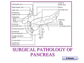
  • 2. SURGICAL PATHOLOGY OF PANCREAS – 4 hours • C 8 – Acute pancteatitis: etiology, phisiopathology, clinical findings, evaluation, diagnosis, differential diagnosis, complications(pancreatic pseudocyst), treatment. – Chronic pancteatitis: etiology, phisiopathology, clinical findings, evaluation, diagnosis, differential diagnosis, complications, treatment. • C 9 – Malignant tumors of the pancreas: etiology, classification, pathology, clinical findings, evaluation, diagnosis, complications, treatment. – Tumors of the Vater's ampulla: etiology, classification, pathology, clinical findings, evaluation, diagnosis, complications, treatment; – Tumors of the endocrine pancreas. 2
  • 3. 3 SURGICAL PATHOLOGY OF PANCREAS Penetrating Injuries • Penetrating injuries to the pancreas are usually diagnosed in the operating room. • The pancreas is located in the retro peritoneum, surrounded by other viscera and major vascular structures. • An isolated injury to the pancreas is unusual, major vascular injuries are seen in 40% to 50% of patients with penetrating pancreatic injuries, PANCREATIC TRAUMA Penetrating Injuries of Pancreas Pancreatic injury involving the ductal system Retroperitoneal location of pancreas
  • 4. 4 SURGICAL PATHOLOGY OF PANCREAS • Patients with penetrating pancreatic trauma have obvious indications for abdominal exploration. • Preoperative serum amylase concentrations are not helpful; (elevated in 30% of patients with penetrating pancreatic injuries). Abdominal exploration, • Signs of pancreatic injury include: – knife or a projectile path that passes in proximity to the pancreas, – a central hematoma in the upper abdomen, and – injuries to the duodenum, vena cava, suprarenal aorta, or mesenteric vessels. • In all these instances, the pancreas should be thoroughly explored: • The anterior surface of the pancreas is visualized by entry into the lesser sac of the peritoneal cavity by division of the gastrocolic ligament in a relatively avascular area to the left of the midline. • The tail of the pancreas can be more fully visualized, especially in its posterior aspect, by mobilization of the spleen and the tail of the pancreas as a unit. • The posterior aspect of the body of the pancreas is visualized by development of the avascular area at the inferior margin of the body and tail of the pancreas with a combination of sharp and blunt dissection. • The posterior aspect of the head of the pancreas can be exposed by an extensive Kocher maneuver. In combination with entry into the lesser sac, this also allows for bimanual palpation of the pancreatic head, with one hand placed on the anterior surface of the pancreas through the hole in the lesser sac and the other hand placed behind the pancreas in the plane developed by the Kocher maneuver. PANCREATIC TRAUMA Penetrating Injuries of Pancreas
  • 5. 5 SURGICAL PATHOLOGY OF PANCREAS • In the evaluation of penetrating pancreatic injuries, the key to operative management is the determination of whether a ductal injury is present. – Transduodenal intraoperative pancreatography has been recommended, but the use of this technique in the identification of ductal injuries is controversial or dangerous. – In certain circumstances, the use of intraoperative ERCP (endoscopic retrograde cholangiopancreatography) eliminates this problem. • Penetrating pancreatic injuries can be classified according to both location and severity. • With respect to location, injuries can be subdivided into those of the – head, – body, and – tail of the pancreas. • With respect to severity, classification systems must be satisfactory to research applications but also in the determination of the best treatment. Classification – Class I injuries are simple contusions of the pancreas; – Class II injuries are lacerations of the parenchyma in the body or tail of the pancreas; – Class III injuries are those with severe disruption of the head or body; – Class IV injuries are those in which there is an associated injury to the duodenum. PANCREATIC TRAUMA Penetrating Injuries of Pancreas
  • 6. 6 SURGICAL PATHOLOGY OF PANCREAS • Class I injuries should be usually observed or simply drained externally. The type of drain used after pancreatic injury is probably not important as long as adequate drainage is effected. This can be accomplished with passive, sump, or closed suction systems. • If drains are used, they should be left in place for at least 5 to 7 days to ensure that a drain tract develops. The timing of drain removal should be based on both the amount and character of the pancreatic drainage: – Drain outputs in excess of 150 to 200 mL/day are suggestive of pancreatic fistula. • The morbidity rate for patients with undrained pancreatic secretions is much greater than that for those with drained pancreatic secretions. • Class II injuries The treatment depends on the presence or absence of a ductal injury, a determination that can be difficult to make. • The argument for intraoperative pancreatography is made for class II injuries. • The presence of a ductal injury in the head of the pancreas does not usually make a difference with respect to treatment, because most of these injuries should be drained regardless. • If a ductal injury is present in the body or tail of the pancreas, the appropriate treatment is resection of the distal pancreas, whereas if no ductal injury is present, simple drainage is adequate. • the difficulty lies in determining whether injuries to the body or tail of the pancreas do in fact include ductal injury. • Intraoperative pancreatography is helpful, regardless of the technique employed. • Class III injuries of the body or tail of the pancreas, should be treated with a distal pancreatectomy. Distal resection can include up to 80% of the gland if necessary. PANCREATIC TRAUMA Penetrating Injuries of Pancreas
  • 7. 7 SURGICAL PATHOLOGY OF PANCREAS • Class III injuries of the head of the pancreas should be drained. Resection of these injuries requires internal drainage, near-total pancreatectomy, or pancreaticoduodenectomy. If the patient develops a pancreatic fistula, the fistula can be controlled by the drains. If the fistula does not resolve with time, the pancreas can be drained internally at a later date. • Class IV injuries of the pancreas involve injuries to the duodenum as well as the pancreas. – If the injuries to the duodenum and pancreas are simple, the duodenum can be repaired primarily, and the pancreas can be drained, or a distal resection can be carried out if the pancreatic injury is in the body or tail. – For more complicated, combined injuries, pyloric exclusion can be done to minimize pancreatic stimulation and protect the duodenal repair (see Duodenum). – For massive injuries to the duodenum and head of the pancreas, pancreaticoduodenectomy with reconstruction should be reserved for cases in which débridement of devitalized tissue results in a de facto removal of the duodenum and head of the pancreas. – Penetrating injuries to the ampulla of Vater may also require formal pancreaticoduodenectomy. • Internal drainage of the pancreas has been suggested as a means of treating ductal injuries without the need for resection of viable and functional pancreatic tissue. • Distal pancreatectomy for traumatic injuries should be performed only after the pancreas has been thoroughly mobilized and exposed. • It is possible to perform a distal pancreatectomy without a concomitant splenectomy. This adds to operative time and can increase the risk of bleeding, particularly if there is an associated injury to the spleen treated with splenorrhaphy. • Splenic salvage should be attempted, therefore, only in hemodynamically stable patients with minimal or no associated intraabdominal or extraabdominal injuries. PANCREATIC TRAUMA Penetrating Injuries of Pancreas
  • 8. 8 SURGICAL PATHOLOGY OF PANCREAS PANCREATIC TRAUMA Penetrating Injuries of Pancreas Internal drainage of a pancreatic injury involving the ductal system with the creation of a Roux-en-Y jejunoileal limb and construction of a pancreaticojejunal anastomosis.
  • 9. 9 SURGICAL PATHOLOGY OF PANCREAS Complications of pancreatic injuries: • Pancreatic fistulas, after trauma are characterized by persistent drainage of pancreatic enzymes and secretions from the pancreatic injury for a number of weeks after injury. Most of these fistulas close spontaneously, especially if there is no proximal obstruction of the pancreatic ductal system. In the rare instances in which fistulas fail to close spontaneously, surgical intervention may be required. • Pseudocysts, that develop after pancreatic trauma often resolve on their own or with percutaneous aspiration. If, after 4 to 6 weeks of observation with serial ultrasound or CT scanning, the pseudocyst does not show signs of resolution, it should be drained internally with a defunctionalized limb of jejunum. • Bleeding in the area of the pancreatic bed, is usually an early complication that is caused by inadequate drainage with resultant autodigestion of the pancreas and surrounding tissue. Bleeding can be avoided by identifying all injuries at the time of exploration and ensuring adequate drainage. If massive bleeding does occur, it should be dealt with through operative intervention. • Pancreatitis, another complication of pancreatic injury, is also related to inadequate drainage of pancreatic secretions. Treatment consists of provision of adequate drainage and supportive care. If the pancreatitis is localized to the distal pancreas and a trial of conservative management fails, distal pancreatectomy should be considered. PANCREATIC TRAUMA Penetrating Injuries of Pancreas
  • 10. 10 SURGICAL PATHOLOGY OF PANCREAS Blunt Injuries • The major difference between penetrating and blunt injuries of the pancreas concerns the diagnosis. • Penetrating injuries are usually discovered on abdominal exploration for associated injuries, but blunt injuries may occur in isolation, and the preoperative diagnosis can be difficult. • Blunt pancreatic injuries are relatively rare, which increases the difficulty of diagnosis. So delays in diagnosis of blunt injuries ranged up to several days, which induce an increased morbidity. • The body of the pancreas lies directly anterior to the vertebral column and is vulnerable to crush injuries when the anterior abdominal wall is forceably compressed, as can occur from a seat belt or a sharp blow to the epigastrium. In such instances, the pancreas may be the only intraabdominal organ injured. • A number of different means are available to make the diagnosis of blunt pancreatic injury: – Physical examination of the abdomen is useful, but because of the retroperitoneal location of the pancreas, the results can be misleadingly benign until a number of hours after injury. – This emphasizes the importance of serial examinations. – In most cases, the abdomen becomes progressively more tender to palpation during the first 24 to 48 hours after injury, and the need for abdominal exploration becomes more obvious. – The physical examination of the abdomen is much less reliable in young children and in patients with head injuries. • The serum amylase concentration is elevated on admission in about 70% of patients with blunt pancreatic injury. • However, elevated serum amylase has a poor positive predictive value and also occurs in many patients without pancreatic injury. The amylase concentration can be elevated because of trauma to other organs, including the salivary glands and the ovaries. • DPL(diagnostic peritoneal lavage) is of little help in the early diagnosis of pancreatic injury unless there have been associated intraperitoneal injuries. The retroperitoneal location of the pancreas results in minimal findings in the lavage fluid, and obtaining amylase concentrations in the lavage fluid is not helpful. PANCREATIC TRAUMA Blunt Injuries of Pancreas
  • 11. 11 SURGICAL PATHOLOGY OF PANCREAS • CT scan of the abdomen allows for visualization of the retroperitoneum, including the pancreas. In the case of isolated injury to the pancreas, the sensitivity of the CT scan is at its lowest shortly after injury. Although it may be a good test for the diagnosis of pancreatic injury after a number of hours have passed, immediate CT of the abdomen will miss some pancreatic injuries, particularly if expert interpretation is not available. • Finally, ERCP is a means of diagnosing pancreatic injury. ERCP is an attractive diagnostic method because it is less invasive than abdominal exploration and also provides information about the status of the ductal system, but there are several practical disadvantages of the technique. – Most of the studies that have reported successful use of ERCP have involved stable patients studied hours to days after injury and sent to a referral center specifically because of suspicion of a pancreatic injury. – These patients are a selected group, quite different from patients who are freshly injured. – ERCP is not universally available and, even in large centers, is often unavailable at the odd hours necessary for early diagnosis in acutely injured patients. – Many endoscopists are fearful of inducing an exacerbation of pancreatitis in patients with mild pancreatic injuries lacking ductal involvement. • To summarize, • the early diagnosis of blunt pancreatic injuries, particularly if they occur in isolation, can be extremely difficult, and • no single test allows for an easy and reliable diagnosis. • A combination of serial abdominal examinations and serum amylase determinations, CT scan, and ERCP in selected patients is the best diagnostic strategy available. • These studies should be combined, with a low threshold for operative intervention if a pancreatic injury is suspected. PANCREATIC TRAUMA Blunt Injuries of Pancreas
  • 12. 12 SURGICAL PATHOLOGY OF PANCREAS • Basic principles of exposure and operative management of blunt injuries of the pancreas are the same as for penetrating injuries. • In many instances of severe injury, the pancreas has already been transected by the trauma, making the pancreatic resection somewhat simpler to carry out. • Isolated injuries of the pancreas from blunt trauma also lend themselves to distal pancreatectomy with splenic preservation. • As in penetrating injury, splenic salvage should be attempted only in stable patients without associated splenic rupture or severe associated intraabdominal or extraabdominal injuries. • Complications of pancreatic injury are similar to those outlined for penetrating injuries. PANCREATIC TRAUMA Blunt Injuries of Pancreas
  • 13. 13 SURGICAL PATHOLOGY OF PANCREAS DEFINITION • Acute pancreatitis is a complex disorder of the exocrine pancreas characterized by acute acinar cell injury and both regional and systemic inflammatory responses. • It is a common disease with a broad spectrum of clinical and pathologic findings that contribute to considerable morbidity and mortality. • Because pathogenic mechanisms are unclear, specific treatment is not available. Empiric supportive care remains standard, and clinical outcomes have improved only to the extent that critical care has evolved in recent years. Most patients with acute pancreatitis have simple edematous pancreatitis, a self-limited and reversible process. In a small number of patients, fulminant or progressive disease develops, with pancreatic necrosis that can lead to multiorgan system failure or death. ACUTE PANCREATITIS The problems related to acute pancreatitis pose a formidable challenge to both the clinical surgeon and the basic scientist.
  • 14. 14 SURGICAL PATHOLOGY OF PANCREAS PATHOLOGY ACUTE PANCREATITIS Normal acinar cell ultrastructure. Cytoplasmic processing of the proenzymes is depicted, with apical discharge into the acinar ductule by means of zymogen granule exocytosis.
  • 15. 15 SURGICAL PATHOLOGY OF PANCREAS PATHOLOGY • Acute pancreatitis is characterized by alterations in acinar cell structure and function as well as by the development of acute regional and systemic inflammatory responses. • The fundamental pathologic event is injury to the acinar cell. • Acute pancreatitis is characterized by: – Interstitial edema formation. – Grossly, the gland becomes enlarged and edematous, with small areas of focal necrosis involving either the pancreas or areas of adjacent retroperitoneal fat. – Microscopically, edema is both interlobular and intralobular, and the acinar units appear dispersed within the relatively sparse fibrous matrix. ACUTE PANCREATITIS The histologic features of acute edematous pancreatitis. There is extensive edema formation, acinar cell cytoplasm vacuolization, and polymorphonuclear cell (PMN) infiltration.
  • 16. 16 SURGICAL PATHOLOGY OF PANCREAS • Acute inflammation occurs rapidly, experimentally, the process begins within minutes. • The initial cellular response involves the infiltration of polymorphonuclear leukocytes into the perivascular regions of the pancreas. • Within hours, mononuclear cells, including macrophages and lymphocytes, accumulate. • Experimental evidence suggests that phagocyte-derived oxygen radicals and possibly other phagocytic products are involved in a primary injury to pancreatic capillary endothelial cells. • The resulting increase in microvascular permeability facilitates access to the acinar cell microenvironment for circulating formed elements (additional neutrophils, monocytes, platelets) and humoral factors, such as complement products and cytokines. – The relative importance of this inflammatory injury to the pancreatic microvasculature is unclear, but it provides one explanation for the process of local edema formation as well as for the systemic microvascular sequelae of acute pancreatitis. • Although clinical – Edematous pancreatitis usually is a reversible disease characterized by acinar cell injury and edema formation, – frank pancreatic necrosis develops in 5% to 10% of patients, which can lead to irreversible regional injury or multiorgan system failure. Predictably, this group of patients is the primary source of morbidity and mortality associated with acute pancreatitis. • Histologic characteristics of advanced disease include – extensive acinar cell necrosis, – interstitial microabscess formation, – extensive peripancreatic fat necrosis, – microvascular thrombosis, and – local hemorrhage. • Pathologically, all these features appear to represent progression of processes already established with acute edematous pancreatitis. ACUTE PANCREATITIS
  • 17. 17 SURGICAL PATHOLOGY OF PANCREAS PATHOPHISIOLOGY • The cellular events that lead to acute pancreatitis may be initiated by a variety of different stimuli, and the process has been considered a final common pathway. Current data offer considerably more insight into the relevant pathogenic events than the simple historic concept that pancreatic autolysis occurs. • Acinar cell proteases (such as trypsin, chymotrypsin, carboxypeptidase, and elastase) and phospholipases are normally synthesized in an inactive zymogen form. • Peptide synthesis is accomplished and the proenzymes are packaged into cytoplasmic zymogen granules. • After apical exocytosis into the acinar ductal lumen, these precursors are transported with water and bicarbonate through the pancreatic duct into the duodenum, where they are converted enzymatically into active forms by enterokinase, a brush-border enzyme. • A variety of endogenous protease inhibitors (α1-antitrypsin, β 2-macroglobulin, and pancreatic secretory trypsin inhibitor) are normally found in pancreatic tissue and pancreatic secretions, and in plasma in quantities sufficient to protect against premature or inappropriate activation of these digestive enzymes. • The specific mechanisms that initiate human disease are not known, the normal orderly secretory sequence appears to be disrupted in acute pancreatitis. • Inappropriate protease activation overcomes endogenous antiprotease defenses. • In experimental acute pancreatitis, zymogen granules become localized with lysosomes and fuse to form autophagic cytoplasmic vacuoles (zymogen lakes). These vacuoles move preferentially to the basolateral acinar cell cytoplasm, rather than to the luminal apex. ACUTE PANCREATITIS
  • 18. 18 SURGICAL PATHOLOGY OF PANCREAS • Disordered discharge of the acinar cell contents through the basolateral cell membrane occurs. • The mechanisms by which this cytoplasmic colocalization of zymogen granules and lysosomes occurs are not known, • Although evidence suggests that cytoskeletal alterations are associated with the loss of normal acinar cell polarity and loss of the ability to achieve apical exocytosis. • Trypsinogen is normally a major constituent of the zymogen granules, whereas cathepsin B is usually abundant within the lysosomal fraction. Cathepsin B–induced trypsinogen cleavage is known to generate activated trypsin, which, in turn, is capable of further cytoplasmic proenzyme conversion. • The cytoplasmic liberation of activated proteases causes cell membrane injury, followed by the disordered discharge of acinar cell contents through the basolateral cell membranes. For example, intracellular trypsinogen cleavage has been demonstrated after either hypoxia- or acidosis-induced acinar cell injury. Collectively, these findings suggest that the outcome of acinar cell injury involves intracellular activation of endogenous proteases, leading to further injury and the local extracellular discharge of acinar cell contents. ACUTE PANCREATITIS Diagram illustrating the loss of acinar cell polarity and the process of cytoplasmic fusion of lysosomes and zymogen granules. Disordered basolateral discharge of activated proteases from the acinar cell follows.
  • 19. 19 SURGICAL PATHOLOGY OF PANCREAS • Once initiated, this process of protease release perpetuates acinar cell injury and initiates a regional acute inflammatory response, generating additional injury. • The endogenous inflammatory system participates early in the development of acute pancreatitis. • Both humoral and cellular factors / elements appear to be involved: – Complement activation, – Histamine release, and – Bradykinin generation are demonstrable. Studies have suggested a role for – Cytokines in the acute inflammatory process of pancreatitis. – Serum levels of tumor necrosis factor (TNF) α, interleukin-6 (IL-6), and IL-1 are elevated in animals with experimental pancreatitis and in patients with systemic complications of acute pancreatitis. Administration of an IL-1 receptor antagonist, even after the onset of acute pancreatitis, limited the degree of pancreatic inflammation. The administration of anti – TNF antibody has both beneficial and deleterious effects on the development of local inflammation and may reflect the different experimental models used. – Neutrophil-mediated pancreatic capillary endothelial injury appears to occur early, when the process is still reversible. – NADPH oxidase–dependent oxygen radicals are directly implicated, whereas other phagocyte products, such as elastase, collagenase, cathepsin G and D, phospholipases A2 and C, DNAase, RNAase, glycosidases and other lysosomal hydrolases, platelet-activating factor, and myeloperoxidase, are all potential participants in both the acinar and endothelial cell injury processes. – Chronic inflammatory cells, particularly macrophages, are recruited to the pancreas within hours and share many of the proinflammatory products noted earlier. The microvascular injury appears to amplify the inflammatory process ACUTE PANCREATITIS
  • 20. 20 SURGICAL PATHOLOGY OF PANCREAS ACUTE PANCREATITIS Schematic diagram illustrating the inflammatory response in acute pancreatitis. Inflammatory effector cells and plasma and tissue mediators are depicted. Increased microvascular permeability results from capillary endothelial cell injury.
  • 21. 21 SURGICAL PATHOLOGY OF PANCREAS • In addition to this localized pancreatic inflammation, evidence of a systemic response exists. • Considerable data implicate the inflammatory process in the pathogenesis of pancreatitis-induced multiorgan system failure. • The systemic distribution of activated neutrophils, mononuclear cells and macrophages, complement activation products, and other factors is clearly linked to remote organ dysfunction. • A common event appears to be microvascular endothelial cell injury in diverse target organs. • Pancreatitis-induced, polymorphonuclear leukocyte–dependent microvascular lung injury is one such example This pathologic process is a likely explanation for the frequent pulmonary symptoms and the occasional adult respiratory distress syndrome in patients with acute pancreatitis. • Other target organs at risk for acute pancreatitis-induced injury are the liver, kidneys, and heart, although the mechanisms involved are less clear. • Complications of acute pancreatitis : Early – Shock – Multiorgan failure – Encephalopathy – Coagulopathy – Sepsis – Hypocalcemia Late – Pseudocyst – Diabetes – Abscess ACUTE PANCREATITIS One consequence of the systemic inflammatory response associated with acute pancreatitis is neutrophil- dependent, oxygen radical–mediated alveolar capillary endothelial cell injury.
  • 22. 22 SURGICAL PATHOLOGY OF PANCREAS • Common clinical associations with acute pancreatitis include: • Ethanol ingestion acute pancreatitis – Acetaldehyde, the hepatic product of ethanol metabolism, is known to induce acinar cell cytoskeletal changes (microtubule disruption) as well as to increase acinar cell membrane permeability. – Ethanol-induced elevations in plasma triglycerides provide a potential source of cytotoxic free fatty – In addition, endogenous trypsin-inhibiting capacity in both the pancreas and in pancreatic duct fluid is diminished after ethanol exposure acids. – It appears that regional pancreatic blood flow is diminished after ethanol exposure, potentially adding ischemia to other acinar cell insults – The cytoskeletal disruption induced by acetaldehyde is consistent with the concept that the loss of acinar cell polarity and associated proteinprocessing abnormalities lead directly to ethanol-induced acute pancreatitis. – Potentially important indirect ethanol effects include reduction in the level of protection afforded by the endogenous antiprotease system and hypoxic stress induced by diminished pancreatic microvascular blood flow. • Pancreatic ductal obstruction • Obstruction of the ampulla of Vater associated with pancreatic duct hypertension or increased permeability may also lead to extravasation of acinar cell contents into the pancreatic interstitium. – In this circumstance, the initial source of proenzyme cleavage is unknown because luminal small bowel contents have no access to this compartment. – Neutrophil and macrophage products do provide a potential source of proteases for proenzyme cleavage and therefore represent a possible triggering mechanism. – Experimentally, acute pancreatitis is reliably induced by bile duct or duodenal occlusion or by retrograde injection of substances such as bile, duodenal aspirate, drugs (including ethanol), and other substances into the pancreatic duct under pressure. – Given the common clinical association of acute pancreatitis and gallstones or other occlusive lesions of the pancreatic duct, it is likely that these are relevant experimental observations. ACUTE PANCREATITIS
  • 23. 23 SURGICAL PATHOLOGY OF PANCREAS • The role of the microcirculation in the development of acute pancreatitis is poorly understood. – In addition to increased permeability, microvascular thrombosis or obstruction by leukoaggregates may lead to local or regional tissue hypoxia. – In nonacinar cell populations, it is well known that hypoxia-induced adenosine triphosphate depletion is associated with cytoskeletal (microtubule and microfilament) disruption. – A similar finding in acinar cells would be consistent with the pathogenic scheme summarized earlier. Diminished microvascular blood flow occasionally initiates the process of acute pancreatitis. – Clinically, hypoxic acinar cell injury is thought to be associated with events such as cardiopulmonary bypass, thromboembolic disease, and myocardial infarction. ACUTE PANCREATITIS Ethanol- and acetaldehyde-induced changes in the pancreas include acinar cell microtubule disruption, increased cell membrane permeability, diminished trypsin-inhibiting capacity, and reduced regional blood flow. Pancreatic duct hypertension or reflux through the ampulla of Vater may lead to increased permeability of the pancreatic duct with leakage of activated proteases into the pancreatic interstitium. The result is self-perpetuating acinar cell injury.
  • 24. 24 SURGICAL PATHOLOGY OF PANCREAS Incidence • The precise incidence of AP is difficult to determine. • Variations among populations are highly dependent on social factors such as ethanol use and on environmental and hereditary determinants such as the incidence of gallstones. Age • AP can occur at any age but is most common in adults between 30 and 70 years of age. • Patients with gallstone-induced pancreatitis are older (age 40 to 60 years), whereas those with alcohol-associated pancreatitis are younger (age 30 to 40 years). Sex ratio • The sex distribution of acute pancreatitis depends on the clinical cause of the disease: – women represents 68% of patients with gallstone-associated pancreatitis – when alcohol is the primary association, most patients are men. Mortality • The mortality rates associated with AP range from 6% to 20.5%. • Acute hemorrhagic or necrotizing pancreatitis is associated with mortality rates of 50% or more. Necrotizing pancreatitis occurs in 5% to 10% of patients in most series of acute pancreatitis. • More recent studies have reported lower mortality rates in AP, reflecting advances in critical care, nutritional support, and antibiotic therapy. ACUTE PANCREATITIS CLINICAL FEATURES
  • 25. 25 SURGICAL PATHOLOGY OF PANCREAS ACUTE PANCREATITIS CLINICAL FEATURES CLINICAL ASSOCIATIONS WITH ACUTE PANCREATITIS  BILIARY TRACT STONE DISEASE  ETHANOL  OTHER: • Trauma • Postprocedural • Postoperative • Post-ERCP • Direct • Mechanical (nongallstone) obstruction • Tumors of the pancreas, duodenum, or bile duct • Duodenal obstruction • Pancreas divisum • Infection • Hyperlipidemia • Hyperparathyroidism • Drugs • Steroids • Estrogen • Glucocorticoids • Diuretics • Furosemide • Thiazides • Ethacrynic acid • Diazoxide • Calcium • Coumadin • Cimetidine • Quinidine • Phenformin • Azothioprine • Mercuric chloride • Paracetamol • Sulfonamides • Tetracyclines • L-Asparaginase • Methyldopa • Clonidine • Pregnancy • Idiopathic
  • 26. 26 SURGICAL PATHOLOGY OF PANCREAS Etiology and Clinical Associations • Clinical associations with AP can be divided into three broad categories: – biliary stones, – ethanol, and – others • Biliary tract stone disease and ethanol-induced pancreatitis account for most cases of acute pancreatitis reported worldwide. • The high incidence of idiopathic and other causes in India and Hong Kong may be related to endemic infestation with Opisthorchis sinensis in these geographic areas. Biliary Tract Stone Disease • Because the intramural portion of the distal common bile duct is shared with the pancreatic duct pancreatic ductal hypertension may result when a gallstone obstructs the ampulla of Vater during passage into the duodenum, the classic common channel concept. • If transient obstruction results in edema formation and ampullary dysfunction, reflux of bile or duodenal contents into the pancreatic duct may then initiate acute pancreatitis. If gallstone passage is arrested within the ampulla, pancreatic ductal hypertension may additionally contribute. • Choledocholithiasis is not always demonstrable in cases of suspected biliary pancreatitis. ACUTE PANCREATITIS CLINICAL FEATURES The common channel concept
  • 27. 27 SURGICAL PATHOLOGY OF PANCREAS • In one review of 1450 patients with gallstone-associated pancreatitis shows the following aspects: – gallstone impaction at the ampulla of Vater was identified in only 2%. – Simple cholelithiasis was found in 72%, – choledocholithiasis in 20%, and – cholecystitis without apparent gallstones in 8%. • Presumably, passage of a gallstone before diagnostic evaluation is routine. When stools are carefully screened in patients with suspected biliary pancreatitis, gallstones can be demonstrated in 85% to 94% of the patients within 10 days of the onset of disease. Ethanol • Ethanol use is the most common cause of AP in the United States. • In addition to the pathophysiologic mechanisms proposed earlier, it is likely that genetic, dietary, and environmental factors contribute. • Pancreatic ductal hypertension is probable after ethanol ingestion and this may occur by several different mechanisms: – The production and precipitation of protein (stone protein) within the pancreatic duct ACUTE PANCREATITIS CLINICAL FEATURES Ethanol induced protein formation (stone protein) within the pancreatic ductules.
  • 28. 28 SURGICAL PATHOLOGY OF PANCREAS – Ethanol-induced increase in ampullary tension and therefore resistance – Ethanol-induced gastric acid secretion increases pancreatic secretion indirectly through acid mediated secretin release. • The combination of increased ampullary resistance and increased ductal flow results in pancreatic ductal hypertension and appears to enhance protease entry into the pancreatic interstitium. Others • A wide variety of other clinical conditions may be associated with AP Postprocedural Pancreatitis • Many surgical procedures in the upper abdomen are associated with postoperative AP. – After gastric resection the incidence of AP is between 0.6 -1.23%. – After biliary tract surgery, particularly after common bile duct exploration, AP occurs with an incidence of 0.5% to 3%. – Direct manipulation or retraction of the pancreas or pancreatic duct appears to be the most common cause. – After endoscopic retrograde cholangiopancreatography (ERCP), AP develops in about 1% of patients This is a predictable event, and the risk can be minimized by limiting the pressure used for contrast injection of the pancreatic duct. – After coronary artery bypass surgery AP also occurs Although pancreatitis in this circumstance is thought to result from ischemia. Abdominal Trauma • AP occurs in about 1% to 2% of patients with abdominal trauma, whether blunt or penetrating. Contusion of the pancreatic parenchyma and pancreatic ductal injury both lead to extravasation of activated enzymes to initiate the process. ACUTE PANCREATITIS CLINICAL FEATURES Ethanol-induced increases in ampullary resistance
  • 29. 29 SURGICAL PATHOLOGY OF PANCREAS Hyperlipoproteinemia • Rare causes of AP are the hyperlipoproteinemias, types I and V. The initial acinar cell injury is thought to result from the liberation of free fatty acids from circulating triglycerides by the local action of lipases within the pancreatic microcirculation. Physical disruption of the microvascular endothelium by cholesterol crystals may also occur. Hyperparathyroidism • AP is reported in 1% to 19% of patients with hyperparathyroidism. It is unclear whether increased circulating plasma levels of calcium or parathyroid hormone are primarily responsible. Hypercalcemia may be associated with the precipitation of calcium phosphate within the pancreatic duct as well as with pancreatic hypersecretion. Parathyroid hormone elevated levels may have a direct cytotoxic effect on acinar cells. Drugs • Drug-induced acute pancreatitis is relatively common and has been reported in association with many pharmacologic agents. The mechanisms involved are largely unknown. Infections • The development of acute pancreatitis has been reported after a variety of bacterial, fungal, parasitic, and viral infections. Postulated mechanisms include direct cytotoxic effects, coexisting immunosuppression, and alterations in pancreatic blood flow. Vascular Disease • Impaired pancreatic blood flow arising from either anatomic lesions or functional events can induce acute pancreatitis. IE: embolization of the pancreaticoduodenal artery after translumbar aortography, celiac artery stenosis, ruptured abdominal aortic aneurysm, and myocardial infarction. Immunologic Factors • AP has been associated with systemic lupus erythematosus, rheumatoid arthritis, polyarteritis nodosa, and Be87het syndrome. Systemic vasculitis, the associated glucocorticoid use, and the presence of circulating antibodies to acinar cells are potentially related findings. ACUTE PANCREATITIS CLINICAL FEATURES
  • 30. 30 SURGICAL PATHOLOGY OF PANCREAS Obstruction of the Duodenum or the Pancreatic Duct • Intrapancreatic or duodenal tumors, parasitic obstruction of the pancreatic duct, other obstructions of the duodenum, and pancreas divisum are also associated with the development of pancreatic duct hypertension and AP. Pregnancy • Acute pancreatitis has been linked to pregnancy with an incidence of 0.01% to 0.1%. Because most reports include patients with gallstones, ethanol use, or other risk factors, it is unclear whether pregnancy is an independent risk factor. Presentation • The cardinal clinical symptom for acute pancreatitis is epigastric pain that is visceral in character and radiates into the back. • The degree of discomfort is variable. The pain may be similar to that of acute peritonitis and may mimic a perforated viscus. At the other extreme, pain may be minimal. Other common signs and symptoms are nonspecific. ACUTE PANCREATITIS CLINICAL FEATURES COMMON SIGNS AND SYMPTOMS OF UNCOMPLICATED ACUTE EDEMATOUS PANCREATITIS Sign or Symptom Frequency (%) Abdominal pain 85–100 Nausea and vomiting 54–92 Anorexia 83 Fever 12–80 Abdominal mass 6–20 Ileus 50–80
  • 31. 31 SURGICAL PATHOLOGY OF PANCREAS • Clinical signs of complex disease or necrotizing pancreatitis include – jaundice – hypotension – retroperitoneal hemorrhage associated with pancreatic necrosis may become apparent as blood dissects into the subcutaneous tissues, producing blue discoloration of the flanks (Grey Turner sign), the umbilicus (Cullen sign), or the inguinal ligament (Fox sign). None of these signs is common. • The clinical course of patients with acute pancreatitis is exceedingly variable. – The spectrum includes patients with minimal symptoms, few signs, and only mild hyperamylasemia who require little or no treatment and have few, if any, complications. – The opposite end of the spectrum, without apparent differentiating risk factors, is fulminant pancreatic necrosis with hemorrhage and progressive, inexorable multisystem organ failure and death. • Several grading systems have been developed, to estimate risks and outcomes based on the presenting clinical features, and the Ranson criteria are widely adopted. • A patient with three or more of these findings has a mortality rate of about 30%. • Pancreatitis-associated morbidity and mortality are directly correlated with the number of these criteria present. • The mortality rate is 28% if three criteria are met, 40% for five or six, and 100% for seven or eight. • The APACHE II (Acute Physiology Score and Chronic Health Evaluation) scoring system for critically ill patients has also been applied. • Although not specific for acute pancreatitis, the APACHE score has some utility by predicting mortality rates in critically ill patients. ACUTE PANCREATITIS CLINICAL FEATURES
  • 32. 32 SURGICAL PATHOLOGY OF PANCREAS On Admission • Age above 55 years • White blood cell count above 16,000/E6L • Glucose level above 200 mg/dL • Lactase dehydrogenase level above 350 IU/L • Serum glutamic-oxaloacetic transaminase value above 250 IU/L After 48 Hours • Hematocrit decrease of 10% • Blood urea nitrogen level increase of 5 mg/dL • Ca2+ level below 8 mg/dL • PaO2 level below 60 mmHg • Base deficit value above 4 mEq/L • Fluid sequestration greater than 6 L ACUTE PANCREATITIS CLINICAL FEATURES RANSON GRAVE PROGNOSTIC SIGNS ASSOCIATED WITH ACUTE PANCREATITIS
  • 33. 33 SURGICAL PATHOLOGY OF PANCREAS DIAGNOSIS • The diagnosis of AP ultimately depends on a clinical judgment, based on the finding of epigastric abdominal pain and tenderness with the laboratory finding of hyperamylasemia. • No single laboratory or physical finding is pathognomonic. Imaging • Plain abdominal roentgenograms may show : – A diffuse ileus and – A solitary left upper abdominal sentinel loop are classic and are often seen on, – The psoas muscle margins may be obscured by retroperitoneal edema; – Pancreatic ascites may be apparent; – Pancreatic calcifications imply preexisting chronic disease. • Chest radiographs abnormalities are present at one third of patients with AP at the time of diagnosis. – Segmental atelectasis, – An elevated hemidiaphragm, – Pleural effusions, or the presence of – Early pulmonary parenchymal infiltrates. • Barium studies of the gastrointestinal tract An upper gastrointestinal contrast study often demonstrates – narrowing or spasm of the duodenum, with – widening of the C loop secondary to pancreatic inflammation and edema formation. • Ultrasound scanning provides a readily available, rapid, and noninvasive imaging of the pancreas. – In the case of simple acute pancreatitis, an enlarged, echolucent gland is typically seen. – Ultrasound examination yields information regarding cholelithiasis, choledocholithiasis, and the status of the intrahepatic and extrahepatic biliary ducts. – The technique is also valuable for assessing and sequentially evaluating peripancreatic fluid collections or pancreatic pseudocysts. ACUTE PANCREATITIS CLINICAL FEATURES
  • 34. 34 SURGICAL PATHOLOGY OF PANCREAS • Computed tomography (CT) has similar capabilities to ultrasound, although sensitivity for detecting cholelithiasis is lower. Intraluminal contrast in the duodenum and small bowel is necessary to optimize CT imaging. Dynamic CT scanning with simultaneous intravenous contrast enhancement can give valuable information in evaluating regional pancreatic perfusion and may provide an estimate of the extent of pancreatic necrosis. CT may show gas bubbles in the pancreatic necrosis or peri pancreatic fluid collections assessing the infection though the indication for surgical approach • Needle aspiration either CT- or ultrasound-guided, allows access to peri pancreatic or pancreatic fluid collections for diagnostic sampling and/or therapeutic drainage. Cultures may be obtained by this route. Contrast injections can assess the relation of a pseudo cyst or abscess cavity to the pancreatic duct. This latter information is important for any preoperative planning. Placement of therapeutic drainage catheters is routinely combined with CT- and ultrasound-directed imaging procedures. • ERCP The role in the diagnostic evaluation of the patient with AP is severely limited because of the high risk of exacerbating existing inflammation. ERCP is generally reserved for the evaluation of a patient with a suspected obstructive lesion and is timed to follow resolution of the acute phase of the illness. In addition, it is appropriate to obtain ERCP in a patient with idiopathic acute pancreatitis after a first recurrence of the disease. This strategy is designed to identify promptly anatomically correctable causes of acute pancreatitis. ERCP is also useful to delineate the pancreatic duct after injury, pseudo cyst drainage, or the development of pancreatic ascites. Lastly, ERCP combined with urgent therapeutic sphincterotomy and gallstone extraction has been used for patients with impacted ampullary gallstones. ACUTE PANCREATITIS CLINICAL FEATURES
  • 35. 35 SURGICAL PATHOLOGY OF PANCREAS Biochemical Markers • Amylase. Amylase is released from the acinar cell into the pancreatic microcirculation in conjunction with the pathophysiologic events described earlier. • The laboratory finding of hyperamylasemia in a patient with clinical signs and symptoms of AP is the usual means of confirming the diagnosis of AP. • Efforts to correlate the degree of hyperamylasemia with disease severity or prognosis have been consistently unsuccessful, and Ranson criteria are notable for the absence of serum amylase levels. An important reason for this relates to: – the relatively rapid clearance of amylase from plasma, – the half-life being about 130 minutes. – Pancreatitis resulting from a discrete event such as transient pancreatic duct obstruction with gallstone passage is characterized by a single serum amylase peak with a rapid rise and prompt clearance, both measured in terms of hours. • A normal or minimally elevated serum amylase level may also be found in a patient with necrotizing pancreatitis or with CP; in these instances, complete or nearly complete destruction of the acinar cell population may have occurred, reducing the plasma amylase level. • Additionally, a number of nonpancreatic sources of amylase exist, so that hyperamylasemia may result from other pathology: – Salivary glands, – fallopian tubes, and – the small bowel are important alternative amylase sources. ACUTE PANCREATITIS CLINICAL FEATURES
  • 36. 36 SURGICAL PATHOLOGY OF PANCREAS Clinical conditions associated with hyperamylasemia include the following: ACUTE PANCREATITIS CLINICAL FEATURES • Salivary gland injury • Burns • Cerebral trauma • Multiple trauma • Diabetic ketoacidosis • Macroamylasemia • Renal transplantation • Renal dysfunction • Pneumonia • Pregnancy • Fallopian tube pathology • Drugs • Afferent loop syndrome • Acute appendicitis • Dissecting aortic aneurysm • Small bowel injury • Perforated ulcer • Small bowel obstruction • Mesenteric infarction
  • 37. 37 SURGICAL PATHOLOGY OF PANCREAS • In the case of salivary gland disease, plasma amylase isoenzyme determinations differentiate the source. An accurate and more rapid amylase assay using a monoclonal antibody specific to salivary isoamylase has been described. • Gastrointestinal tract pathology other than pancreatitis may lead to increased amylase absorption through the intestine or peritoneum and relatively mild elevations of pancreatic amylase in the circulation. • Lipase. Lipase is derived primarily from pancreatic acinar cells, and its elevation is also taken as evidence of acinar cell injury in acute pancreatitis. Lipase is also nonspecific and has not proved more useful than serum amylase determinations in clinical use. • Other Serum Enzymes. Other acinar cell products, such as immunoreactive trypsin, chymotrypsin, elastase, ribonuclease, and phospholipase A2, may be detectable in plasma after the onset of acute pancreatitis. Measurement techniques are not in wide clinical use, but institutional enthusiasm for one particular assay or another has led to an abundant literature. • Ribonuclease and phospholipase A2 plasma elevations may correlate with more complex disease. • Methemalbumin. Methemalbumin results from the proteolytic conversion of hemoglobin into oxidized hematin that is conjugated with plasma albumin. Acinar cell protease release into the circulation increases red blood cell exposure to proteases and accelerates this process, so that methemalbumin plasma levels may be elevated with acute pancreatitis. A correlation between levels of methemalbumin and severity of pancreatic disease is proposed but unproved. • Other Serum Abnormalities. Other characteristic but nonspecific biochemical features commonly associated with acute pancreatitis are summarized in the following Table These may have both therapeutic and diagnostic relevance. ACUTE PANCREATITIS CLINICAL FEATURES
  • 38. 38 SURGICAL PATHOLOGY OF PANCREAS Other characteristic but nonspecific biochemical features commonly associated with acute pancreatitis ACUTE PANCREATITIS CLINICAL FEATURES INCREASED Hematocrit, hemoglobin (hemoconcentration) White blood cell count Blood urea nitrogen Creatinine Bilirubin Lipid, triglyceride levels Glucose Alkaline phosphatase SGOT, SGPT DECREASED Hematocrit, hemoglobin (hemorrhage) Calcium Magnesium Pao2 OTHER Respiratory alkalosis (early) Metabolic alkalosis (early) Consumptive coagulopathy Metabolic acidosis (late) Respiratory acidosis (late)
  • 39. 39 SURGICAL PATHOLOGY OF PANCREAS DEFINITION • Acute pancreatitis is a complex disorder of the exocrine pancreas characterized by acute acinar cell injury and both regional and systemic inflammatory responses. • It is a common disease with a broad spectrum of clinical and pathologic findings that contribute to considerable morbidity and mortality. • Because pathogenic mechanisms are unclear, specific treatment is not available. Empiric supportive care remains standard, and clinical outcomes have improved only to the extent that critical care has evolved in recent years. Most patients with acute pancreatitis have simple edematous pancreatitis, a self-limited and reversible process. In a small number of patients, fulminant or progressive disease develops, with pancreatic necrosis that can lead to multiorgan system failure or death. ACUTE PANCREATITIS The problems related to acute pancreatitis pose a formidable challenge to both the clinical surgeon and the basic scientist.
  • 40. 40 SURGICAL PATHOLOGY OF PANCREAS MANAGEMENT • Neither medical nor surgical treatment strategies provide specific therapy for the acinar cell injury characteristic of acute pancreatitis. • Modern approaches provide general supportive care in the form of: – appropriate resuscitation, – nutrition, and – ventilation in the expectation that the cellular pathophysiologic processes will be self-limited. Medical Treatment • Conventional medical therapy for AP consists fundamentally of: – intravenous fluid resuscitation, – nasogastric decompression, and – monitoring of hematocrit, electrolytes, and blood gases. • All are empiric and are not known to shorten or favorably alter the course of the disease. • Regional retroperitoneal inflammation and the systemic microvascular injury contribute to the loss of intravascular plasma volume. • Hypovolemia may be mild or profound to the point of shock. • A relation exists between the magnitude of extravascular fluid sequestration and the severity of the pancreatitis. • This is recognized by inclusion of an estimated plasma volume deficit exceeding 6 L in the Ranson scoring system (see earlier list) as a grave prognostic sign. • More than half of patients with acute pancreatitis have clinical evidence of inadequate end-organ perfusion. • Resuscitation requires the intravenous administration of large volumes of isotonic crystalloid solution and the aggressive use of invasive hemodynamic monitoring devices. ACUTE PANCREATITIS MANAGEMENT
  • 41. 41 SURGICAL PATHOLOGY OF PANCREAS Pulmonary dysfunction occurs in about two thirds of patients with AP. • More than 70% of patients have evidence of transient hypoxemia, defined as a single PaO2 of less than 70 mmHg. • Progression to acute respiratory failure requiring endotracheal intubation and mechanical ventilation carries a mortality rate of up to 75% but is a relatively uncommon event (about 5% of all cases). • Monitoring of pulmonary gas exchange with periodic arterial blood gases is standard in all patients with AP. Renal dysfunction is demonstrable in 40% to 80% of patients with acute pancreatitis. Therefore, renal function must be sequentially evaluated biochemically, and the urine output must be carefully monitored during the acute resuscitative phase of the illness. • Early recognition of oliguria or azotemia allows correction. • Hypovolemia is almost invariably the cause of the renal dysfunction and is therefore simply treated if recognized. • The reported incidence of acute renal failure secondary to acute pancreatitis ranges from 2% to 20%. If required, dialysis, hemofiltration, and similar supportive measures are all associated with substantially increased mortality rates (up to 80%). Patients with AP share many hemodynamic and metabolic characteristics with SEPTIC patients. – Glucose production is exaggerated, – Insulin resistance increased – Pancreatic endocrine insufficiency may develop, – Protein catabolism is marked. ACUTE PANCREATITIS MANAGEMENT
  • 42. 42 SURGICAL PATHOLOGY OF PANCREAS – Nitrogen losses of as much as 40 g/dl have been measured in patients with acute pancreatitis. – Nutritional support is a necessary feature of their care. Nutritional support To be initiated immediately after the acute resuscitation phase because of the unpredictable return of intestinal function and the extraordinary metabolic requirements. • Considerable debate has surrounded the selection of a nutritional route. • The choice of route is less important than the need to provide adequate calories and to establish positive nitrogen balance. Antibiotic therapy is reserved for specific infectious complications such as pneumonia or pancreatic abscess. • Antibiotic use for simple acute edematous pancreatitis IS CONTRAINDICATED. Prospective randomized studies in patients with simple acute pancreatitis using ampicillin, lincomycin, and cephalothin have shown neither improvement in the clinical course nor a reduced likelihood of septic complications. Specific metabolic complications such as hypokalemia, hypocalcemia, hemorrhage, and consumptive coagulopathy are treated with appropriate replacement products, such as potassium chloride, intravenous calcium gluconate or chloride, red blood cells, and fresh-frozen plasma. • Hyperglycemia and glycosuria are the manifestations of altered carbohydrate metabolism in these patients. Hyperglycemia occurs in about 10% of patients and is generally a transient phenomenon. Permanent residual diabetes mellitus is much less frequent, occurring in fewer than 2% of patients. • Treatment for the acute illness consists of the carefully titrated administration of exogenous glucose and insulin to maintain a euglycemic state. ACUTE PANCREATITIS MANAGEMENT
  • 43. 43 SURGICAL PATHOLOGY OF PANCREAS Encephalopathy Clinical evidence of encephalopathy is discernible in 4% to 20% of patients with AP. • Symptoms may include: – disorientation, – confusion, – delirium, – delusions, or – hallucinations. – transient acute psychosis is also reported. Because ethanol ingestion also produce all these symptoms, it is often difficult to distinguish the underlying cause. • Cerebral edema, hemorrhage, and focal necrosis, presumably secondary to microvascular blood flow alterations, have all been associated with AP. • Treatment is nonspecific and supportive. Disorders of the coagulation system, particularly microvascular thrombosis and disseminated intravascular coagulopathy, are common in acute pancreatitis. • At later times (6 or 7 days), clinical studies have shown hyperfibrinogenemia and increased platelet counts, suggesting enhanced thrombotic potential. • Heparin(low-molecular-weight), dextran, and fibrinolytic therapy have been shown to prevent microvascular thrombosis in experimental acute pancreatitis in dogs. In this favorable and controlled setting, these agents prevent the development of acute hemorrhagic pancreatitis. Hypocalcemia is relatively common (3% to 30%) in AP. • The magnitude of the hypocalcemia correlates with the degree of illness. • It appears that large quantities of calcium are bound in the tissues during the process of peripancreatic fat saponification. Other factors, such as changes in plasma levels of parathyroid hormone, glucagon, and calcitonin, may also contribute. Unattended, the hypocalcemia may progress to tetany. Periodic monitoring of serum calcium is standard practice during the acute illness. Treatment consists of intravenous replacement to maintain the serum calcium level within a normal range. ACUTE PANCREATITIS MANAGEMENT
  • 44. 44 SURGICAL PATHOLOGY OF PANCREAS A variety of pharmacologic agents that directly or indirectly reduce acinar cell enzyme release or ductal secretion have undergone clinical evaluation for the treatment of acute pancreatitis, generally with unimpressive results. • Among the first were anticholinergic drugs, pancreatic anti enzymes, a somatostatin analogue. • Despite the theoretic appeal, it has not been possible to demonstrate that pancreatic anti enzymes and somatostatin alters the natural history or prognosis of simple acute pancreatitis, although it diminishes pancreatic secretion. Surgical Therapy Surgical therapy for AP is reserved for specific complications and for those situations in which a correctable anatomic cause can be identified. Endoscopic sphincterotomy is usually the initial therapeutic procedure for relief of biliary ductal obstruction when acute pancreatitis occurs in association with choledocholithiasis. • The procedure reliably decompresses the ampulla of Vater, with overall clinical success for gallstone disimpaction or passage in 90% of patients. • ERCP has greatly reduced the need for operative procedures designed to either divert or open and explore the common bile duct. • In the more usual circumstance, a patient who has simple cholelithiasis and an episode of acute pancreatitis is treated nonoperatively(ERCP) with resolution of the AP but gallbladder litiasis must be operated. Cholecystectomy is often performed after the resolution of acute pancreatitis but before hospital discharge. ACUTE PANCREATITIS MANAGEMENT
  • 45. 45 SURGICAL PATHOLOGY OF PANCREAS Surgery of Anatomically correctable lesions that can cause acute pancreatitis include: – pancreas divisum, – choledochal cysts (particularly a type III cyst or choledochocele), and – pancreatic duct obstruction related to tumor, stricture, or injury. • In the obstructive category, many patients have CP or recurrent acute pancreatitis. • Surgical therapy is directed at achieving adequate pancreatic duct drainage, usually by diversion into the jejunum for benign obstructions, or tumor resection in malignancies if appropriate. • Symptomatic pancreas divisum is best treated with operative transduodenal sphincteroplasty, • The type III choledochal cyst should be marsupialized into the duodenum. • Acute pancreatitis may be clinically indistinguishable at presentation from an acute abdomen related to other pathology : – Perforated duodenal ulcer, – Acute appendicitis, – Ruptured abdominal aortic aneurysms are sources of erroneous diagnoses. • Virtually every experienced surgeon has performed an exploratory laparotomy for clinical evidence of peritonitis only to find simple acute edematous pancreatitis. In this situation, recognition of the correct diagnosis is crucial. It is important to avoid biopsy, resection, or other nontherapeutic procedures that carry significant risks. In the event that devitalized tissue or saponified retroperitoneal fat are present, careful débridement and external drainage may be appropriate. • Peritoneal lavage as a specific therapy for acute pancreatitis was proposed after experimental studies demonstrated improved survival in animals with fulminant pancreatitis. ACUTE PANCREATITIS MANAGEMENT
  • 46. 46 SURGICAL PATHOLOGY OF PANCREAS • The concept was appealing in that activated proteases and other vasoactive substances identifiable in peritoneal aspirates from patients with pancreatitis would be removed, rather than systemically absorbed. • Unfortunately, clinical trials using this approach have produced disappointing results, and the eventual overall mortality rate appears unchanged. • Surgical approach for patients with necrotizing pancreatitis. • When pancreatitis does not resolve spontaneously, diagnostic efforts are directed at distinguishing infected and noninfected areas of pancreatic necrosis. • The presence of an infected sequestrum mandates operative exploration to débride devitalized tissue and to provide external drainage. • Specific antibiotic coverage is essential and should be dictated by intraoperative cultures or aspirates from the necrotic tissue. • Débridement is often required on multiple occasions, usually at 24- to 48-hour intervals, until the necrotic tissue is replaced by a granulating wound. Many strategies related to multiple operations with open and closed peritoneal drainage systems have been devised. • This is often a difficult judgment and is necessarily individualized for each patient. ACUTE PANCREATITIS MANAGEMENT
  • 47. 47 SURGICAL PATHOLOGY OF PANCREAS Pancreatic Pseudocysts • A pancreatic pseudocyst is a fluid-filled cystic structure without a true epithelial lining that is associated with the pancreas or pancreatic duct CT scan showing pancreatic pseudocyst. ACUTE PANCREATITIS Complications of AP Pancreatic Pseudocysts
  • 48. 48 SURGICAL PATHOLOGY OF PANCREAS • True cysts of the pancreas (epithelium-lined) are rare, whereas pseudocysts are relatively common. • Pancreatic pseudocysts account for 2% to 10% of patients with pancreatic disease. • The most common cause of pancreatic pseudocyst in the United States is ethanol-related CP. Biliary and posttraumatic pancreatitis follow in frequency with regard to cause. • The pseudocyst wall is composed of displaced adjacent viscera (often stomach, small bowel, or colon) and a fibrous capsule that has evidence of both acute and chronic inflammation. • The thickness of this fibrous capsule is variable, depending on how long the pseudocyst has been present. • The presence of a fibrous capsule becomes an important consideration in the timing and selection of drainage procedures. • The fluid within the cyst cavity is usually serous in character and contains pancreatic secretions, including amylase and proteases, as well as albumin and inflammatory cells. The amylase content may be high; it is not unusual to see a level of several thousand international units in pseudocyst aspirates. If recent hemorrhage has occurred, bile pigment may also be present, and bacteria are cultured in about 35% of pseudocysts. The clinical presentation of a pancreatic pseudocyst is usually that of: – Persistent visceral pain – Ileus after an episode of acute pancreatitis. – Fever, – Leukocytosis, – Palpable epigastric mass are common, – Nausea and vomiting – Jaundice suggests common bile duct obstruction from either intrinsic stones or extrinsic distortion. ACUTE PANCREATITIS Complications of AP Pancreatic Pseudocysts
  • 49. 49 SURGICAL PATHOLOGY OF PANCREAS • Most pseudocysts are unilocular and located in the head of the pancreas. • • Pseudocysts can dissect essentially anywhere within the retroperitoneal space. Therefore, a variety of unusual presentations may occur, including intrathoracic or intraabdominal mass lesions. • A left-sided pleural effusion is classic on chest radiograph. Splenic vein or portal vein thrombosis may occur with pseudocyst formation, resulting in so-called left-sided portal hypertension and bleeding esophageal varices. • The management of pancreatic pseudocysts is based on whether the cyst is symptomatic. • If the cyst is small (less than 5 cm), and the patient does not have symptomsthe pseudo cyst can be safely observed; many of these resolve over a period of weeks. • If the pseudocyst size greater than 5 cm the presence of a multilocular or debris-filled pseudocyst cavity, and chronicity (a evolution longer than 6 weeks) associated with concurrent chronic alcoholic pancreatitis are all factors that are associated with a lower probability of spontaneous resolution. • ERCP is indicated to determine the pancreatic ductal anatomy: – A pancreatic duct that communicates with the pseudocyst generally requires operative management. In some institutions, endoscopic placement of pancreatic stents to bridge the ductal disruption into the pseudocyst has been tried with moderate success. LOCATION OF PANCREATIC PSEUDOCYSTS Location Incidence (%) Head 45 Body 28 Tail 27 ACUTE PANCREATITIS Complications of AP Pancreatic Pseudocysts
  • 50. 50 SURGICAL PATHOLOGY OF PANCREAS – If no ductal communication can be demonstrated by ERCP, a percutaneous drainage may be attempted. • For patients with infected pseudocysts, percutaneous drainage and intravenous antibiotics should be the initial management. • Definitive management, based on ductal anatomy, is performed after resolution of the infection. • Pseudocysts that present with bleeding or that develop bleeding as a complication during conservative treatment should undergo emergent angiography with embolization. If tumor is suspected, either percutaneous or intraoperative biopsy is indicated, with operative management based on the biopsy results and the location of the tumor. • Timing for operative management is important. In general, a waiting period of 4 to 6 weeks from the onset of symptoms is appropriate to allow the pseudocyst wall to develop a thick fibrous capsule. • The wall thickness may be estimated from an imaging study such as ultrasound or CT. • Generally, a pancreatic pseudocyst can be observed for a period of weeks or months in an effort to allow for spontaneous resolution. • Half or more of pseudocysts resolve spontaneously without complication. • Resolution may occur as a result of: – The reabsorption of cyst fluid, – Resolution of obstruction of the pancreatic duct, or – Rupture into an adjacent hollow viscus. • Concurrent chronic alcoholic pancreatitis, pseudocyst size greater than 5 cm, the presence of a multilocular or debris-filled pseudocyst cavity, and chronicity (longer than 6 weeks) are all associated with a lower probability of spontaneous resolution. • Indications for drainage include – increasing size, – infection, – gastrointestinal tract obstruction, – hemorrhage, – spontaneous rupture, and – failure to resolve. ACUTE PANCREATITIS Complications of AP Pancreatic Pseudocysts
  • 51. 51 SURGICAL PATHOLOGY OF PANCREAS • Percutaneous ultrasound or CT-directed aspiration or drainage catheter placement is an initial treatment option for these lesions, but in the following conditions: – If the initial aspirate is sterile Simple aspiration is performed; – If the aspirate is infected, a drainage-catheter or a open drainage procedure is appropriate. • Determination of pancreatic ductal anatomy is important whenever possible. Therefore, contrast injection into the pseudocyst at the time of aspiration should be considered to assess the possibility of pancreatic ductal communication and obstruction, or multiple cysts. • The pseudocyst recurrence rate after simple aspiration is about 20% to 25%. Open surgical approaches: Drainages or Pancreatic Resections Open surgical drainage is usually reached when a complication develops or if the pseudocyst recurre after repeated aspirations. • The decision of an open surgical drainage depends on: – Size of the pseudocyst, – Location of the pseudocyst,, – The underlying cause of the cyst – Pseudocyst wall. Generally, a waiting period of at least 6 weeks is needed for the pseudocyst to resolve or for the wall to develop a thick fibrous capsule. If it is impossible to judge the date of pseudocyst formation, 6 weeks from the time of initial diagnosis is generally chosen; the wall thickness may be estimated from imaging studies such as ultrasound or CT. – The status of the pancreatic duct which should be assessed preoperatively, preferably by ERCP. Knowledge of the anatomy of the pancreatic duct allows the design of an appropriate drainage plan. • Operative drainage can be either external or internal. – External drainage is chosen in the presence of infection or an immature capsule. The disadvantages of external drainage include the risk of pancreatic fistula formation and a pseudocyst recurrence. External drainage has been associated with a higher mortality rate, probably because it is used in patients at higher risk, especially those with sepsis, pancreatic abscesses, or ruptured pseudocysts. ACUTE PANCREATITIS Complications of AP Pancreatic Pseudocysts
  • 52. 52 SURGICAL PATHOLOGY OF PANCREAS • The internal drainage procedure selected depends on the location of the pseudocyst and whether there is associated pancreatic ductal pathology. • Cystogastrostomy is the simplest and safest alternative if the pseudocyst is appropriately adjacent to the posterior wall of the stomach. • Cystoduodenostomy, using a transduodenal approach • Cystojejunostomy using a Roux-en-Y or loop jejunostomy may also be appropriate, depending on the location and specific anatomy of the pseudocyst. Pancreatic resection is associated with the lowest recurrence rate (3%), but is limited to pseudocysts occurring in the tail of the pancreas. ACUTE PANCREATITIS Complications of AP Pancreatic Pseudocysts Cystojejunostomy Roux en Y Roux en Y technique for multiple pancreatic pseudocysts.
  • 53. 53 SURGICAL PATHOLOGY OF PANCREAS Pancreatic Abscess • The common causes of pancreatic abscess are: – an infected pancreatic pseudocyst and – necrotizing pancreatitis. • The diagnosis is suggested by – Persistent fever, – Leukocytosis, – A palpable abdominal mass. – Bacteremia and systemic toxicity are late clinical features. – On imaging, Ultrasounds, and/or CT scans: debris within a cyst is more suggestive of an abscess; – Ultrasound-guided needle aspiration of suspicious peripancreatic fluid collections. – Percutaneous aspiration with positive cultures is the definitive preoperative test. • The treatment of choice is wide surgical debridement with removal of all infected and devitalized tissues. Generous drainage is mandatory. • Whether to leave the abdomen open and the choice of drainage systems remain controversial. – Advocates of closed drainage (ie, placement of large, dependent drains and abdominal closure) report mortality rates of about 30%. – Use of open drainage or marsupialization with frequent dressing changes has a reported mortality rate of 10% to 15%. • The important principles are: – to employ aggressive (often sequential) débridement, – appropriate antibiotics, and – effective external drainage. ACUTE PANCREATITIS Complications of AP Pancreatic Abscess
  • 54. 54 SURGICAL PATHOLOGY OF PANCREAS • If the pancreatic abscess is correlated with the Ranson criteria: – if three risk factors are present the mortality rate is 14%, – if five are present 100% mortality rate. ACUTE PANCREATITIS Complications of AP Pancreatic Abscesses
  • 55. 55 SURGICAL PATHOLOGY OF PANCREAS Definition CP is a disease characterized by progressive and permanent destruction of the pancreatic exocrine parenchyma associated with fibrosis of the gland. • In acute pancreatitis the lesions such as edema, hemorrhage, and fat necrosis, may regress completely when the underlying cause is eliminated. • Although both acute and CP may be categorized into relapsing and nonrelapsing forms depending on their clinical presentation, • Progressive morphologic and functional derangement is demonstrated only by CP. • In CP, fibrotic destruction of the exocrine gland is often also accompanied by endocrine dysfunction. Classification • The classification of pancreatitis is usually reduced to include only acute and chronic disease. • Within CP, calcifying CP and obstructive CP may be distinguished, with important functional and therapeutic implications. Incidence • The incidence and prevalence of CP, is not known with precision. • In the United States and Western Europe, the incidence of new cases approximates 5 to 10 per 100,000 population per year, with a prevalence of about 25 cases per 100,000 inhabitants. • Because alcohol consumption is the most important risk factor for the development of CP, countries with low alcohol consumption rates generally have lower incidence rates of CP. Correlation of alcohol intake with incidence of CP within various populations has often been discrepant, however, suggesting that environmental or hereditary factors may also influence susceptibility to the disease. Cronic Pancreatitis General Aspects
  • 56. 56 SURGICAL PATHOLOGY OF PANCREAS Alcohol Consumption • Alcohol consumption is the major cause of CP, with about 70% of cases attributable to this factor. • Most patients have consumed large volumes of alcohol for long periods of time. • The average daily intake of alcohol was 150 to 175 g, and the risk for development of CP increased with increasing alcohol intake. • The mean duration of alcoholism before recognition of CP was 18 years for men and 11 years for women. • Because only 10% of alcoholics develop CP, however, factors other than long-term alcohol exposure may influence susceptibility. • Diet may be important in this regard, the risk of alcohol-induced CP is increased by high-protein, high- fat diets. Heredity • The hereditary form of CP is transmitted as an autosomal dominant trait of incomplete penetrance. • Affected patients usually become symptomatic in childhood at an average age of 10 to 12 years. • The clinical and histologic features of hereditary pancreatitis differ little from nonhereditary forms of the disease. • The diagnosis may be made if several members of a family develop CP in the absence of alcohol consumption or other known causes. Hyperparathyroidism • Calcifying CP may occur in the presence of long-standing untreated hyperparathyroidism. • Hyperparathyroidism is detected and treated at an early stage in Western countries,so the incidence of associated CP is decreasing, currently accounting for not more than 1% to 2% of cases. • The pathogenesis of CP in hyperparathyroidism is presumed to be due to injury caused by: – The acute elevation of serum calcium which is a potent secretagogue for human pancreatic enzymes. – Intraductal precipitation of calcium in pancreatic secretions Cronic Pancreatitis Etiology
  • 57. 57 SURGICAL PATHOLOGY OF PANCREAS Tropical Pancreatitis • Tropical CP is a nutritional disease of importance in tropical Africa and Southeast Asia. The disease develops among juveniles and young adults in the setting of chronic malnutrition. Protein-calorie malnutrition and deficiencies of copper, zinc, and selenium have been associated with the disease. The precise cause remains elusive. Duct Obstruction • Obstruction of the main pancreatic duct can cause a distinctive form of chronic pancreatic disease known as obstructive pancreatitis. Occlusion may be caused by: – tumors, – congenital anomalies, – scars from prior injury or – inflammatory disease, or fibrosis of the ampulla of Vater. • Although the gross alterations of the pancreas observed in obstructive pancreatitis are similar to those of other forms of CP, microscopic changes are different : – Obstruction causes diffuse atrophy of the exocrine tissue, whereas patchy atrophy is more common in early forms of nonobstructive CP. – Alternating areas of ductal stenosis and dilation are not seen; instead, the ductal system behind the obstruction is uniformly dilated. – The ductal epithelium is preserved, and protein plugs and calcifications are unusual. – The relief of obstruction can be followed by reversal of parenchymal fibrosis and atrophy. A restitution of pancreatic structure and function is not observed in other forms of CP. • Obstructive CP is unusual, accounting for not more than 5% of cases. Idiopathic Causes • The most common form of nonalcoholic calcifying pancreatitis is idiopathic, a designation given to those cases with an unrecognized cause. Idiopathic pancreatitis accounts for about 15% of cases. • Idiopathic pancreatitis has two peaks in incidence, suggesting that differing underlying causes may exist: – The first peak occurs in young adulthood; – The second type, termed senile pancreatitis, has a peak occurrence at 60 years of age. Cronic Pancreatitis General Aspects
  • 58. 58 SURGICAL PATHOLOGY OF PANCREAS • The mechanisms by which alcohol consumption causes CP are unknown. • Investigations have focused on: – direct toxic effects of alcohol on pancreatic exocrine cells, – the effects of chronic alcohol intake on pancreatic protein secretion, and – the role of calcium ions secreted into pancreatic juice. • Most investigators believe that the initial lesion in CP is acinar cell injury. • A characteristic feature of early alcoholic CP is: a patchy distribution of normal acinar tissues in the midst of abnormal lobules. • Microscopic examination shows: – irregular dilation of ductules, – loss of ductal epithelium and acinar tissue, – localized obstruction, and formation of small intraparenchymal cysts. – Typically, precipitates of proteinaceous material are observed in intercalated and canalicular ducts. As the disease progresses, these protein plugs cause increasing ductal obstruction. – Infiltration of the interstitium by inflammatory cells is followed by the deposition of fibrous tissue within and between lobules. – In advanced stages, exocrine tissue is replaced by fibrosis. – Endocrine islets survive in isolated nests in the midst of broad areas of scar tissue. • Increasing evidence indicates that abnormalities in pancreatic protein and calcium secretion may influence the development of CP. • In humans, chronic alcohol consumption is associated with a marked increase in total protein concentration in pancreatic secretions. The increased concentration of secretory enzymes is probably due to increased biosynthesis within acinar cells. • In humans with CP, abnormal proteins are detected in pancreatic secretions. Lactoferrin, not normally found in pancreatic secretions, may constitute up to 0.3% of total protein. This anionic molecule strongly associates with acidic molecules to form intraductal protein precipitates, postulated to be the basis for formation of pancreatic concretions. Cronic Pancreatitis Pathogenesis
  • 59. 59 SURGICAL PATHOLOGY OF PANCREAS • A novel protein has been identified in the pancreatic secretions of normal controls and patients with CP. • The association of this protein with pancreatic calculi led to its original designation as pancreatic stone protein. • Pancreatic stone protein is now referred to as lithostathine. This phosphoglycoprotein has a molecular weight of 14,000. It has been localized immunohistochemically to zymogen granules in acinar cells, suggesting an exocrine secretory pathway paralleling digestive enzymes. • Lithostathine is hydrolyzed by trypsin and cathepsin to lithostathine H1 and H2. • Lithostathine H1 acts to inhibit pancreatic stone formation. Lithostathine has the unique property of suppressing nucleation of calcium carbonate. Pancreatic secretions normally contain calcium at supersaturated concentrations, and the function of lithostathine is presumed to be inhibition of calcium carbonate crystal formation. Low levels of this protein could thus have a major influence on the development of calcific CP. • Some patients with CP have an elevated pancreatic ductal pressure. Sphincter of Oddi manometry is usually normal in such patients, suggesting that sphincteric dysfunction is not the cause of ductal hypertension. Cronic Pancreatitis Pathogenesis
  • 60. 60 SURGICAL PATHOLOGY OF PANCREAS Pain • Pain is the predominant symptom in most patients with CP. Pain associated with CPis usually localized to the epigastrium, with radiation to the back in the region of the upper lumbar vertebrae (Fig.). • The pain is usually dull rather than sharp and constant rather than intermittent or colicky. Radiation to areas other than the back is distinctly unusual. • The discomfort may occasionally be alleviated by bending forward and is worsened by the supine position. Cronic Pancreatitis Clinical Presentation Topographic locations of pancreatic pain. • Ingestion of food or alcohol exacerbates the pain in many patients, usually immediately after eating. • Most patients experience pain daily; occasionally, painful attacks are interposed by several pain-free days. • The mechanisms responsible for pain in CP are incompletely understood. Possibilities include: – inflammation of the gland, – damage to intrapancreatic nerves, – increased pancreatic interstitial and intraductal pressure – associated conditions such as pseudocysts, bile duct stenosis, or duodenal obstruction. • Intrapancreatic neural inflammation may be observed histologically in CP. • Intrapancreatic nerves usually remain viable while the parenchyma is replaced by fibrosis,
  • 61. 61 SURGICAL PATHOLOGY OF PANCREAS • The nerves are retained in an abnormal condition: – The nerves are larger and more numerous, – The organization of intraneural organelles is disturbed. – The perineural sheath is disrupted so that it no longer forms a barrier to noxious substances. – Foci of inflammatory cells are associated with nerves and ganglia so it has been postulated that degranulation of inflammatory cells in contact with nerves generates pain. – Nerve bundles are edematous. – The amount of neurotransmitters, such as substance P, in afferent neurons is increased, and it has been postulated that altered afferent nerves generate sustained painful signals. • Clinical studies suggest that continued pancreatic secretion in the presence of ductal obstruction by stricture or intraductal stone can result in elevated intrapancreatic pressure and thus pain. • Cessation of pancreatic pain occurs eventually in most patients with CP. Pain relief correlates closely with development of pancreatic insufficiency in these patients. • These observations indicate that loss of exocrine function relieves pancreatic pain by reducing secretory ductal pressures. • Direct measurement of ductal pressures, made in limited numbers of patients with CP and dilated pancreatic ducts, supports this concept: – In normal individuals,pancreatic intraductal pressures average 7 to 10 mmHg. – In patients with CP, intraoperatively measured pancreatic tissue pressures averaged 17 mmHg. – In patients with dilated pancreatic ducts, direct-puncture pressure measurements made at surgery ranged from 18 to 48 mmHg. • Surgical decompression of dilated pancreatic ducts normalizes intrapancreatic pressure and is associated with immediate (but not necessarily permanent) pain relief in most patients. • Peripancreatic inflammation involving contiguous organs can cause painful symptoms localized to the affected region. The most commonly affected organs are the common bile duct and the duodenum. No evidence suggests that pancreatic ductal stones are a cause of pain in CP. Cronic Pancreatitis Clinical Presentation
  • 62. 62 SURGICAL PATHOLOGY OF PANCREAS Malabsorption and Weight Loss • Malabsorption occurs when loss of functioning exocrine tissue is advanced, usually greater than 90%. • Because pancreatic lipase secretion usually decreases before proteolytic enzymes, steatorrhea is often clinically apparent before azotorrhea. • Steatorrhea occurs when lipase secretion falls below 5% to 10% of normal. – Bulky, oily bowel movements and abdominal bloating are common complaints. – Weight loss is nearly always observed. – Deficiencies of fat-soluble vitamins are occasionally of clinical importance in patients with pancreatic insufficiency. • Syndromes associated with fat-soluble vitamins deficiencies include: – coagulopathy (vitamin K), – osteomalacia (vitamin D), – neuropathy (vitamin E), – night blindness (vitamin A), and – dermatitis (essential fatty acids). • In most patients with CP, deficiencies of fat-soluble vitamins are subclinical. • Coincident with reduction of enzyme production, pancreatic bicarbonate secretion is diminished. • As a result, postprandial duodenal pH may be decreased for prolonged periods. If duodenal pH is less than 4, acid denaturation of pancreatic enzymes may exacerbate malabsorption. • Postprandial abdominal pain is common in patients with CP, and the fear of this pain can lead to a further reduction in food intake. Endocrine Insufficiency • Although the pathogenesis of CP centers around the progressive destruction of exocrine tissues. • The endocrine portion of the gland is inevitably affected as well. • Exocrine insufficiency always precedes endocrine deficits in CP. Cronic Pancreatitis Clinical Presentation
  • 63. 63 SURGICAL PATHOLOGY OF PANCREAS • In fact, subclinical endocrine defects are common in the early stages of CP: – Altered insulin secretion has been consistently observed in these patients. – Abnormal glucose tolerance can be demonstrated in 50% to 70% of patients with CP; – overt diabetes is present in 32% to 40%. • Deficits are progressive; if individual patients are repetitively tested, progressive deterioration is observed. Cronic Pancreatitis Clinical Presentation
  • 64. 64 SURGICAL PATHOLOGY OF PANCREAS Routine Laboratory Tests • Routine tests of blood or serum are not helpful in making a diagnosis of CP: – Anemia is common but nonspecific. – Leukocytosis is not observed unless acute disease is superimposed on CP. – Deficiencies of fat-soluble vitamins occur but are unpredictable after steatorrhea develops. – Elevation of alkaline phosphatase may be observed if chronic fibrosis or compression by pseudocyst causes intrapancreatic biliary ductal obstruction. – Serum amylase levels may be normal, elevated, or subnormal in CP. Low values have been attributed to advanced loss of exocrine tissue, although total serum amylase is usually maintained near normal as a result of salivary amylase secretion. Persistently elevated amylase values in patients with CPshould suggest a superimposed attack of acute pancreatitis or the development of a complication, such as pseudocyst. • Determination of urine tests, urinary amylase secretion does not increase sensitivity, nor does assay for other pancreatic enzymes, such as lipase, elastase, isoamylase, or trypsin. Tests of Pancreatic Exocrine Function • Tests of exocrine function may be divided into: – Tests that directly measure pancreatic secretion of enzymes or bicarbonate and – Tests that indirectly measure the effects of secreted enzymes by assaying compounds that require pancreatic digestion before absorption. • Direct measurement of exocrine function requires collection of pancreatic secretions. This may be accomplished by direct cannulation of the pancreatic duct using ERCP or by using double-balloon tubes that isolate portions of the duodenum. Collection of pancreatic secretion is thus relatively invasive and uncomfortable. Appropriate corrections must be made for the effects of incomplete collection or dilution by gastric or intestinal secretions. Because basal pancreatic output in humans is variable, examination of secretion stimulated by intravenous cholecystokinin or secretin is necessary. Cronic Pancreatitis Diagnosis
  • 65. 65 SURGICAL PATHOLOGY OF PANCREAS • Demonstration of subnormal pancreatic enzyme or bicarbonate secretion is diagnostic of CP if pancreatic cancer has been excluded. • Because of the inability to detect early disease reliably and because of the attendant patient discomfort, direct measurements of exocrine function are not commonly performed. • Indirect tests of pancreatic function measure the absorption of some nutrient that first requires pancreatic digestion. • Because clinically detectable malabsorption does not occur until 90% of exocrine function is lost, it is apparent that indirect tests of pancreatic function do not detect early stages of CP. • For all such tests, sensitivity is 90% to 100% for advanced disease but less than 50% for early CP. • False-positive tests may occur in other states that cause malabsorption (Crohn’s disease, sprue, postgastrectomy states) or in association with diabetes mellitus, cirrhosis, or renal disease. In the bentiromide test, N-benzoyl-L-tyrosyl-p-aminobenzoic acid is digested by trypsin to release paraaminobenzoic acid. Free paraaminobenzoic acid, absorbed by the small intestine, is secreted by the kidney and assayed in urine. • Fat malabsorption can be tested by feeding (14C)olein and measuring exhaled 14CO2. Pancreatic Imaging Studies • Radiologic studies to define pancreatic structure have largely supplanted pancreatic functional tests in confirming the clinical diagnosis of CP. • The simplest confirmatory test for CP is a plain abdominal film demonstrating calcification of the pancreas. This finding, present in 32% of patients, is pathognomonic of CP. Cronic Pancreatitis Diagnosis
  • 66. 66 SURGICAL PATHOLOGY OF PANCREAS Ultrasound is useful in initial evaluation of patients with suspected CP. Supportive findings include: – atrophy of the gland, – reduced echogenicity, – dilation of the pancreatic duct to greater than 4 mm, and – associated cystic lesions. • Ultrasound examination of the pancreas may sometimes be compromised by overlying intestinal gas. • Ultrasound has a reported sensitivity rate of about 60% for CP and a specificity rate of 80% to 90%. Computed tomographic (CT) examination of the pancreas is more sensitive than ultrasound in CP, although CT examination involves ionizing radiation and is more expensive. • CT findings consistent with this diagnosis include glandular atrophy, irregularity of the pancreatic outline, calcification, and ductal dilation (Fig. 32-3 and Fig. 32-4). Small cystic lesions are well demonstrated by CT. Sensitivity for CT approaches 75% to 90%, with a specificity of 85%. Cronic Pancreatitis Diagnosis Abdominal CT scan in a patient with chronic pancreatitis, illustrating dilation of main pancreatic duct Abdominal CT scan demonstrating pancreatic calcifications (arrow in A) and associated cystic lesions (arrows in B)
  • 67. 67 SURGICAL PATHOLOGY OF PANCREAS ERCP has become widely recognized as the most sensitive and reliable method for diagnosing CP. • The sensitivity of ERCP approaches 90%, with equal specificity: – In earliest CP, ductal changes are limited to secondary and tertiary ducts that show irregular dilation. – In moderate disease, the main pancreatic duct may be dilated with alternating areas of stenosis. – In advanced CP, marked ductal changes may form a chain-of-lakes appearance. • ERCP is also useful in demonstrating associated anatomic abnormalities, such as common bile duct stenosis or pancreatic pseudocyst. Most studies comparing the sensitivity of ERCP and pancreatic secretory tests have found good correlation in advanced disease. In earlier stages of CP, correlation of morphologic changes and pancreatic function is often poor. Cronic Pancreatitis Diagnosis ERCP illustrating moderate dilation of the main pancreatic duct and ectasia of secondary ducts associated with moderately advanced chronic pancreatitis. Arrows indicate intraductal pancreatic stones. ERCP illustrating early changes of chronic pancreatitis with ductal ectasia confined to the pancreatic tail (arrows).
  • 68. 68 SURGICAL PATHOLOGY OF PANCREAS Pain Abstinence • Management of pain in patients with CPshould begin with abstinence. With elimination of alcohol, 50% to 75% of patients have some decrease in pain, although most do not become pain free. Because alcohol is a secretagogue for pancreatic enzymes, pain relief is more likely in patients who retain some exocrine function. Enzyme Replacement • Exogenous enzyme administration as a treatment for pain has been proposed, based on the concept of negative-feedback inhibition of pancreatic secretion. • In humans, the intraduodenal administration of trypsin or chymotrypsin inhibits pancreatic secretion, and diversion of pancreatic secretion from the duodenum stimulates secretion of digestive enzymes. • It has been postulated that patients with CP may have continuous stimulation by hormonal or neural pathways because of diminished secretion of digestive enzymes. Cronic Pancreatitis Treatment of Complications Pain Schematic diagram of stimulatory and inhibitory influences on pancreatic exocrine secretion. CCK, cholecystokinin; PP, pancreatic polypeptide.
  • 69. 69 SURGICAL PATHOLOGY OF PANCREAS • The increased stimulatory signals are presumed to cause or exacerbate pain. • If this contention is correct, effective delivery of pancreatic enzymes to the duodenum will reduce chronic stimulation, decrease ductal pressure, and relieve pain. Although initial controlled trials suggested that improvement in pain can occur as a result of enzyme replacement, especially in patients with idiopathic pancreatitis, disappointing results have also been reported. • Enzyme replacement trials are difficult to interpret due to a high placebo effect rate (30%) in patients with CP. Further studies are needed to evaluate this novel therapy. • Cholecystokinin receptor antagonist and the somatostatin analogue octreotide have also been proposed for treatment of pain in CP, but their efficacy is still unproved. Endoscopic Therapy • The use of endoprostheses or stents placed into the pancreatic duct endoscopically has been proposed as a treatment for CPcomplicated by ductal stenosis. • About 30% to 75% of patients treated in this way had symptomatic improvement when observed for 14 to 36 months. • The use of pancreatic duct stents, however, is associated with a risk of pancreatic ductal injury and fibrosis. Because of this concern, many investigators recommend short-term use of stents to identify patients most likely to benefit from surgical drainage. • The role of endoscopic removal of pancreatic ductal stones is unsettled. • Fragmented by extracorporeal shock wave lithotripsy, stones can be extracted after sphincterotomy of the pancreatic duct. When stones are cleared, half of patients report long-term improvement in pancreatic pain. Analgesics • Analgesics remain the mainstay for nonoperative treatment of pain in CP. • Nonnarcotic analgesics should be used initially. If pain is progressive, increases in dose or frequency of these agents should be attempted before narcotics are prescribed. Cronic Pancreatitis Treatment of Complications Pain
  • 70. 70 SURGICAL PATHOLOGY OF PANCREAS • Eventually, most patients with chronic pancreatitis require narcotic pain relief; addiction is common and makes evaluation of treatments aimed at pain relief difficult. Percutaneous, radiologically guided injection of the celiac ganglia with neural ablative agents has been used in patients with CP, based on the success of this approach in patients with pancreatic cancer. The procedure is not usually effective long-term in CP, with pain relief lasting 6 months in fewer than half of treated patients. Repeated injection is not usually successful. Surgical Treatment • Intractable pain is the most frequent indication for operation in patients with CP. • Operation may be considered when pain is severe enough: – to interfere substantially with quality of life, – to interrupt employment or normal family life, – to affect general health by interfering with nutrition, or – to cause narcotic addiction. • All patients being considered for operative treatment should undergo CT examination of the pancreas to exclude pancreatic carcinoma and ERCP to evaluate pancreatic ductal anatomy. • Operative treatment for chronic pain associated with CPcan be broadly divided into: – ductal drainage procedures and – resection procedures. Ductal drainage procedures • Pancreaticojejunostomy. When patients with chronic pancreatitis have pancreatic ducts dilated to more than 8 mm, ductal decompression using pancreaticojejunostomy (Puestow procedure) may be employed for relief of pain. Cronic Pancreatitis Treatment of Complications Pain
  • 71. 71 SURGICAL PATHOLOGY OF PANCREAS • Pancreatic Resection. Pancreatic drainage is not feasible when pancreatic ducts are small or normal in diameter. Pancreatic resection may be considered when patients have small ducts, when the disease process involves primarily one portion of the gland and the remainder of the gland is nearly normal, or after a failed pancreaticojejunostomy. The rationale for pancreatic resection is that pain and risk of complications are reduced by removing the diseased portion of the gland. • Distal pancreatectomy may be performed when pathologic changes are confined to the tail or body of the pancreas. • Resection of the pancreatic head by pancreaticoduodenectomy may be appropriate in selected patients with disease confined predominantly to the head of the gland. • Indications for pancreaticoduodenectomy include: (1) a chronic inflammatory mass involving primarily the head of the gland and the uncinate process, (2) a chronic inflammatory mass in the head of the pancreas associated with duodenal stenosis, (3) multiple pseudocysts confined to the head of the pancreas, and failure of pancreaticojejunostomy secondary to inadequate drainage of the uncinate process. Cronic Pancreatitis Treatment of Complications Pain
  • 72. 72 SURGICAL PATHOLOGY OF PANCREAS Cronic Pancreatitis Treatment of Complications Pain Pancreatico-jejunostomy
  • 73. 73 SURGICAL PATHOLOGY OF PANCREAS Cronic Pancreatitis Treatment of Complications Pain Points of parenchymal transection for 50% and 85% distal pancreatectomies. Reconstruction after standard pancreaticoduodenectomy.