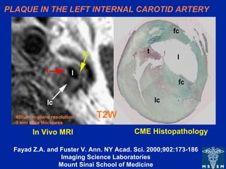
1. PLAQUE IN THE LEFT INTERNAL CAROTID ARTERY
T2W
CME HistopathologyIn Vivo MRI
450µm in-plane resolution
3 mm slice thickness
Fayad Z.A. and Fuster V. Ann. NY Acad. Sci. 2000;902:173-186
Imaging Science Laboratories
Mount Sinai School of Medicine
2. TEE
T2W
4.5 mm plaque
AHA TYPE Va (fibroatheroma) AORTIC PLAQUE
Fayad ZA et al.
Circ 2000;101;2503-2509
Imaging Science Laboratories
Mount Sinai School of Medicine
Plaque
Fibrous
Cap
3. Fayad ZA et al. Circ. 2000;102;506-510
Imaging Science Laboratories
Mount Sinai School of Medicine
X-ray Angiogram
high grade stenosis
LAD
LAD Wall
MR CORONARY WALL IMAGING
Notas do Editor
In vivo transverse T2-weighted fast SE MR imaging of a left internal carotid artery. Plaque characterization was based on information obtained from T1-, intermediate-, and T2-weighted MR images. Left: T2-weighted MR image (repetition time, two R-R intervals; echo time, 55 msec; 3-mm section thickness; 450-µm in-plane resolution) shows low-signal-intensity lipid core (lc), high-signal-intensity fibrous cap (fc), and very high–signal-intensity thrombus (t). l = arterial lumen. Right: Corresponding histopathologic section. (Mason-eosin stain; original magnification, X10).
Fayad ZA, Fuster V. Characterization of atherosclerotic plaques by magnetic resonance imaging. Ann N Y Acad Sci. 2000;902:173-86.
• In vivo magnetic resonance images of a 4.5 mm thick plaque in the descending thoracic aorta: A) T1-weighted; B) Proton density-weighted; C) T2-weighted; with the corresponding transesophageal echocardiography (TEE) image (panel D). The MR images show an example of an AHA type Va plaque with a dark area in the center (arrow) identified on the T2-weighted image as a lipid rich core (panel C). The lipid rich core is separated from the lumen by a fibrous cap. Plaque characterization was based on the information obtained from T1-, proton-density-, and T2- weighted MR images. Image resolution is 0.8 mm.