Gram positive and gram negative
•Transferir como DOCX, PDF•
32 gostaram•26,336 visualizações


Recomendados
Mais conteúdo relacionado
Mais procurados
Mais procurados (20)
Destaque
Destaque (20)
Semelhante a Gram positive and gram negative
Semelhante a Gram positive and gram negative (20)
Último
Último (20)
Gram positive and gram negative
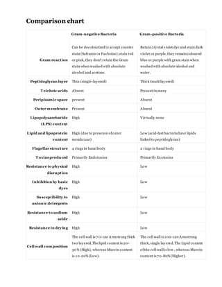
- 1. Comparison chart Gram-negative Bacteria Gram-positive Bacteria Gram reaction Can be decolourized to accept counter stain (Safranin or Fuchsine);stain red or pink, they don't retain the Gram stain when washed with absolute alcohol and acetone. Retain crystal violet dye and stain dark violet or purple, they remain coloured blue or purple with gram stain when washed with absolute alcohol and water. Peptidoglycan layer Thin (single-layered) Thick (multilayered) T eichoic acids Absent Present in many Periplasmic space present Absent Outer membrane Present Absent Lipopolysaccharide (LPS) content High Virtually none Lipid and lipoprotein content High (due to presence ofouter membrane) Low (acid-fast bacteria have lipids linked to peptidoglycan) Flagellar structure 4 rings in basal body 2 rings in basal body T oxins produced Primarily Endotoxins Primarily Exotoxins Resistance to physical disruption High Low Inhibition by basic dyes High Low Susceptibility to anionic detergents High Low Resistance to sodium azide High Low Resistance to drying High Low Cell wall composition The cell wall is 7 0-120 Armstrong thick two layered.Thelipid content is 20- 30% (High), whereas Murein content is 10-20%(Low). The cell wall is 100-120 Armstrong thick, single layered. The Lipid content of the cell wall is low , whereas Murein content is 70-80%(Higher).
- 2. Gram-negative Bacteria Gram-positive Bacteria Mesosome Mesosome is less prominent. Mesosome is more prominent. Antibiotic Resistance More Resistant to antibiotics. More Susceptibleto antibiotics GRAM POSITIVECELL WALL Carbohydrate backbone is composed of alternating N-acetylmuramic acid (NAM) and N- acetylglucosamine (NAG) molecules. Attached to each of the muramic acid molecules are a tetrapeptide consisting of both D- and L- amino acids, the precise composition of which differs between bacteria.
- 3. Special Aminoacids found in Peptidoglycan layer: Diaminopimelic acid: Unique to bacterial cells. D- alanine: Involved in the cross links between tetrapeptides and in the action of penicillin. Functions of Peptidoglycan Layer 1. It provides rigid support to bacterial cells and maintains the characteristic shape of the cell. 2. Allows bacterial cell to withstand media of low osmotic pressure, such as water. Medical Importance of Peptidoglycan Layer Peptidoglycan is a good target for antibacterial drugs. Eg. Penicillins, cephalosporins etc inhibit transpeptidase reaction which makes cross-links between the two adjacent tetrapeptides. Lysozyme enzyme present in human tears, mucus, and saliva cleave peptidoglycan backbone breaking its glycosyl bonds. plasmid (ˈplæzmɪd) — n a small circle of bacterial DNA that is independent of the main bacterial chro mosome. Plasmids oftencontain genes for drug resistances and can be trans mitted between bacteria of the same and differentspecies: used in genetic e ngineering Difference Between Endotoxin and Exotoxin Exotoxins are toxic substances secreted by bacteria and released outside the cell. Endotoxins are bacterial toxins consisting of lipids that are located within a cell.
- 4. Gram-negative bacteria display the following characteristics: 1. Cell membrane (cytoplasmic). 2. Thin peptidoglycan layer (which is much thicker in gram-positive bacteria) 3. Outer membrane containing lipopolysaccharide (LPS, which consists oflipid A, core polysaccharide, and O antigen) in its outer leaflet andphospholipids in the inner leaflet 4. Porins exist in the outer membrane, which act like pores for particular molecules 5. There is a space between the peptidoglycan layer and the secondary cell membrane called the periplasmic space 6. The S-layer is directly attached to the outer membrane rather than the peptidoglycan 7. If present, flagella have four supporting rings instead of two 8. No teichoic acids or lipoteichoic acids are present 9. Lipoproteins are attached to the polysaccharide backbone.
- 5. 10.Some of them contain Braun's lipoprotein, which serves as a link between the outer membrane and the peptidoglycan chain by a covalent bond 11.Most, with very few exceptions, do not form spores. 12.Release some endotoxin Lipopolysaccharide (LPS) of Gram Negative Bacteria, characteristics and functions Outer layer of cellwall of gram negative bacteria also called endotoxin. It is a characteristics feature of Gram Negative Bacteria. As in peptidoglycan biosynthesis, LPS molecules are assembled at the plasma or inner membrane. Exception: Only one Gram Positive Bacteria, i.e. Listeria monocyotogenes has been found to contain an authentic Lipopolysaccharide. Lipopolysaccharide is Pyrogenic (Responsible for fever), and also causes endotoxic shock etc.The LPS is composed of three distinct units.
- 6. A phospholipid called Lipid A, which is responsible for toxic effects A core polysaccharide of fiver sugars linked through ketodeoxyoctulonate (KDO) to lipid A Structural Unit of Lipopolysaccharide Source: South Carolina School of Medicine An outer polysaccharide consisting of up to 25 repeating units of 3-5 sugars, also called somatic or O antigen. O antigens are used to identify certain organisms in microbiology laboratory.
- 8. DanishscientistHansChristianGramdevisedamethodtodifferentiate twotypesof bacteriabased onthe structural differencesintheircell walls.Inhistest,bacteriathatretainthe crystal violetdye dosobecause of a thicklayerof peptidoglycanandare called Gram-positive bacteria.Incontrast,Gram-negative bacteriadonotretainthe violetdye and are coloredredor pink.ComparedwithGram-positive bacteria,Gram-negative bacteriaare more resistant againstantibodiesbecauseof theirimpenetrablecell wall.These bacteriahave awide varietyof applicationsranging frommedical treatmentto industrial use andSwisscheese production.