OSCE 12-lead ECG protocol
•Transferir como DOC, PDF•
6 gostaram•6,519 visualizações

This document provides instructions for performing a 12-lead electrocardiogram (ECG). It describes introducing yourself to the patient, obtaining consent, preparing the patient by removing chest hair or washing the skin, and properly placing the 10 electrodes on the chest and limbs in their designated positions. The student is asked to explain each step out loud as they perform the procedure. Once the ECG is obtained, the electrodes are removed and the patient is covered. Key details like the patient's name, date and time are documented, along with any presence of chest pain. The clinical area is then tidied and hand hygiene is performed.

Recomendados
Mais conteúdo relacionado
Mais procurados
Mais procurados (20)
Destaque
Destaque (9)
Semelhante a OSCE 12-lead ECG protocol
Semelhante a OSCE 12-lead ECG protocol (17)
Mais de Christiane Riedinger
Mais de Christiane Riedinger (20)
OSCE 12-lead ECG protocol
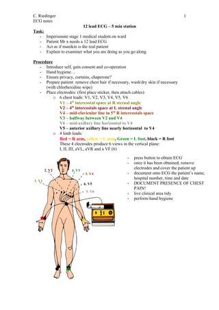
- 1. C. Riedinger 1 ECG notes 12 lead ECG – 5 min station Task: - Impersonate stage 1 medical student on ward - Patient Mr x needs a 12 lead ECG - Act as if manikin is the real patient - Explain to examiner what you are doing as you go along Procedure - Introduce self, gain consent and co-operation - Hand hygiene… - Ensure privacy, curtains, chaperone? - Prepare patient: remove chest hair if necessary, wash/dry skin if necessary (with chlorhexidine wipe) - Place electrodes: (first place sticker, then attach cables) o 6 chest leads: V1, V2, V3, V4, V5, V6 V1 – 4th intercostal space at R sternal angle V2 – 4th intercostals space at L sternal angle V4 – mid-clavicular line in 5th R intercostals space V3 – halfway between V2 and V4 V6 – mid-axillary line horizontal to V4 V5 – anterior axillary line nearly horizontal to V4 o 4 limb leads: Red = R arm, yellow = L arm, Green = L foot, black = R foot These 4 electrodes produce 6 views in the vertical plane: I, II, III, aVL, aVR and a VF (6) - press button to obtain ECG - once it has been obtained, remove 2. V2 electrodes and cover the patient up 4. V3 3. V4 - document onto ECG the patient’s name, hospital number, time and date 1. V1 6. V5 - DOCUMENT PRESENCE OF CHEST PAIN! 5. V6 - live clinical area tidy - perform hand hygiene