
Recomendados
Recomendados
Mais conteúdo relacionado
Mais procurados
Mais procurados (20)
Semelhante a Carotid intima-media thickness
Semelhante a Carotid intima-media thickness (20)
Mais de Samir Haffar
Mais de Samir Haffar (20)
Último
Último (20)
Carotid intima-media thickness
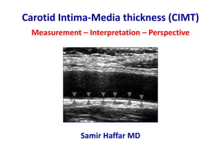
- 1. Carotid Intima-Media thickness (CIMT) Measurement – Interpretation – Perspective Samir Haffar MD
- 2. Number of PubMed publications using “CIMT” in title or abstract CIMT: carotid intima-media thickness Bots ML et al. Chin Med J 2016;129:215‐26.
- 3. Carotid intima-media thickness (CIMT) Good correlation of CIMT between histology & ultrasound
- 4. Measurement of CIMT • Distal common carotid artery • Carotid bulb • Proximal internal carotid artery Different sites of measure CIMT: carotid intima-media thickness
- 5. Standardization of CIMT measurement • Longitudinal scan of CCA • Far wall of CCA • At least 5 mm from carotid bulb • 10 mm segment of far wall CCA: common carotid artery – CIMT: carotid intima-media thickness Touboul PJ et al. Cerebrovasc Dis 2012;34:290–296.
- 6. CCA, bifurcation & origin of ICA & ECA CCA: common carotid artery – ECA: external carotid artery – ICA: internal carotid artery Touboul PJ et al. Cerebrovasc Dis 2012;34:290–296. Double arrow line corresponds to end of CCA where near & far walls start diverging
- 7. Manual measurement of CIMT Double-line pattern CIMT: carotid intima-media thickness
- 8. Automated measurement of CIMT Radiofrequency signals QI 0.94 – Maximal 0.713 mm – Mean 0.648 mm – SD 0.047 mm Quality Index: 94% of 150 measures could be performed Maximal: highest value obtained from all the measures Mean: average of all measurements that could be performed CIMT: carotid intima-media thickness Touboul PJ et al. Cerebrovasc Dis 2012;34:290–296
- 9. Carotid intima-media thickness Imaging pitfalls Over-gained image
- 10. Image not horizontal Carotid intima-media thickness Imaging pitfalls
- 11. Correlation between CIMT & age healthy asymptomatic individuals Systematic review: 17 studies – 32 unique study populations 10,124 healthy asymptomatic individuals free from CVD risk factors CIMT: carotid intima media thickness – CVD: cardio-vascular disease van den Munckhof ICL et al. Clinical Cardiology 2018;41:698–704.
- 12. Age-related quartiles of risk factors Touboul PJ et al. Cerebrovasc Dis 2012;34:290–296.
- 13. CIMT & plaque
- 14. CIMT & plaque 1. Focal encoarching into arterial lumen of at least 0.5 mm 2. > 1.5 mm intima-media thickness Mannheim IMT consensus. Cerbrovasc Dis 2012;34:290 –296. Plaques
- 15. Indications of CIMT measurement • Screening for subclinical atherosclerosis • Risk stratification for future CVD-related events • Assessment of cardio-vascular drug efficacy
- 16. CIMT as predictor of future CV events One-time CIMT measurement 14 studies & 45,828 asymptomatic individuals Median follow-up 10.8 years Hazard ratio per 0.10 mm mean common CIMT difference: - First myocardial infarction 1.08 (95% CI: 1.05 – 1.10) - First stroke 1.12 (95% CI: 1.10 – 1.15) Individual participant data meta-analysis CI: confidence interval Den Ruijter HM et al. JAMA 2012; 308:796.
- 17. • 16 studies, 36,984 patients without known CV disease • Mean follow-up 7 years with yearly progression rates of CIMT (mean & maximum CIMT of CCA, bifurcation & ICC) • Baseline CIMT was independent predictors of future CVE • Progression of CIMT was not associated with future events CIMT as predictor of future CV events Serial CIMT measurement Individual participant data meta-analysis CCA: common carotid artery – CVE: cardio-vascular events – ICC: internal carotid artery Lorenz MW et al. Lancet 2012; 379:2053.
- 18. Technical issues in CIMT measurement Touboul JP. Front Neurol Neurosci 2015;36:31-39. Patient Age Sex Race Tissue echogenicity Neck anatomy Risk factors Device Frequency Gray-scale Depth settings Gain settings Frame rate Sonographer Education Far wall CCA/Bif/ICA Left/right Plaque/no plaque Measurement Manual Semi-automatic Software Mean/max Cardiac cycle
- 19. • Low risk individuals <10% • Intermediate risk individuals 11-20% • High risk individuals >20% Framingham coronary heart disease risk score Most commonly used score 10-year risk of coronary heart disease events
- 20. https://www.mdcalc.com/framingham-coronary-heart-disease-risk-score Framingham coronary heart disease risk score
- 21. FRS: Framingham risk score http://www.cscc.unc.edu/aricnews/CIMTCHD/RiskCalc2.html ARIC study CIMT & carotid plaque included in FRS 13,145 participants without CVD 7% improvement in all subjects 17% improvement in intermediate risk subjects
- 22. Assessment of cardio-vascular drug efficacy • CIMT used to test efficacy of CV drugs in clinical trials • Lipid-lowering: statins, niacin, ezetimibe Anti-hypertensive, anti-obesity, antioxidants • Modification of therapy based on CIMT not shown to alter end points (death, myocardial infarction, stroke) CIMT is not used in clinical practice to monitor effects of medical therapy in individual patient
- 23. CIMT measurement in primary prevention Guidelines differ in their recommendations 1 Naghavi M et al. Am J Cardiol 2006;98:2H–15H. 2 Greenland P et al. Circulation 2010;122:2748–2764. 3 Society of Atherosclerosis Imaging and Prevention. Atherosclerosis 2011;214:43–46. 4 Goff DC Jr et al. J Am Coll Cardiol 2014; 63:2935. Year Guidelines Recommendations 2006 Screening for Heart Attack Prevention and Education (SHAPE)1 All individuals 2010 American College of Cardiology (ACC)2 Individuals at intermediate risk 2011 Society of Atherosclerosis Imaging and Prevention (SAIP)3 Intermediate-risk patients Patient with metabolic syndrome Older patients 2014 American College of Cardiology (ACC)4 No routine use in clinical practice
- 24. Toward better evaluation of atherosclerosis Guidelines of ACC/AHA 2014 • Family history • High sensibility CRP (hs-CRP) • Coronary artery calcium score (CACS) • Ankle-brachial index (ABI) ACC/AHA: American college of cardiology/American heart association 4 Goff DC Jr et al. J Am Coll Cardiol 2014; 63:2935. Newer risk markers for quantitative risk assessment
- 25. Ankle Brachial Index (ABPI) Continuous wave Doppler (takes 10 - 15 min) Posterior tibial artery Dorsalis pedis artery Thrush A, Hartshorne T. Peripheral vascular ultrasound: How, why and when. Elsevier Churchill Livingstone, London, 2nd edition, 2005. Peroneal artery Highest ankle pressure / highest brachial pressure
- 26. Grading arterial disease using ankle brachial index ABPI Comment > 1.3 Falsely high value (suspicion of medial sclerosis) 0.9 – 1.3 Normal finding 0.75 – 0.9 Mild peripheral arterial disease 0.4 – 0.75 Moderate peripheral arterial disease < 0.4 Severe peripheral arterial disease ABPI: Ankle Brachial Pressure Index Stiegler H & Brandl R. Ultraschall in Med 2009;30:334–363.
- 27. Carotid hemodynamics parameters PSV: pic systolic velocity – EDV: end diastolic velocity – RI: resistivity index Kim GH et al. Korean Circ J 2017;47(1):1-8. Decreased PSV Decreased EDV Increased RI More predictive of CV events than CIMT in several preliminary studies
- 28. Conclusion • CIMT is indicator of subclinical atherosclerosis • CIMT can predict the risk of future CV events • Other single or combined parameters could be more predictive of future CV events than CIMT
- 29. Thank You
Notas do Editor
- Typically, normal common carotid CIMT at age 10 is approximately 0.4 to 0.5 mm, while from the fifth decade of life onward this progresses to 0.7 to 0.8 mm or more [3].
- Measurement in diastole: systolic expansion of lumen causes the CIMT to become thinner. Measurement
- Until additional data are available that directly correlate CIMT measurement with improvements in hard outcomes (ie, death, myocardial infarction, stroke), we do not recommend routine CIMT measurement.
- While the highest ankle pressure is used in most studies, the sensitivity for the detection of a relevant arterial occlusion disease of 68% was able to be increased to 93% with a comparable specificity of almost 100% in a current study for an ABI < 0.9 by using the lowest foot artery pressure value.