SlideShare uma empresa Scribd logo
1 de 8
Baixar para ler offline
Recent advances in resin based adhesives and restorative
materials together with the improvements in formulation
and simplification of the adhesive procedures have stimu-
lated an increase in the use of resin based composites in the
posterior region.3,5 Direct posterior resin restorations con-
tinue to become more and more popular as functional, es-
thetic alternatives to amalgam.25 Adhesively bonded resin
composites have the advantage of conserving tooth structure
with the potential for tooth reinforcement, while at the same
time providing a cosmetically acceptable restoration.11
During the last decade, new formulations have been pre-
sented; the average filler size was reduced drastically, and
altered size distribution of submicrometer filler particles
were used to optimize the filler load in order to improve the
mechanical and wear characteristics.6 However, no one com-
posite material has been able to meet both the functional
needs of a posterior Class I or II restoration and the superior
esthetics required for anterior restorations.8
The revolutionary development of nanotechnology has
gained great interest in dentistry, as well as in the other
fields of science and medicine. Nanotechnology is based on
the production of functional materials and structures in the
range of 1 to 100 nanometers using various physical and
chemical methods. One of the most distinguished and sig-
nificant contributions of this technology to dentistry has
been the development of resin-based composite materials.
In nanotechnology, some particles are produced using a
Clinical Performance of Novel Resin Composites in
Posterior Teeth: 18-Month Results
Zeynep Ergücüa/L. Sebnem Türkünb
Purpose: The aim of this study was to evaluate the clinical success potential of two nanocomposites placed in poste-
rior teeth using an antibacterial adhesive system over 18 months.
Methods: A total of 49 Class I and 47 Class II restorations were placed in the permanent teeth of thirty adult pa-
tients. The carious lesions were restored with Grandio (Voco) or Filtek Supreme (3M ESPE) using a two-step self-etch-
ing antibacterial adhesive system Clearfil Protect Bond (Kuraray). The restorations were finished with fine-grit
diamond burs, Enhance polishing system, and Sof-Lex finishing brushes. The restorations were evaluated at baseline,
6, 12, and 18 months after placement using modified Ryge criteria for color stability, marginal discoloration, marginal
adaptation, caries formation, anatomic form, postoperative sensitivity, surface roughness, and retention.
Results: The changes in the parameters were assessed using the Cochran Q test and the McNemar test at a signifi-
cance level of p < 0.05. All restorations were classified as clinically satisfactory after 18 months. Statistical analysis
demonstrated differences only in superficial roughness, with Grandio exhibiting more surface roughness than Filtek
Supreme (p < 0.05).
Conclusion: Posterior restorations built up with the novel nanocomposites using an antibacterial self-etching system
showed satisfactory results at the 18-month recall appointment relative to all criteria except the surface texture in the
case of Grandio. Further evaluations are necessary for a more in-depth analysis.
Keywords: nanofill, nanohybrid, antibacterial adhesive, posterior restorations.
J Adhes Dent 2007; 9: xx-xx. Submitted for publication: 02.05.06; accepted for publication: 13.09.06.
Vol 9, No 2, 2007 209
a Assistant Professor, Ege University School of Dentistry, Department of
Restorative Dentistry, Izmir, Turkey.
b Associate Professor, Ege University School of Dentistry, Department of
Restorative Dentistry, Izmir, Turkey.
Reprint requests: Dr. Zeynep Ergücü, Ege University School of Dentistry, De-
partment of Restorative Dentistry, 35100 Izmir, Turkey. Tel: +90-232-388-03-
28, Fax: +90-232-388-03-25. e-mail: zergucu@yahoo.com
This study, with its 12-month results, was presented at the Joint Meeting of the
CED/NOF of the IADR, 14-17 September 2005, Amsterdam, the Netherlands.
new technique, and grit size is no longer determined by sim-
ple milling. The milling procedures usually cannot reduce the
filler particle size below 100 nm. To circumvent this barrier,
the particles are created from different components using
Sol-gel chemistry.24
Currently, there is a conflict in the terminology used to de-
fine the resin composites that contain nanoparticles. In re-
cent studies, the terms “nanofilled”, “nanohybrid,” or “nano-
structured” were used by various authors in order to classi-
fy the type of some resin composites.4,2,35 Therefore, to
avoid confusion, these novel materials are named nanocom-
posites in this study. Nanocomposites combine the esthetic
properties required for cosmetic restorations and the me-
chanical properties necessary for posterior restorations.
Due to the reduced dimension of the particles and to a wide
size distribution, an increased filler load can be achieved
with the consequence of reducing the polymerization shrink-
age27 and increasing the mechanical properties, such as
tensile strength, compressive strength, and resistance to
fracture. These properties seem to be equivalent to or even
sometimes higher than those of universal composites, and
significantly higher when compared to microfilled compos-
ites.24,26,27 Besides, the optical properties of these resin
composites are improved, as the diameters of the filler par-
ticles are a fraction of the wavelength of visible light (0.4 to
0.8 μm).24 Additionally, better gloss retention and diminished
wear rate have also been reported in some studies.24,32
One of these materials is Filtek Supreme, which contain
zirconia-silica particles consisting of 5- to 75-nm fillers and
0.6- to 1.4-μm nanoclusters.1 Another one, Grandio, intro-
duced in early 2003, contains glass-ceramic particles of
1 μm and silicium dioxide particles of 20 to 50 nm.37 Be-
sides the innovations in the field of resin composites, there
have been important developments in dentin adhesive sys-
tems as well.
Most of the current adhesive systems require two or few-
er steps and can be divided into two main groups: total-etch
and self-etching systems. The total-etch systems are offered
as three- or two-step systems (self-priming or one-bottle). The
self-etching systems are divided into two- or one-step sys-
tems (all-in-one).14 Self-etching adhesives, which are finding
increasing use,7,23 do not require a separate acid etching
step and only modify the smear layer. Considering that there
may be some bacteria left inadvertently in the smear layer,
an antibacterial self-etching adhesive system may be of
great benefit in eliminating the residual bacteria in the cav-
ity after operative procedures. The use of antibacterial self-
etching systems subsequently increases the success rate of
minimally invasive treatments.18 Since 1995, Imazato et
al16,17 have conducted investigations on the use of the an-
tibacterial monomer methacryloxy dodecyl pyridinium bro-
mide (MDPB) for dentin bonding systems. In 2004, an an-
tibacterial self-etching adhesive system containing MDPB
(Clearfil Protect Bond) was introduced with the aim of ex-
hibiting strong antibacterial activity against residual bacte-
ria in the cavity when applied, as well as inhibition of the bac-
teria that invade through microleakage after being cured by
a “contact active” effect.15 Türkün34 investigated the clini-
cal performance of Clearfil Protect Bond and Xeno III
(Dentsply De Trey; Konstanz, Germany) in noncarious cervi-
cal lesions. The one-year results demonstrated that Clearfil
Protect Bond performed well during this period without caus-
ing any postoperative sensitivity or side effects.
Currently, there is no clinical trial comparing the perfor-
mance of two different nanocomposites placed with this
new antibacterial system in posterior restorations. It is well
established that clinical trials are crucial for assessing all
the potential variables which influence the overall perfor-
mance of a restorative. Therefore, the aim of this university
dental hospital-based study was to evaluate the clinical suc-
cess potential of two nanocomposites placed in posterior
teeth using an antibacterial adhesive system.
MATERIALS AND METHODS
A total of 30 patients (23 female, 7 male patients, mean age
23.9 years with a range of 13 to 48 years) participated in this
study. Patients were selected for this study according to the
following criteria:
• No allergy to resin materials or components of adhesives.
• Absence of pain related to restored teeth.
• No pathological periodontal or pulpal diagnosis.
• No need of deep caries treatment among the selected
teeth.
• An opposing or neighboring tooth to the tooth to be re-
stored exists.
• There are at least two comparable cavities to be restored
with a resin composite.
The exclusion criteria were clinical symptoms of pulpitis,
such as spontaneous pain or sensitivity to pressure. En-
dodontically treated teeth were also excluded. It was not
obligatory for the patients to return for recalls; however, pa-
tients who would not be able to return for the controls were
not accepted for this clinical trial. The patients were not se-
lected from a certain pool; they were asked whether they
wanted to participate in the study. Participants were routine
patients of a large university dental hospital serving a so-
cioeconomically less advantaged part of a greater metro-
politan area. The study design was approved by the Human
Ethical Research Committee of Ege University (Izmir, Turkey).
Written informed consent was obtained from every patient
in this investigation. The indications for treatment were pri-
mary caries or replacement of existing insufficient restora-
tions. The clinicians, having experience with adhesive com-
posite restorations, were calibrated in terms of using the ma-
terials.
In the course of the present controlled prospective clini-
cal study, 96 cavities in 64 molars and 32 premolars were
restored with one nanohybrid resin composite, Grandio (Vo-
co; Cuxhaven, Germany), or one nanofilled resin composite,
Filtek Supreme (3M ESPE; St Paul, MN, USA), in 30 patients
in a split-mouth design. Forty-nine cavities were Class I and
47 cavities were Class II; 52 cavities were in the maxillary
and 44 were in the mandibular arch.The self-etching, an-
tibacterial dentin adhesive system, Clearfil Protect Bond (Ku-
raray; Osaka, Japan), was used according to the manufac-
turer’s instructions. The properties of the materials are pre-
sented in Table 1. Equal numbers of both materials were
210 The Journal of Adhesive Dentistry
Ergücü/Türkün
Vol 9, No 2, 2007 211
Ergücü/Türkün
placed in each patient. The numerical vitality test scores of
the teeth before any preparations were recorded using a vi-
tality tester. The cavity preparation was achieved with a con-
servative adhesive design. The margins received no bevel
preparations. The Class I caries extended one-fourth to one-
third of the way up one or more of the cuspal slopes. The
proximal portion of the Class II caries extended into the in-
terproximal embrasures. The restorations were mostly qual-
ified as small to moderate (80%),29 while the remaining 20%
larger restorations were also included. The adhesive prepa-
ration of teeth where amalgam was replaced did not result
in a transformation of the existing undercuts to a nonreten-
tive form. In those cases, only a trimming of the margins was
conducted to cut enamel in the right direction. Teeth were
isolated with cotton rolls, while rubber-dam was used for 4
teeth with deep proximal cavities.
The percentages of Grandio and Filtek Supreme restora-
tions in the maxillary and mandibular arches are shown in
Table 2. Fifty-two percent of the Grandio restorations and
50% of the Filtek Supreme restorations were one-sided,
while 44% of the Grandio and 46% of the Filtek Supreme
restorations were two-sided. Only 4% of both Grandio and Fil-
tek Supreme restorations were three-sided.
The protocol was the same for all restorations. The primer
of Clearfil Protect Bond was applied to the cavity, left undis-
turbed for 20 s, and evaporated with an air syringe for a few
seconds. Then the bonding agent was applied with a brush,
gently spread with an air syringe and polymerized for 10 s
with the Degulux light-activating unit with an output in ex-
cess of 450 mW/cm2 (Degussa; Düsseldorf, Germany). The
proximal portion of the Class II cavity peparations extended
into the interproximal embrasures. Pre-wedging with an
anatomically contoured wooden wedge (Kerr Hawe; Bioggio,
Switzerland) was done before Class II cavity preparation.
This procedure allowed slow separation and served as a
guide to determine the proper height of the gingival floor. A
sectional precontoured matrix system (Palodent, Dentsply
De Trey; Konstanz, Germany) was used to restore the Class
II cavities. For all the cavities, the composite resins were in-
serted with hand instruments using an incremental tech-
nique. The first increment, 1 to 1.5 mm thick, was con-
densed in the opposite corner and light cured on that side.
Material
Adhesive
Clearfil Protect Bond
(Kuraray, Osaka, Japan)
Resin based composite
Filtek Supreme
(3M ESPE, St Paul, MN, USA)
Grandio
(Voco, Cuxhaven, Germany)
Table 1 Composition of materials tested*
Composition/property
Primer: 10-methacryloyloxydodecyl dihydrogen phosphate (MDP),
12-methacryloyloxydodecylpyridinium bromide (MDPB), 2-hydroxyethyl methacrylate
(HEMA), hydrophilic dimethacrylate, water
Bond: 10-methacryloyloxydodecyl dihydrogen phosphate (MDP),
bis-phenol A diglycidylmethacrylate (bis-GMA), 2-hydroxyethyl methacrylate (HEMA),
hydrophobic dimethacrylate, di-camphorquinone, N,N- diethanol-p-toluidine
Matrix: bis-phenol A diglycidylmethacrylate (bis-GMA), triethylene
glycol dimethacrylate (TEG-DMA), urethane dimethacrylate (UDMA), bisphenol A
polyethylene glycol diether dimethacylate
Filler: silica nanofillers (5-75 nm) zirconia/silica nanoclusters (0.6-1.4μm)
Filler content: 78.5% (w/w) 59% (v/v)
Matrix: bis-GMA, dimethacrylate, urethane dimethacrylate
(UDMA), triethylene glycol dimethacrylate (TEG-DMA)
Filler: silicium dioxide nanofillers (20-50 nm) glass ceramic
microfillers (1 μm)
Filler content: 87% (w/w) (71.4% (v/v)
* All composition and property information is from the resp. manufacturer’s technical manual.
Resin Maxillary arch Mandibular arch Total
composites Premolar Molar Premolar Molar
Grandio 23% 31% 35% 11% 100%
Filtek Supreme 19% 35% 31% 15% 100%
Table 2 Distribution of restorations by localization
Ergücü/Türkün
212 The Journal of Adhesive Dentistry
Succeeding increments 1.5 to 2 mm thick were placed and
sequentially light cured until the proximal and occlusal con-
tours were restored fully. Each increment was polymerized
for 20 s using the same visible light-curing device. After re-
moval of the matrix band, the proximal regions of the Class
II restorations were separately polymerized buccally and lin-
gually for 20 s each. The restorations were finished under
water-spray cooling with fine-grit diamond burs to remove
gross excess (No.859 EF.314.014, Komet Dental; Lemgo
Germany), followed by Enhance points (Dentsply DeTrey) and
Sof-Lex finishing brushes (3M ESPE; St Paul, MN, USA) to ob-
tain a smooth, reflective occlusal surface. For approximal fin-
ishing and polishing, finishing strips (3M ESPE) and Sof-Lex
disks (3M ESPE) were used.
Two volunteer clinicians, trained in the technique and not
involved with the treatment procedures, examined the
restorations at baseline (one week after placement), and at
6,12, and 18 months, and evaluated them using the modi-
fied Ryge criteria, commonly known as US Public Health Ser-
vice (USPHS) criteria.21,30 Evaluation parameters included
color matching ability, marginal adaptation, anatomical
form, cavosurface margin discoloration, secondary caries
formation, and surface texture (Table 3). For each of the cri-
teria, Alpha indicates the highest degree of clinical accept-
ability; Bravo and Charlie indicate progressively lessening
degrees of clinical acceptability. When disagreement oc-
curred during evaluation, the final decision was made by
consensus of the evaluators, who were calibrated before the
study by a joint examination of 20 resin-based composite
restorations each. The evaluators recorded numerical vital-
ity scores at each recall. Both cavities were photographed
(Canon Power Shot S45; Kyushu, Japan) at baseline and at
Criterion
Color-matching ability
Cavosurface margin
discoloration
Marginal adaptation
(occlusal and proximal)
Secondary caries
formation
Surface texture
Anatomical form
(occlusal and proximal)
Inspection method
Visual inspection with a mirror at
18 inches
Visual inspection with a mirror at
18 inches
Visual inspection with explorer and
mirror, if needed
Visual inspection with explorer, mir-
ror and radiographs
Visual inspection with explorer and
mirror, if needed
Visual inspection with explorer and
mirror, if needed
Rating scale
A: The restoration matches the adjacent tooth structure in color
and translucency.
B: Light mismatch in color, shade or translucency between the
restoration and the adjacent tooth.
C: The mismatch in color and translucency is outside the ac-
ceptable range of tooth color and translucency.
A: No discoloration anywhere along the margin between the
restoration and the adjacent tooth.
B: Slight discoloration along the margin between the restoration
and the adjacent tooth.
C: The discoloration penetrated along the margin of the restora-
tive material in a pulpal direction.
A: No visible evidence of a crevice along the margin
B: Visible evidence of a crevice along the margin into which the
explorer will penetrate.
C: The dentin or base is exposed.
D:The restoration is fractured, mobile or missing.
A: No evidence of caries.
B: Evidence of caries along the margin of the restoration.
A: The restoration surface is as smooth as the surrounding
enamel.
B: The restoration surface is rougher than the surrounding
enamel.
C: There is a crevice and fracture on the surface of the restora-
tion.
A: The restoration is continuous with existing anatomical form.
B: The restoration is discontinuous with existing anatomical
form, but the material is not sufficient to expose the dentin or
base.
C: Sufficient material lost to expose the dentin or the base.
Table 3 Modified Ryge direct evaluation criteria*
* Source: Ryge
A: Highest degree of clinical acceptability; B and C: less clinical acceptability, D: clinically unacceptable
Vol 9, No 2, 2007 213
Ergücü/Türkün
each recall appointment. For each subject, the number of
teeth restored using each nanocomposite was equal, so that
each restoration could be compared with its own same-sub-
ject control.
The Cochran-Q test was used to evaluate the changes
across the four time points for each of the criteria. The two
resin composites were compared at the same recall period
for each criterion using the McNemar test. For all of the sta-
tistical analyses, p was set at 0.05.
RESULTS
Thirty patients requiring posterior resin composite restora-
tions were included in this study; however, two patients were
unavailable for recall appointments because they had
moved. Of the 96 restorations placed, 90 novel resin com-
posite (Grandio and Filtek Supreme) restorations that were
placed with the antibacterial two-step adhesive system
(Clearfil Protect Bond) in 28 patients were evaluated at the
18-month recall appointment. The clinical assessment re-
sults are shown in Table 4.
Esthetics and tooth vitality were all rated satisfactory for
the two nanocomposites investigated (Figs 1 and 2). Gingi-
val response was evaluated with a periodontal probe using
a gingival index.31 There was no inflammation, color change,
or bleeding around the restorations, and gingival status was
normal. None of the restorations showed evidence of sec-
ondary caries formation (Figs 3 and 4), and no postoperative
sensitivity was reported by any of the patients at any recall.
One restoration of each resin composite showed slightly dis-
colored margins at the 6-, 12-, and 18-month recalls (p >
0.05) (Figs 5 and 6).
Baseline 6 months 12 months 18 months
Criterion Filtek Grandio Filtek Grandio Filtek Grandio Filtek Grandio
Supreme Supreme Supreme Supreme
Color matching ability
A 48 48 48 48 48 48 45 45
B 0 0 0 0 0 0 0 0
C 0 0 0 0 0 0 0 0
Cavosurface margin
discoloration
A 48 48 47 47 47 47 44 44
B 0 0 1 1 1 1 1 1
C 0 0 0 0 0 0 0 0
Marginal adaptation
A 48 48 48 48 48 48 45 45
B 0 0 0 0 0 0 0 0
C 0 0 0 0 0 0 0 0
Secondary caries
formation
A 48 48 48 48 48 48 45 45
B 0 0 0 0 0 0 0 0
Surface texture
A 48 48 48 0 48 0 45 0
B 0 0 0 48* 0 48* 0 45*
C 0 0 0 0 0 0 0 0
Anatomical form
A 48 48 48 48 48 48 45 45
B 0 0 0 0 0 0 0 0
C 0 0 0 0 0 0 0 0
1Source: Ryge
Alpha (A) indicates excellent restorations, while Bravo (B) and Charlie (C) indicate progressively worsening degrees of clinical acceptability.
*p < 0.05 Statistically significant difference between the materials.
Table 4 Ryge criteria obtained at each recall1
Ergücü/Türkün
214 The Journal of Adhesive Dentistry
Fig 5 Occlusal restoration of Grandio in the maxillary right second
molar exhibited a marginal discoloration after 18 months. (G):
Grandio.
Fig 6 Marginal discoloration at the cavosurface margin of an oc-
clusal restoration of Filtek Supreme in the maxillary left second mo-
lar at the 18-month recall. (FS): Filtek Supreme.
Fig 1 Class II restorations in maxillary left second premolar and
first molar after 18 months. The disto-occlusal composite of the
second premolar was Filtek Supreme and the mesio-occlusal com-
posite in the first molar was Grandio. (FS): Filtek Supreme (G):
Grandio.
Fig 2 Class II restorations in maxillary right premolars after 18
months. Color match, anatomical form and marginal adaptation
were clinically excellent. (FS): Filtek Supreme (G): Grandio.
Fig 3 Occlusal restoration of Grandio in the maxillary right first mo-
lar at the 18-month recall appointment. (G): Grandio.
Fig 4 Occlusal restoration of Filtek Supreme in the maxillary left
molar after 18 months. (FS): Filtek Supreme.
Grandio exhibited a statistically significantly rougher sur-
face texture than Filtek Supreme (p < 0.05). Nevertheless,
there were no patient complaints regarding the surface
roughness of these restorations. All restorations were clas-
sified as clinically acceptable after 18 months.
DISCUSSION
The two novel resin composite restorative materials evalu-
ated in this study were used in combination with a two-step
self-etching antibacterial adhesive system. The clinical per-
formance of adhesives has improved significantly, allowing
adhesive restorations to be placed with a highly predictable
level of clinical success. It is considered that modern adhe-
sive systems, being superior to their predecessors especial-
ly in terms of retention, are no longer the main cause of pre-
mature clinical failure.33,36
A clinically effective adhesive system should keep the
restoration in place for a significant time and completely seal
the restoration margins against the ingress of oral fluids and
microorganisms. Incomplete marginal sealing will result in
postplacement sensitivity, marginal staining, and recurrent
caries, which are still the most common symptoms associ-
ated with clinical failure of adhesive restorations.28 Postop-
erative sensitivity was the most frequent complication of ear-
ly posterior composite restorations. With the introduction of
self-etcihng adhesives that are able to completely penetrate
into decalcified dentin and/or to obdurate dentinal tubules,
the incidence of postoperative sensitivity has dropped. In
our study, Clearfil Protect Bond was used as the dentin ad-
hesive system for both nanocomposites instead of the rec-
ommended proprietary dentin adhesives, because this an-
tibacterial adhesive system has not been previously used in
combination with novel resin composites in the posterior re-
gion and it might be of benefit to use this system in order to
eliminate the risk of secondary caries in deep cavities. Fur-
thermore, since the main focus of this investigation was to
compare the clinical success of two novel resin composites,
we wanted to standardize the type of the adhesive system.
The results of this clinical study demonstrated that both nov-
el composites placed in combination with Clearfil Protect
Bond performed well after 18 months. In a previous study,
Türkün34 investigated the clinical performance of Clearfil
Protect Bond and Xeno III in combination with the resin com-
posite Esthet-X (Dentsply DeTrey) in noncarious cervical le-
sions after 1 year, and reported that Clearfil Protect Bond
was clinically successful during this period without causing
any postoperative sensitivity or side effects.
The modified Ryge criteria (USPHS) for direct evaluation
remains the preferred system for evaluating important char-
acteristics of dental restorations, such as color matching,
secondary caries, cavosurface margin discoloration, and
postoperative sensitivity. Regarding the loss of anatomical
form, the direct evaluation method is able to generate data
that are of clinical significance and are compatible with clin-
ical treatment.13,19,20 Compared with the baseline observa-
tions, direct evaluations made using the modified Ryge cri-
teria demonstrated that color match, cavosurface margin
discoloration, and caries formation remained essentially un-
changed at the end of 18-month period for the two nanocom-
posites.
Polishability is one of the desirable features of tooth-col-
ored dental restoratives. High quality finishing and polishing
can improve the esthetics and longevity of these materials.
Nowadays, several restorative materials with finer organic
particles are produced by means of advanced technology.
The latest innovations are the nanocomposites. In vitro tests
showed that nanocomposites displayed high polish reten-
tion after toothbrush abrasion. During the abrasion tests, on-
ly nanosized particles are plucked away, leaving the sur-
faces with smaller defects than the wavelength of light, and
better surface characteristics than the conventional hybrid
composites are obtained.9,24
In regard to surface texture, direct evaluations of Grandio
restorations at the 6- and 12-month recall appointment were
scored as Bravo for all the restorations. At the 18-month re-
call, all of the Grandio restorations were again slightly rough
and thus were scored as Bravo (p < 0.05). The slight rough-
ness indicated that Bravo scores were perceptible only with
a sharp explorer and none of the patients noticed or com-
plained about the situation.
Mesquita et al22 demonstrated that Grandio showed a
rigidity approaching that of dentin. Concerning surface
roughness, the special hardness of Grandio can be seen as
a main cause. In the present authors’ opinion, some special
polishing systems may be of use in optimizing the surface
texture of this material after finishing procedures.
In the present study, both nanocomposites showed ex-
cellent marginal integrity at the end of the 18-month recall
period. Frankenberger et al12 compared the performance of
Grandio and Tetric Ceram in posterior teeth and reported
that no deterioration regarding the modified Ryge criteria
was found except for marginal integrity. According to their
findings, Grandio showed an equal outcome to Tetric Ceram
after one year of clinical service.
Ernst et al10 compared the clinical performance of Filtek
Supreme and Tetric Ceram placed in posterior teeth in a two-
year clinical study, finding that there were no statistically sig-
nificant differences between the two materials and that Fil-
tek Supreme could be used successfully in large stress-bear-
ing posterior cavities.
Since adhesive dentistry is technique sensitive, the suc-
cess of an adhesive restoration is mostly determined by the
ability of the dentists, beyond the performance of the mate-
rial. Even with the improved materials, the performance of
the material cannot be overestimated to exceed that of the
dentist. Therefore, it is still important for the clinician to
practice appropriately and meticulously when using adhe-
sive materials. Careful case selection and meticulous at-
tention to the restorative technique are recognized as es-
sential requirements for achieving longer-term clinical suc-
cess with posterior composite restorations.
Clinical trials are limited in number and require several
years with regular recall appointments to achieve sufficient
clinical validation. Owing to the rapid evolution in resin com-
posite technology and the higher turnover of novel resin
composites, these materials are released onto the dental
market without sufficient proof of their clinical performance.
Long-term quantitative observations are essential to deter-
Vol 9, No 2, 2007 215
Ergücü/Türkün
Ergücü/Türkün
216 The Journal of Adhesive Dentistry
mine the clinical performance of new posterior resin com-
posite materials, and selection should be primarily based on
these results. This study was planned to be a long-term clin-
ical trial; however, due to the shading problems of Filtek
Supreme encountered by some opinion leaders, the manu-
facturer decided to take the material off the market, and we
had to terminate the study, although both materials per-
formed well in our 18-month clinical trial. Furthermore, we
did not use the layering technique for the posterior restora-
tions, and used only one shade for the incremental tech-
nique. Thus, the shading problems with the restorations
were not seen, and we replaced only the ones that had dis-
colorations after 30 months. Similar to the present results,
Ernst et al10 reported that Filtek Supreme restorations
showed heavy staining at the fissures, while Tetric Ceram
restorations showed none after 2 years. Currently, the shad-
ing system of Filtek Supreme has been optimized and the
product is now being marketed as Filtek Supreme XT.
The findings of the current study demonstrated that both
nanocomposites performed equally well after 18 months in
spite of the surface roughness observed in Grandio restora-
tions. Although an 18-month observation period is not long,
the clinical problems encountered in this study were very
few. Selecting limited-sized cavities and paying strict atten-
tion to the restorative technique are essential for achieving
longer-term clinical success with posterior resin based com-
posite restorations.
CONCLUSION
It may be concluded that, with the improvement in materi-
als, careful case selection and application of the restorative
techniques, the novel resin composites can be used for pos-
terior restorations. Evaluations still need to be carried out to
reveal longer-term clinical performance of such materials.
ACKNOWLEDGMENTS
The authors would like to thank the companies Voco, 3M ESPE, and Ku-
raray Dental for their generous donation of the materials used in this
study.
REFERENCES
1. 3M Dental Products Filtek Supreme Plus Universal Restorative System. Tech-
nical Product Profile. St Paul MN, USA, 2005:9-12.
2. Baseren M. Surface roughness of nanofill and nanohybrid composite resin
and ormocer-based tooth-colored restorative materials after several finishing
and polishing procdures. J Biomater Appl 2004;19:121-34.
3. Bayne SC, Heymann HO, Swift Jr ER. Update on dental composite restora-
tions. J Am Dent Assoc 1994;125:687-701.
4. Beun S, Glorieux T, Devaux J, Vreven J, Leloup G. Characterization of
nanofilled compared to universal and microfilled composites. Dent Mater Jan
16. (Epub of ahead of print)
5. Brunthaler A, König F, Lucas T, Sperr W, Schedle A. Longevity of direct resin
composite restorations in posterior teeth. Clin Oral Invest 2003;7:63-70.
6. Bryant RW. Direct posterior composite resin restorations: a review. 1. Factors
influencing case selection. Aust Dent J 1992;37:81-7.
7. Clinician’s preferences 2001. CRA Newsletter 2001;25:1-4.
8. Denehy GE. A direct approach to restore anterior teeth. Am J Dent
2000;13(special issue):55-9.
9. Ergücü Z, Türkün LS. Surface Roughness of Novel Resin Composites Polished
with One-step Systems. Oper Dent 2007;32:185-192.
10. Ernst CP, Brandenbusch M, Meyer F, Canbek K, Gottschalk F, Willershausen
B. Two year clinical performance of a nanofiller vs a fine-particle hybrid resin
composite. Clin Oral Invest 2006;10:119-125.
11. Fortin D, Vargas MA. Two spectrums of composites: new techniques and ma-
terials. J Am Dent Assoc 2000;131(supplement):26S-30S.
12. Frankenberger R, Reinelt C, Eimer F, Krämer N. Posterior restorations with a
nanofilled resin composite- results after 12 months. J Dent Res 2005;(ab-
stracts Baltimore):579.
13. Geurtsen W, Schoeler U. A 4-year retrospective clinical study of Class 1 and
Class 2 composite restorations. J Dent 1997;25:229-32.
14. Haller B. Recent development in dentin bonding. Am J Dent 2000;13:44-50.
15. Imazato S, Ebi N, Tarumi H, Russell RRB, Kaneko T, Ebisu S. Bactericidal ac-
tivity and cytotoxicity of antibacterial monomer MDPB. Biomaterials
1999;20:899-903.
16. Imazato S, Kinomoto Y, Tarumi H, Ebisu S, Tay FR. Antibacterial activity and
bonding characteristics of an adhesive resin containing antibacterial
monomer MDPB. Dent Mater 2003;19:313-319.
17. Imazato S, Kinomoto Y, Tarumi H, Russell RR, McCabe JF. Incorporation of
antibacterial monomer MDPB in dentin primer. J Dent Res1997;76:768-
772.
18. Imazato S. Antibacterial properties of resin composites and dentin bonding
systems. Dent Mater 2003;19:449-457.
19. Leinfelder KF, Taylor DF, Barkmeier WW, Goldberg AJ. Quantitative wear mea-
surement of posterior composite resins. Dent Mater 1986;2:198-201.
20. Leinfelder KF, Wilder AD Jr., Teixeira LC. Wear rates of posterior composite
resins. J Am Dent Assoc 1986;112:829-833.
21. Leinfelder KF. Evaluation of criteria used for assessing the clinical perfor-
mance of composite resins in posterior teeth. Quintessence Int 1987;
18:531-536.
22. Mesquita RV, Axmann D, Geis-Gerstofer J. Dynamic visco-elastic properties
of dental composite resins. Dent Mater 2005;Sep 2 (Epub ahead of
print)xxAuthor: Can’t be, if published in 2005!!.
23. Miller MB. Self-etching adhesives: solving the sensitivity conundrum. Pract
Proced Aesthet Dent 2002;14:406.
24. Mitra SB, Wu D, Holmes BN. An application of nanotechnology in advanced
dental materials. J Am Dent Assoc 2003;134:1382-90.
25. Morgan M. Finishing and polishing of direct posterior resin restorations.
Pract Proced Aesthet Dent 2004;16:211-217.
26. Moszner N, Klapdohr S. Nanotechnology for dental composites. Int J Nan-
otechnol 2004;1:130-156.
27. Moszner N, Salz U. New developments of polymeric dental composites. Prog
Polym Sci 2001;26:535-576.
28. Qvist V, Qvist J, Mjör IA. Placement and longevity of tooth-colored restorations
in Denmark. Acta Odontol Scand 1990;48:305-311.
29. Raskin A, Michette-Theall B, Vreven J, Wilson NH. Clinical evaluation of a pos-
terior composite 10-year report. J Dent 1999;27:13-19.
30. Ryge G. Clinical criteria. Int Dent J 1980;30:347-358.
31. Silness P, Löe H. Periodontal disease in pregnancy . Acta Odontol Scand
1964;2:121-135.
32. Terry DA. Direct applications of a nanocomposite resin system. Part 1. The
evoluotion of contemporary composite materials. Pract Proced Aesthet Dent
2004;16:417-422.
33. Türkün LS. Clinical evaluation of a self-etching and a one-bottle adhesive sys-
tem at two years. J Dent 2003;31:527-534.
34. Türkün LS. The clinical performance of one-and two-step self-etching adhe-
sive systems at one year. J Am Dent Assoc2005;136:656-664.
35. Turssi CP, Ferracane JL, Ferracane LL. Wear and fatigue behavior of nano-
structured dental composites. J Biomed Mater Res B Appl Biomater 2006
Jan 30. (Epub ahead of print)
36. Van Meerbeek B, Perdigão J, Lambrechts P, Vanherle G. The clinical perfor-
mance of adhesives. J Dent 1998;26:1-20.
37. Voco Grandio. Light-curing Nano-hybrid Filling Material, Corresponds to EN
24049/ ISO 4049 Instructions for use. April 2003.
Clinical relevance: Posterior restorations built up with the
novel nanocomposites using an antibacterial self-etching
system showed satisfactory results at the 18-month recall
appointment relative to all criteria except the surface tex-
ture in the case of Grandio (p < 0.05). The surface rough-
ness of this material was not found to be clinically impor-
tant, as the patients did not have any complaints and
some were not even aware of it.

Mais conteúdo relacionado

Mais procurados

JofIMAB_2016-22-2p1099-1110.pdf
JofIMAB_2016-22-2p1099-1110.pdfJofIMAB_2016-22-2p1099-1110.pdf
JofIMAB_2016-22-2p1099-1110.pdfHawzheen Saeed
 
New materials for a post-Minamata era (Jirun Sun)
New materials for a post-Minamata era (Jirun Sun)New materials for a post-Minamata era (Jirun Sun)
New materials for a post-Minamata era (Jirun Sun)fdiworlddental
 
Effect Of Solvent Type On Microtensile Bond Strength Of TotalEtch One-Bottle ...
Effect Of Solvent Type On Microtensile Bond Strength Of TotalEtch One-Bottle ...Effect Of Solvent Type On Microtensile Bond Strength Of TotalEtch One-Bottle ...
Effect Of Solvent Type On Microtensile Bond Strength Of TotalEtch One-Bottle ...iosrjce
 
Critical review on glass ionomer seal under composite resin of obturated root...
Critical review on glass ionomer seal under composite resin of obturated root...Critical review on glass ionomer seal under composite resin of obturated root...
Critical review on glass ionomer seal under composite resin of obturated root...Ziad Abdul Majid
 
Effects of filler size on the mechanical properties of polymer filled dental ...
Effects of filler size on the mechanical properties of polymer filled dental ...Effects of filler size on the mechanical properties of polymer filled dental ...
Effects of filler size on the mechanical properties of polymer filled dental ...Fathie Kundie
 
Duddeck edi journal-1-2015_surface-analysis_
Duddeck edi journal-1-2015_surface-analysis_Duddeck edi journal-1-2015_surface-analysis_
Duddeck edi journal-1-2015_surface-analysis_datapro_cl
 
Amalgam/ rotary endodontic courses by indian dental academy
Amalgam/ rotary endodontic courses by indian dental academyAmalgam/ rotary endodontic courses by indian dental academy
Amalgam/ rotary endodontic courses by indian dental academyIndian dental academy
 
Biodentine (newer material in dentistry)
Biodentine   (newer material in dentistry)Biodentine   (newer material in dentistry)
Biodentine (newer material in dentistry)jhansi mutyala
 
Implant impressions- journal club - Accuracy of implant impressions using var...
Implant impressions- journal club - Accuracy of implant impressions using var...Implant impressions- journal club - Accuracy of implant impressions using var...
Implant impressions- journal club - Accuracy of implant impressions using var...Partha Sarathi Adhya
 
Influence of light and laser activation of tooth bleaching systems on enamel ...
Influence of light and laser activation of tooth bleaching systems on enamel ...Influence of light and laser activation of tooth bleaching systems on enamel ...
Influence of light and laser activation of tooth bleaching systems on enamel ...DR.AJAY BABU GUTTI M.D.S
 
Complementary remineralizing effect of self-assembling peptide (P11-4) with C...
Complementary remineralizing effect of self-assembling peptide (P11-4) with C...Complementary remineralizing effect of self-assembling peptide (P11-4) with C...
Complementary remineralizing effect of self-assembling peptide (P11-4) with C...DR.AJAY BABU GUTTI M.D.S
 
Bioactive Materials Subjected to Erosion/Abrasion and Their Influence on Dent...
Bioactive Materials Subjected to Erosion/Abrasion and Their Influence on Dent...Bioactive Materials Subjected to Erosion/Abrasion and Their Influence on Dent...
Bioactive Materials Subjected to Erosion/Abrasion and Their Influence on Dent...DR.AJAY BABU GUTTI M.D.S
 
A Comparative Study of Light Transmission by Various Dental Restorative Mater...
A Comparative Study of Light Transmission by Various Dental Restorative Mater...A Comparative Study of Light Transmission by Various Dental Restorative Mater...
A Comparative Study of Light Transmission by Various Dental Restorative Mater...DR.AJAY BABU GUTTI M.D.S
 
The bond strength of endodontic sealers to root /prosthodontic courses
The bond strength of endodontic sealers to root /prosthodontic coursesThe bond strength of endodontic sealers to root /prosthodontic courses
The bond strength of endodontic sealers to root /prosthodontic coursesIndian dental academy
 
Bioceramic in dentistry ( Endodontics)
Bioceramic in dentistry ( Endodontics)Bioceramic in dentistry ( Endodontics)
Bioceramic in dentistry ( Endodontics)Nakul Patidar
 

Mais procurados (18)

JofIMAB_2016-22-2p1099-1110.pdf
JofIMAB_2016-22-2p1099-1110.pdfJofIMAB_2016-22-2p1099-1110.pdf
JofIMAB_2016-22-2p1099-1110.pdf
 
New materials for a post-Minamata era (Jirun Sun)
New materials for a post-Minamata era (Jirun Sun)New materials for a post-Minamata era (Jirun Sun)
New materials for a post-Minamata era (Jirun Sun)
 
Effect Of Solvent Type On Microtensile Bond Strength Of TotalEtch One-Bottle ...
Effect Of Solvent Type On Microtensile Bond Strength Of TotalEtch One-Bottle ...Effect Of Solvent Type On Microtensile Bond Strength Of TotalEtch One-Bottle ...
Effect Of Solvent Type On Microtensile Bond Strength Of TotalEtch One-Bottle ...
 
Critical review on glass ionomer seal under composite resin of obturated root...
Critical review on glass ionomer seal under composite resin of obturated root...Critical review on glass ionomer seal under composite resin of obturated root...
Critical review on glass ionomer seal under composite resin of obturated root...
 
Effects of filler size on the mechanical properties of polymer filled dental ...
Effects of filler size on the mechanical properties of polymer filled dental ...Effects of filler size on the mechanical properties of polymer filled dental ...
Effects of filler size on the mechanical properties of polymer filled dental ...
 
35th publication ijmr - 6th name
35th publication   ijmr - 6th name35th publication   ijmr - 6th name
35th publication ijmr - 6th name
 
Duddeck edi journal-1-2015_surface-analysis_
Duddeck edi journal-1-2015_surface-analysis_Duddeck edi journal-1-2015_surface-analysis_
Duddeck edi journal-1-2015_surface-analysis_
 
Casestudies6nov2013
Casestudies6nov2013Casestudies6nov2013
Casestudies6nov2013
 
Amalgam/ rotary endodontic courses by indian dental academy
Amalgam/ rotary endodontic courses by indian dental academyAmalgam/ rotary endodontic courses by indian dental academy
Amalgam/ rotary endodontic courses by indian dental academy
 
Biodentine (newer material in dentistry)
Biodentine   (newer material in dentistry)Biodentine   (newer material in dentistry)
Biodentine (newer material in dentistry)
 
Implant impressions- journal club - Accuracy of implant impressions using var...
Implant impressions- journal club - Accuracy of implant impressions using var...Implant impressions- journal club - Accuracy of implant impressions using var...
Implant impressions- journal club - Accuracy of implant impressions using var...
 
75th publication sjm- 3rd name
75th publication  sjm- 3rd name75th publication  sjm- 3rd name
75th publication sjm- 3rd name
 
Influence of light and laser activation of tooth bleaching systems on enamel ...
Influence of light and laser activation of tooth bleaching systems on enamel ...Influence of light and laser activation of tooth bleaching systems on enamel ...
Influence of light and laser activation of tooth bleaching systems on enamel ...
 
Complementary remineralizing effect of self-assembling peptide (P11-4) with C...
Complementary remineralizing effect of self-assembling peptide (P11-4) with C...Complementary remineralizing effect of self-assembling peptide (P11-4) with C...
Complementary remineralizing effect of self-assembling peptide (P11-4) with C...
 
Bioactive Materials Subjected to Erosion/Abrasion and Their Influence on Dent...
Bioactive Materials Subjected to Erosion/Abrasion and Their Influence on Dent...Bioactive Materials Subjected to Erosion/Abrasion and Their Influence on Dent...
Bioactive Materials Subjected to Erosion/Abrasion and Their Influence on Dent...
 
A Comparative Study of Light Transmission by Various Dental Restorative Mater...
A Comparative Study of Light Transmission by Various Dental Restorative Mater...A Comparative Study of Light Transmission by Various Dental Restorative Mater...
A Comparative Study of Light Transmission by Various Dental Restorative Mater...
 
The bond strength of endodontic sealers to root /prosthodontic courses
The bond strength of endodontic sealers to root /prosthodontic coursesThe bond strength of endodontic sealers to root /prosthodontic courses
The bond strength of endodontic sealers to root /prosthodontic courses
 
Bioceramic in dentistry ( Endodontics)
Bioceramic in dentistry ( Endodontics)Bioceramic in dentistry ( Endodontics)
Bioceramic in dentistry ( Endodontics)
 

Destaque (8)

Jada doktora
Jada doktoraJada doktora
Jada doktora
 
1194-Turkun Amelogenesis
1194-Turkun Amelogenesis1194-Turkun Amelogenesis
1194-Turkun Amelogenesis
 
JADA protect bond
JADA protect bondJADA protect bond
JADA protect bond
 
Longterm antibacterial effect of CHX glass ionomers
Longterm antibacterial effect of CHX glass ionomersLongterm antibacterial effect of CHX glass ionomers
Longterm antibacterial effect of CHX glass ionomers
 
3year Surefil clinical
3year Surefil clinical3year Surefil clinical
3year Surefil clinical
 
Kompozit rezinler G-mag
Kompozit rezinler G-magKompozit rezinler G-mag
Kompozit rezinler G-mag
 
J Dentistry self etch and one bottle clinic
J Dentistry self etch and one bottle clinicJ Dentistry self etch and one bottle clinic
J Dentistry self etch and one bottle clinic
 
Amelogenesis imperfecta
Amelogenesis imperfectaAmelogenesis imperfecta
Amelogenesis imperfecta
 

Semelhante a Jad-Ergucu-Turkun

1. estudio clinico de 10 años de inlays direcra e indirecto de composite y ce...
1. estudio clinico de 10 años de inlays direcra e indirecto de composite y ce...1. estudio clinico de 10 años de inlays direcra e indirecto de composite y ce...
1. estudio clinico de 10 años de inlays direcra e indirecto de composite y ce...Lady viviana panduro monteiro
 
Evaluation effect of an in office zoom bleaching gel agent on the
Evaluation effect of an in office zoom bleaching gel agent on theEvaluation effect of an in office zoom bleaching gel agent on the
Evaluation effect of an in office zoom bleaching gel agent on theAmir Hamde
 
1. adhesive cementation of indirect composite inlays and onlays. a literature...
1. adhesive cementation of indirect composite inlays and onlays. a literature...1. adhesive cementation of indirect composite inlays and onlays. a literature...
1. adhesive cementation of indirect composite inlays and onlays. a literature...Lady viviana panduro monteiro
 
Esthetics of Class IV Restorations with Composite Resins
Esthetics of Class IV Restorations with Composite ResinsEsthetics of Class IV Restorations with Composite Resins
Esthetics of Class IV Restorations with Composite ResinsAbu-Hussein Muhamad
 
In vitro tests of adhesive and composite dental materials
In vitro tests of adhesive and composite dental materialsIn vitro tests of adhesive and composite dental materials
In vitro tests of adhesive and composite dental materialsSilas Toka
 
Prosthodontics ( inhibition of denture plaque)
Prosthodontics ( inhibition of denture plaque)Prosthodontics ( inhibition of denture plaque)
Prosthodontics ( inhibition of denture plaque)DHANANJAYSHETH1
 
Preference of materials for posterior restorations: A cross-sectional study a...
Preference of materials for posterior restorations: A cross-sectional study a...Preference of materials for posterior restorations: A cross-sectional study a...
Preference of materials for posterior restorations: A cross-sectional study a...Premier Publishers
 
Strip Crowns Technique for Restoration of Primary Anterior Teeth: Case Report
Strip Crowns Technique for Restoration of Primary Anterior Teeth: Case ReportStrip Crowns Technique for Restoration of Primary Anterior Teeth: Case Report
Strip Crowns Technique for Restoration of Primary Anterior Teeth: Case ReportAbu-Hussein Muhamad
 
Restoration of endodontically treated teeth review & treatment recomendations
Restoration of endodontically treated teeth review & treatment recomendationsRestoration of endodontically treated teeth review & treatment recomendations
Restoration of endodontically treated teeth review & treatment recomendationsJinny Shaw
 
Adhesion in restorative dentistry
Adhesion in restorative dentistryAdhesion in restorative dentistry
Adhesion in restorative dentistryIAU Dent
 
A Conservative Approach to Ceramic Veneers: A Case Report
A Conservative Approach toCeramic Veneers: A Case ReportA Conservative Approach toCeramic Veneers: A Case Report
A Conservative Approach to Ceramic Veneers: A Case ReportNadeem Aashiq
 

Semelhante a Jad-Ergucu-Turkun (20)

posterior composite
posterior compositeposterior composite
posterior composite
 
10. indirect resin composites
10. indirect resin composites10. indirect resin composites
10. indirect resin composites
 
1. estudio clinico de 10 años de inlays direcra e indirecto de composite y ce...
1. estudio clinico de 10 años de inlays direcra e indirecto de composite y ce...1. estudio clinico de 10 años de inlays direcra e indirecto de composite y ce...
1. estudio clinico de 10 años de inlays direcra e indirecto de composite y ce...
 
Evaluation effect of an in office zoom bleaching gel agent on the
Evaluation effect of an in office zoom bleaching gel agent on theEvaluation effect of an in office zoom bleaching gel agent on the
Evaluation effect of an in office zoom bleaching gel agent on the
 
Paper
PaperPaper
Paper
 
JADA 2 year surefil
JADA 2 year surefilJADA 2 year surefil
JADA 2 year surefil
 
1. adhesive cementation of indirect composite inlays and onlays. a literature...
1. adhesive cementation of indirect composite inlays and onlays. a literature...1. adhesive cementation of indirect composite inlays and onlays. a literature...
1. adhesive cementation of indirect composite inlays and onlays. a literature...
 
55041413951400
5504141395140055041413951400
55041413951400
 
Esthetics of Class IV Restorations with Composite Resins
Esthetics of Class IV Restorations with Composite ResinsEsthetics of Class IV Restorations with Composite Resins
Esthetics of Class IV Restorations with Composite Resins
 
In vitro tests of adhesive and composite dental materials
In vitro tests of adhesive and composite dental materialsIn vitro tests of adhesive and composite dental materials
In vitro tests of adhesive and composite dental materials
 
Prosthodontics ( inhibition of denture plaque)
Prosthodontics ( inhibition of denture plaque)Prosthodontics ( inhibition of denture plaque)
Prosthodontics ( inhibition of denture plaque)
 
Preference of materials for posterior restorations: A cross-sectional study a...
Preference of materials for posterior restorations: A cross-sectional study a...Preference of materials for posterior restorations: A cross-sectional study a...
Preference of materials for posterior restorations: A cross-sectional study a...
 
In Vitro Evaluation of Shear Bond Strength of Two Different Fluoride-Releasin...
In Vitro Evaluation of Shear Bond Strength of Two Different Fluoride-Releasin...In Vitro Evaluation of Shear Bond Strength of Two Different Fluoride-Releasin...
In Vitro Evaluation of Shear Bond Strength of Two Different Fluoride-Releasin...
 
Strip Crowns Technique for Restoration of Primary Anterior Teeth: Case Report
Strip Crowns Technique for Restoration of Primary Anterior Teeth: Case ReportStrip Crowns Technique for Restoration of Primary Anterior Teeth: Case Report
Strip Crowns Technique for Restoration of Primary Anterior Teeth: Case Report
 
Restoration of endodontically treated teeth review & treatment recomendations
Restoration of endodontically treated teeth review & treatment recomendationsRestoration of endodontically treated teeth review & treatment recomendations
Restoration of endodontically treated teeth review & treatment recomendations
 
articol ISI
articol ISI articol ISI
articol ISI
 
Gingival Recession type defects
Gingival Recession type defectsGingival Recession type defects
Gingival Recession type defects
 
Pit and fissure sealants
Pit and fissure sealantsPit and fissure sealants
Pit and fissure sealants
 
Adhesion in restorative dentistry
Adhesion in restorative dentistryAdhesion in restorative dentistry
Adhesion in restorative dentistry
 
A Conservative Approach to Ceramic Veneers: A Case Report
A Conservative Approach toCeramic Veneers: A Case ReportA Conservative Approach toCeramic Veneers: A Case Report
A Conservative Approach to Ceramic Veneers: A Case Report
 

Jad-Ergucu-Turkun

  • 1. Recent advances in resin based adhesives and restorative materials together with the improvements in formulation and simplification of the adhesive procedures have stimu- lated an increase in the use of resin based composites in the posterior region.3,5 Direct posterior resin restorations con- tinue to become more and more popular as functional, es- thetic alternatives to amalgam.25 Adhesively bonded resin composites have the advantage of conserving tooth structure with the potential for tooth reinforcement, while at the same time providing a cosmetically acceptable restoration.11 During the last decade, new formulations have been pre- sented; the average filler size was reduced drastically, and altered size distribution of submicrometer filler particles were used to optimize the filler load in order to improve the mechanical and wear characteristics.6 However, no one com- posite material has been able to meet both the functional needs of a posterior Class I or II restoration and the superior esthetics required for anterior restorations.8 The revolutionary development of nanotechnology has gained great interest in dentistry, as well as in the other fields of science and medicine. Nanotechnology is based on the production of functional materials and structures in the range of 1 to 100 nanometers using various physical and chemical methods. One of the most distinguished and sig- nificant contributions of this technology to dentistry has been the development of resin-based composite materials. In nanotechnology, some particles are produced using a Clinical Performance of Novel Resin Composites in Posterior Teeth: 18-Month Results Zeynep Ergücüa/L. Sebnem Türkünb Purpose: The aim of this study was to evaluate the clinical success potential of two nanocomposites placed in poste- rior teeth using an antibacterial adhesive system over 18 months. Methods: A total of 49 Class I and 47 Class II restorations were placed in the permanent teeth of thirty adult pa- tients. The carious lesions were restored with Grandio (Voco) or Filtek Supreme (3M ESPE) using a two-step self-etch- ing antibacterial adhesive system Clearfil Protect Bond (Kuraray). The restorations were finished with fine-grit diamond burs, Enhance polishing system, and Sof-Lex finishing brushes. The restorations were evaluated at baseline, 6, 12, and 18 months after placement using modified Ryge criteria for color stability, marginal discoloration, marginal adaptation, caries formation, anatomic form, postoperative sensitivity, surface roughness, and retention. Results: The changes in the parameters were assessed using the Cochran Q test and the McNemar test at a signifi- cance level of p < 0.05. All restorations were classified as clinically satisfactory after 18 months. Statistical analysis demonstrated differences only in superficial roughness, with Grandio exhibiting more surface roughness than Filtek Supreme (p < 0.05). Conclusion: Posterior restorations built up with the novel nanocomposites using an antibacterial self-etching system showed satisfactory results at the 18-month recall appointment relative to all criteria except the surface texture in the case of Grandio. Further evaluations are necessary for a more in-depth analysis. Keywords: nanofill, nanohybrid, antibacterial adhesive, posterior restorations. J Adhes Dent 2007; 9: xx-xx. Submitted for publication: 02.05.06; accepted for publication: 13.09.06. Vol 9, No 2, 2007 209 a Assistant Professor, Ege University School of Dentistry, Department of Restorative Dentistry, Izmir, Turkey. b Associate Professor, Ege University School of Dentistry, Department of Restorative Dentistry, Izmir, Turkey. Reprint requests: Dr. Zeynep Ergücü, Ege University School of Dentistry, De- partment of Restorative Dentistry, 35100 Izmir, Turkey. Tel: +90-232-388-03- 28, Fax: +90-232-388-03-25. e-mail: zergucu@yahoo.com This study, with its 12-month results, was presented at the Joint Meeting of the CED/NOF of the IADR, 14-17 September 2005, Amsterdam, the Netherlands.
  • 2. new technique, and grit size is no longer determined by sim- ple milling. The milling procedures usually cannot reduce the filler particle size below 100 nm. To circumvent this barrier, the particles are created from different components using Sol-gel chemistry.24 Currently, there is a conflict in the terminology used to de- fine the resin composites that contain nanoparticles. In re- cent studies, the terms “nanofilled”, “nanohybrid,” or “nano- structured” were used by various authors in order to classi- fy the type of some resin composites.4,2,35 Therefore, to avoid confusion, these novel materials are named nanocom- posites in this study. Nanocomposites combine the esthetic properties required for cosmetic restorations and the me- chanical properties necessary for posterior restorations. Due to the reduced dimension of the particles and to a wide size distribution, an increased filler load can be achieved with the consequence of reducing the polymerization shrink- age27 and increasing the mechanical properties, such as tensile strength, compressive strength, and resistance to fracture. These properties seem to be equivalent to or even sometimes higher than those of universal composites, and significantly higher when compared to microfilled compos- ites.24,26,27 Besides, the optical properties of these resin composites are improved, as the diameters of the filler par- ticles are a fraction of the wavelength of visible light (0.4 to 0.8 μm).24 Additionally, better gloss retention and diminished wear rate have also been reported in some studies.24,32 One of these materials is Filtek Supreme, which contain zirconia-silica particles consisting of 5- to 75-nm fillers and 0.6- to 1.4-μm nanoclusters.1 Another one, Grandio, intro- duced in early 2003, contains glass-ceramic particles of 1 μm and silicium dioxide particles of 20 to 50 nm.37 Be- sides the innovations in the field of resin composites, there have been important developments in dentin adhesive sys- tems as well. Most of the current adhesive systems require two or few- er steps and can be divided into two main groups: total-etch and self-etching systems. The total-etch systems are offered as three- or two-step systems (self-priming or one-bottle). The self-etching systems are divided into two- or one-step sys- tems (all-in-one).14 Self-etching adhesives, which are finding increasing use,7,23 do not require a separate acid etching step and only modify the smear layer. Considering that there may be some bacteria left inadvertently in the smear layer, an antibacterial self-etching adhesive system may be of great benefit in eliminating the residual bacteria in the cav- ity after operative procedures. The use of antibacterial self- etching systems subsequently increases the success rate of minimally invasive treatments.18 Since 1995, Imazato et al16,17 have conducted investigations on the use of the an- tibacterial monomer methacryloxy dodecyl pyridinium bro- mide (MDPB) for dentin bonding systems. In 2004, an an- tibacterial self-etching adhesive system containing MDPB (Clearfil Protect Bond) was introduced with the aim of ex- hibiting strong antibacterial activity against residual bacte- ria in the cavity when applied, as well as inhibition of the bac- teria that invade through microleakage after being cured by a “contact active” effect.15 Türkün34 investigated the clini- cal performance of Clearfil Protect Bond and Xeno III (Dentsply De Trey; Konstanz, Germany) in noncarious cervi- cal lesions. The one-year results demonstrated that Clearfil Protect Bond performed well during this period without caus- ing any postoperative sensitivity or side effects. Currently, there is no clinical trial comparing the perfor- mance of two different nanocomposites placed with this new antibacterial system in posterior restorations. It is well established that clinical trials are crucial for assessing all the potential variables which influence the overall perfor- mance of a restorative. Therefore, the aim of this university dental hospital-based study was to evaluate the clinical suc- cess potential of two nanocomposites placed in posterior teeth using an antibacterial adhesive system. MATERIALS AND METHODS A total of 30 patients (23 female, 7 male patients, mean age 23.9 years with a range of 13 to 48 years) participated in this study. Patients were selected for this study according to the following criteria: • No allergy to resin materials or components of adhesives. • Absence of pain related to restored teeth. • No pathological periodontal or pulpal diagnosis. • No need of deep caries treatment among the selected teeth. • An opposing or neighboring tooth to the tooth to be re- stored exists. • There are at least two comparable cavities to be restored with a resin composite. The exclusion criteria were clinical symptoms of pulpitis, such as spontaneous pain or sensitivity to pressure. En- dodontically treated teeth were also excluded. It was not obligatory for the patients to return for recalls; however, pa- tients who would not be able to return for the controls were not accepted for this clinical trial. The patients were not se- lected from a certain pool; they were asked whether they wanted to participate in the study. Participants were routine patients of a large university dental hospital serving a so- cioeconomically less advantaged part of a greater metro- politan area. The study design was approved by the Human Ethical Research Committee of Ege University (Izmir, Turkey). Written informed consent was obtained from every patient in this investigation. The indications for treatment were pri- mary caries or replacement of existing insufficient restora- tions. The clinicians, having experience with adhesive com- posite restorations, were calibrated in terms of using the ma- terials. In the course of the present controlled prospective clini- cal study, 96 cavities in 64 molars and 32 premolars were restored with one nanohybrid resin composite, Grandio (Vo- co; Cuxhaven, Germany), or one nanofilled resin composite, Filtek Supreme (3M ESPE; St Paul, MN, USA), in 30 patients in a split-mouth design. Forty-nine cavities were Class I and 47 cavities were Class II; 52 cavities were in the maxillary and 44 were in the mandibular arch.The self-etching, an- tibacterial dentin adhesive system, Clearfil Protect Bond (Ku- raray; Osaka, Japan), was used according to the manufac- turer’s instructions. The properties of the materials are pre- sented in Table 1. Equal numbers of both materials were 210 The Journal of Adhesive Dentistry Ergücü/Türkün
  • 3. Vol 9, No 2, 2007 211 Ergücü/Türkün placed in each patient. The numerical vitality test scores of the teeth before any preparations were recorded using a vi- tality tester. The cavity preparation was achieved with a con- servative adhesive design. The margins received no bevel preparations. The Class I caries extended one-fourth to one- third of the way up one or more of the cuspal slopes. The proximal portion of the Class II caries extended into the in- terproximal embrasures. The restorations were mostly qual- ified as small to moderate (80%),29 while the remaining 20% larger restorations were also included. The adhesive prepa- ration of teeth where amalgam was replaced did not result in a transformation of the existing undercuts to a nonreten- tive form. In those cases, only a trimming of the margins was conducted to cut enamel in the right direction. Teeth were isolated with cotton rolls, while rubber-dam was used for 4 teeth with deep proximal cavities. The percentages of Grandio and Filtek Supreme restora- tions in the maxillary and mandibular arches are shown in Table 2. Fifty-two percent of the Grandio restorations and 50% of the Filtek Supreme restorations were one-sided, while 44% of the Grandio and 46% of the Filtek Supreme restorations were two-sided. Only 4% of both Grandio and Fil- tek Supreme restorations were three-sided. The protocol was the same for all restorations. The primer of Clearfil Protect Bond was applied to the cavity, left undis- turbed for 20 s, and evaporated with an air syringe for a few seconds. Then the bonding agent was applied with a brush, gently spread with an air syringe and polymerized for 10 s with the Degulux light-activating unit with an output in ex- cess of 450 mW/cm2 (Degussa; Düsseldorf, Germany). The proximal portion of the Class II cavity peparations extended into the interproximal embrasures. Pre-wedging with an anatomically contoured wooden wedge (Kerr Hawe; Bioggio, Switzerland) was done before Class II cavity preparation. This procedure allowed slow separation and served as a guide to determine the proper height of the gingival floor. A sectional precontoured matrix system (Palodent, Dentsply De Trey; Konstanz, Germany) was used to restore the Class II cavities. For all the cavities, the composite resins were in- serted with hand instruments using an incremental tech- nique. The first increment, 1 to 1.5 mm thick, was con- densed in the opposite corner and light cured on that side. Material Adhesive Clearfil Protect Bond (Kuraray, Osaka, Japan) Resin based composite Filtek Supreme (3M ESPE, St Paul, MN, USA) Grandio (Voco, Cuxhaven, Germany) Table 1 Composition of materials tested* Composition/property Primer: 10-methacryloyloxydodecyl dihydrogen phosphate (MDP), 12-methacryloyloxydodecylpyridinium bromide (MDPB), 2-hydroxyethyl methacrylate (HEMA), hydrophilic dimethacrylate, water Bond: 10-methacryloyloxydodecyl dihydrogen phosphate (MDP), bis-phenol A diglycidylmethacrylate (bis-GMA), 2-hydroxyethyl methacrylate (HEMA), hydrophobic dimethacrylate, di-camphorquinone, N,N- diethanol-p-toluidine Matrix: bis-phenol A diglycidylmethacrylate (bis-GMA), triethylene glycol dimethacrylate (TEG-DMA), urethane dimethacrylate (UDMA), bisphenol A polyethylene glycol diether dimethacylate Filler: silica nanofillers (5-75 nm) zirconia/silica nanoclusters (0.6-1.4μm) Filler content: 78.5% (w/w) 59% (v/v) Matrix: bis-GMA, dimethacrylate, urethane dimethacrylate (UDMA), triethylene glycol dimethacrylate (TEG-DMA) Filler: silicium dioxide nanofillers (20-50 nm) glass ceramic microfillers (1 μm) Filler content: 87% (w/w) (71.4% (v/v) * All composition and property information is from the resp. manufacturer’s technical manual. Resin Maxillary arch Mandibular arch Total composites Premolar Molar Premolar Molar Grandio 23% 31% 35% 11% 100% Filtek Supreme 19% 35% 31% 15% 100% Table 2 Distribution of restorations by localization
  • 4. Ergücü/Türkün 212 The Journal of Adhesive Dentistry Succeeding increments 1.5 to 2 mm thick were placed and sequentially light cured until the proximal and occlusal con- tours were restored fully. Each increment was polymerized for 20 s using the same visible light-curing device. After re- moval of the matrix band, the proximal regions of the Class II restorations were separately polymerized buccally and lin- gually for 20 s each. The restorations were finished under water-spray cooling with fine-grit diamond burs to remove gross excess (No.859 EF.314.014, Komet Dental; Lemgo Germany), followed by Enhance points (Dentsply DeTrey) and Sof-Lex finishing brushes (3M ESPE; St Paul, MN, USA) to ob- tain a smooth, reflective occlusal surface. For approximal fin- ishing and polishing, finishing strips (3M ESPE) and Sof-Lex disks (3M ESPE) were used. Two volunteer clinicians, trained in the technique and not involved with the treatment procedures, examined the restorations at baseline (one week after placement), and at 6,12, and 18 months, and evaluated them using the modi- fied Ryge criteria, commonly known as US Public Health Ser- vice (USPHS) criteria.21,30 Evaluation parameters included color matching ability, marginal adaptation, anatomical form, cavosurface margin discoloration, secondary caries formation, and surface texture (Table 3). For each of the cri- teria, Alpha indicates the highest degree of clinical accept- ability; Bravo and Charlie indicate progressively lessening degrees of clinical acceptability. When disagreement oc- curred during evaluation, the final decision was made by consensus of the evaluators, who were calibrated before the study by a joint examination of 20 resin-based composite restorations each. The evaluators recorded numerical vital- ity scores at each recall. Both cavities were photographed (Canon Power Shot S45; Kyushu, Japan) at baseline and at Criterion Color-matching ability Cavosurface margin discoloration Marginal adaptation (occlusal and proximal) Secondary caries formation Surface texture Anatomical form (occlusal and proximal) Inspection method Visual inspection with a mirror at 18 inches Visual inspection with a mirror at 18 inches Visual inspection with explorer and mirror, if needed Visual inspection with explorer, mir- ror and radiographs Visual inspection with explorer and mirror, if needed Visual inspection with explorer and mirror, if needed Rating scale A: The restoration matches the adjacent tooth structure in color and translucency. B: Light mismatch in color, shade or translucency between the restoration and the adjacent tooth. C: The mismatch in color and translucency is outside the ac- ceptable range of tooth color and translucency. A: No discoloration anywhere along the margin between the restoration and the adjacent tooth. B: Slight discoloration along the margin between the restoration and the adjacent tooth. C: The discoloration penetrated along the margin of the restora- tive material in a pulpal direction. A: No visible evidence of a crevice along the margin B: Visible evidence of a crevice along the margin into which the explorer will penetrate. C: The dentin or base is exposed. D:The restoration is fractured, mobile or missing. A: No evidence of caries. B: Evidence of caries along the margin of the restoration. A: The restoration surface is as smooth as the surrounding enamel. B: The restoration surface is rougher than the surrounding enamel. C: There is a crevice and fracture on the surface of the restora- tion. A: The restoration is continuous with existing anatomical form. B: The restoration is discontinuous with existing anatomical form, but the material is not sufficient to expose the dentin or base. C: Sufficient material lost to expose the dentin or the base. Table 3 Modified Ryge direct evaluation criteria* * Source: Ryge A: Highest degree of clinical acceptability; B and C: less clinical acceptability, D: clinically unacceptable
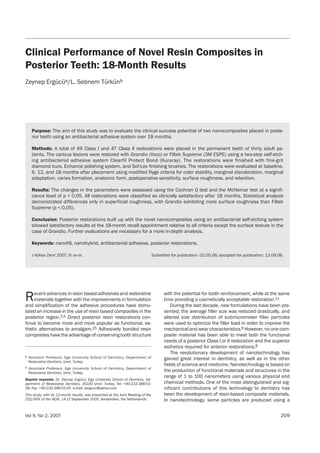
  • 5. Vol 9, No 2, 2007 213 Ergücü/Türkün each recall appointment. For each subject, the number of teeth restored using each nanocomposite was equal, so that each restoration could be compared with its own same-sub- ject control. The Cochran-Q test was used to evaluate the changes across the four time points for each of the criteria. The two resin composites were compared at the same recall period for each criterion using the McNemar test. For all of the sta- tistical analyses, p was set at 0.05. RESULTS Thirty patients requiring posterior resin composite restora- tions were included in this study; however, two patients were unavailable for recall appointments because they had moved. Of the 96 restorations placed, 90 novel resin com- posite (Grandio and Filtek Supreme) restorations that were placed with the antibacterial two-step adhesive system (Clearfil Protect Bond) in 28 patients were evaluated at the 18-month recall appointment. The clinical assessment re- sults are shown in Table 4. Esthetics and tooth vitality were all rated satisfactory for the two nanocomposites investigated (Figs 1 and 2). Gingi- val response was evaluated with a periodontal probe using a gingival index.31 There was no inflammation, color change, or bleeding around the restorations, and gingival status was normal. None of the restorations showed evidence of sec- ondary caries formation (Figs 3 and 4), and no postoperative sensitivity was reported by any of the patients at any recall. One restoration of each resin composite showed slightly dis- colored margins at the 6-, 12-, and 18-month recalls (p > 0.05) (Figs 5 and 6). Baseline 6 months 12 months 18 months Criterion Filtek Grandio Filtek Grandio Filtek Grandio Filtek Grandio Supreme Supreme Supreme Supreme Color matching ability A 48 48 48 48 48 48 45 45 B 0 0 0 0 0 0 0 0 C 0 0 0 0 0 0 0 0 Cavosurface margin discoloration A 48 48 47 47 47 47 44 44 B 0 0 1 1 1 1 1 1 C 0 0 0 0 0 0 0 0 Marginal adaptation A 48 48 48 48 48 48 45 45 B 0 0 0 0 0 0 0 0 C 0 0 0 0 0 0 0 0 Secondary caries formation A 48 48 48 48 48 48 45 45 B 0 0 0 0 0 0 0 0 Surface texture A 48 48 48 0 48 0 45 0 B 0 0 0 48* 0 48* 0 45* C 0 0 0 0 0 0 0 0 Anatomical form A 48 48 48 48 48 48 45 45 B 0 0 0 0 0 0 0 0 C 0 0 0 0 0 0 0 0 1Source: Ryge Alpha (A) indicates excellent restorations, while Bravo (B) and Charlie (C) indicate progressively worsening degrees of clinical acceptability. *p < 0.05 Statistically significant difference between the materials. Table 4 Ryge criteria obtained at each recall1
  • 6. Ergücü/Türkün 214 The Journal of Adhesive Dentistry Fig 5 Occlusal restoration of Grandio in the maxillary right second molar exhibited a marginal discoloration after 18 months. (G): Grandio. Fig 6 Marginal discoloration at the cavosurface margin of an oc- clusal restoration of Filtek Supreme in the maxillary left second mo- lar at the 18-month recall. (FS): Filtek Supreme. Fig 1 Class II restorations in maxillary left second premolar and first molar after 18 months. The disto-occlusal composite of the second premolar was Filtek Supreme and the mesio-occlusal com- posite in the first molar was Grandio. (FS): Filtek Supreme (G): Grandio. Fig 2 Class II restorations in maxillary right premolars after 18 months. Color match, anatomical form and marginal adaptation were clinically excellent. (FS): Filtek Supreme (G): Grandio. Fig 3 Occlusal restoration of Grandio in the maxillary right first mo- lar at the 18-month recall appointment. (G): Grandio. Fig 4 Occlusal restoration of Filtek Supreme in the maxillary left molar after 18 months. (FS): Filtek Supreme.
  • 7. Grandio exhibited a statistically significantly rougher sur- face texture than Filtek Supreme (p < 0.05). Nevertheless, there were no patient complaints regarding the surface roughness of these restorations. All restorations were clas- sified as clinically acceptable after 18 months. DISCUSSION The two novel resin composite restorative materials evalu- ated in this study were used in combination with a two-step self-etching antibacterial adhesive system. The clinical per- formance of adhesives has improved significantly, allowing adhesive restorations to be placed with a highly predictable level of clinical success. It is considered that modern adhe- sive systems, being superior to their predecessors especial- ly in terms of retention, are no longer the main cause of pre- mature clinical failure.33,36 A clinically effective adhesive system should keep the restoration in place for a significant time and completely seal the restoration margins against the ingress of oral fluids and microorganisms. Incomplete marginal sealing will result in postplacement sensitivity, marginal staining, and recurrent caries, which are still the most common symptoms associ- ated with clinical failure of adhesive restorations.28 Postop- erative sensitivity was the most frequent complication of ear- ly posterior composite restorations. With the introduction of self-etcihng adhesives that are able to completely penetrate into decalcified dentin and/or to obdurate dentinal tubules, the incidence of postoperative sensitivity has dropped. In our study, Clearfil Protect Bond was used as the dentin ad- hesive system for both nanocomposites instead of the rec- ommended proprietary dentin adhesives, because this an- tibacterial adhesive system has not been previously used in combination with novel resin composites in the posterior re- gion and it might be of benefit to use this system in order to eliminate the risk of secondary caries in deep cavities. Fur- thermore, since the main focus of this investigation was to compare the clinical success of two novel resin composites, we wanted to standardize the type of the adhesive system. The results of this clinical study demonstrated that both nov- el composites placed in combination with Clearfil Protect Bond performed well after 18 months. In a previous study, Türkün34 investigated the clinical performance of Clearfil Protect Bond and Xeno III in combination with the resin com- posite Esthet-X (Dentsply DeTrey) in noncarious cervical le- sions after 1 year, and reported that Clearfil Protect Bond was clinically successful during this period without causing any postoperative sensitivity or side effects. The modified Ryge criteria (USPHS) for direct evaluation remains the preferred system for evaluating important char- acteristics of dental restorations, such as color matching, secondary caries, cavosurface margin discoloration, and postoperative sensitivity. Regarding the loss of anatomical form, the direct evaluation method is able to generate data that are of clinical significance and are compatible with clin- ical treatment.13,19,20 Compared with the baseline observa- tions, direct evaluations made using the modified Ryge cri- teria demonstrated that color match, cavosurface margin discoloration, and caries formation remained essentially un- changed at the end of 18-month period for the two nanocom- posites. Polishability is one of the desirable features of tooth-col- ored dental restoratives. High quality finishing and polishing can improve the esthetics and longevity of these materials. Nowadays, several restorative materials with finer organic particles are produced by means of advanced technology. The latest innovations are the nanocomposites. In vitro tests showed that nanocomposites displayed high polish reten- tion after toothbrush abrasion. During the abrasion tests, on- ly nanosized particles are plucked away, leaving the sur- faces with smaller defects than the wavelength of light, and better surface characteristics than the conventional hybrid composites are obtained.9,24 In regard to surface texture, direct evaluations of Grandio restorations at the 6- and 12-month recall appointment were scored as Bravo for all the restorations. At the 18-month re- call, all of the Grandio restorations were again slightly rough and thus were scored as Bravo (p < 0.05). The slight rough- ness indicated that Bravo scores were perceptible only with a sharp explorer and none of the patients noticed or com- plained about the situation. Mesquita et al22 demonstrated that Grandio showed a rigidity approaching that of dentin. Concerning surface roughness, the special hardness of Grandio can be seen as a main cause. In the present authors’ opinion, some special polishing systems may be of use in optimizing the surface texture of this material after finishing procedures. In the present study, both nanocomposites showed ex- cellent marginal integrity at the end of the 18-month recall period. Frankenberger et al12 compared the performance of Grandio and Tetric Ceram in posterior teeth and reported that no deterioration regarding the modified Ryge criteria was found except for marginal integrity. According to their findings, Grandio showed an equal outcome to Tetric Ceram after one year of clinical service. Ernst et al10 compared the clinical performance of Filtek Supreme and Tetric Ceram placed in posterior teeth in a two- year clinical study, finding that there were no statistically sig- nificant differences between the two materials and that Fil- tek Supreme could be used successfully in large stress-bear- ing posterior cavities. Since adhesive dentistry is technique sensitive, the suc- cess of an adhesive restoration is mostly determined by the ability of the dentists, beyond the performance of the mate- rial. Even with the improved materials, the performance of the material cannot be overestimated to exceed that of the dentist. Therefore, it is still important for the clinician to practice appropriately and meticulously when using adhe- sive materials. Careful case selection and meticulous at- tention to the restorative technique are recognized as es- sential requirements for achieving longer-term clinical suc- cess with posterior composite restorations. Clinical trials are limited in number and require several years with regular recall appointments to achieve sufficient clinical validation. Owing to the rapid evolution in resin com- posite technology and the higher turnover of novel resin composites, these materials are released onto the dental market without sufficient proof of their clinical performance. Long-term quantitative observations are essential to deter- Vol 9, No 2, 2007 215 Ergücü/Türkün
  • 8. Ergücü/Türkün 216 The Journal of Adhesive Dentistry mine the clinical performance of new posterior resin com- posite materials, and selection should be primarily based on these results. This study was planned to be a long-term clin- ical trial; however, due to the shading problems of Filtek Supreme encountered by some opinion leaders, the manu- facturer decided to take the material off the market, and we had to terminate the study, although both materials per- formed well in our 18-month clinical trial. Furthermore, we did not use the layering technique for the posterior restora- tions, and used only one shade for the incremental tech- nique. Thus, the shading problems with the restorations were not seen, and we replaced only the ones that had dis- colorations after 30 months. Similar to the present results, Ernst et al10 reported that Filtek Supreme restorations showed heavy staining at the fissures, while Tetric Ceram restorations showed none after 2 years. Currently, the shad- ing system of Filtek Supreme has been optimized and the product is now being marketed as Filtek Supreme XT. The findings of the current study demonstrated that both nanocomposites performed equally well after 18 months in spite of the surface roughness observed in Grandio restora- tions. Although an 18-month observation period is not long, the clinical problems encountered in this study were very few. Selecting limited-sized cavities and paying strict atten- tion to the restorative technique are essential for achieving longer-term clinical success with posterior resin based com- posite restorations. CONCLUSION It may be concluded that, with the improvement in materi- als, careful case selection and application of the restorative techniques, the novel resin composites can be used for pos- terior restorations. Evaluations still need to be carried out to reveal longer-term clinical performance of such materials. ACKNOWLEDGMENTS The authors would like to thank the companies Voco, 3M ESPE, and Ku- raray Dental for their generous donation of the materials used in this study. REFERENCES 1. 3M Dental Products Filtek Supreme Plus Universal Restorative System. Tech- nical Product Profile. St Paul MN, USA, 2005:9-12. 2. Baseren M. Surface roughness of nanofill and nanohybrid composite resin and ormocer-based tooth-colored restorative materials after several finishing and polishing procdures. J Biomater Appl 2004;19:121-34. 3. Bayne SC, Heymann HO, Swift Jr ER. Update on dental composite restora- tions. J Am Dent Assoc 1994;125:687-701. 4. Beun S, Glorieux T, Devaux J, Vreven J, Leloup G. Characterization of nanofilled compared to universal and microfilled composites. Dent Mater Jan 16. (Epub of ahead of print) 5. Brunthaler A, König F, Lucas T, Sperr W, Schedle A. Longevity of direct resin composite restorations in posterior teeth. Clin Oral Invest 2003;7:63-70. 6. Bryant RW. Direct posterior composite resin restorations: a review. 1. Factors influencing case selection. Aust Dent J 1992;37:81-7. 7. Clinician’s preferences 2001. CRA Newsletter 2001;25:1-4. 8. Denehy GE. A direct approach to restore anterior teeth. Am J Dent 2000;13(special issue):55-9. 9. Ergücü Z, Türkün LS. Surface Roughness of Novel Resin Composites Polished with One-step Systems. Oper Dent 2007;32:185-192. 10. Ernst CP, Brandenbusch M, Meyer F, Canbek K, Gottschalk F, Willershausen B. Two year clinical performance of a nanofiller vs a fine-particle hybrid resin composite. Clin Oral Invest 2006;10:119-125. 11. Fortin D, Vargas MA. Two spectrums of composites: new techniques and ma- terials. J Am Dent Assoc 2000;131(supplement):26S-30S. 12. Frankenberger R, Reinelt C, Eimer F, Krämer N. Posterior restorations with a nanofilled resin composite- results after 12 months. J Dent Res 2005;(ab- stracts Baltimore):579. 13. Geurtsen W, Schoeler U. A 4-year retrospective clinical study of Class 1 and Class 2 composite restorations. J Dent 1997;25:229-32. 14. Haller B. Recent development in dentin bonding. Am J Dent 2000;13:44-50. 15. Imazato S, Ebi N, Tarumi H, Russell RRB, Kaneko T, Ebisu S. Bactericidal ac- tivity and cytotoxicity of antibacterial monomer MDPB. Biomaterials 1999;20:899-903. 16. Imazato S, Kinomoto Y, Tarumi H, Ebisu S, Tay FR. Antibacterial activity and bonding characteristics of an adhesive resin containing antibacterial monomer MDPB. Dent Mater 2003;19:313-319. 17. Imazato S, Kinomoto Y, Tarumi H, Russell RR, McCabe JF. Incorporation of antibacterial monomer MDPB in dentin primer. J Dent Res1997;76:768- 772. 18. Imazato S. Antibacterial properties of resin composites and dentin bonding systems. Dent Mater 2003;19:449-457. 19. Leinfelder KF, Taylor DF, Barkmeier WW, Goldberg AJ. Quantitative wear mea- surement of posterior composite resins. Dent Mater 1986;2:198-201. 20. Leinfelder KF, Wilder AD Jr., Teixeira LC. Wear rates of posterior composite resins. J Am Dent Assoc 1986;112:829-833. 21. Leinfelder KF. Evaluation of criteria used for assessing the clinical perfor- mance of composite resins in posterior teeth. Quintessence Int 1987; 18:531-536. 22. Mesquita RV, Axmann D, Geis-Gerstofer J. Dynamic visco-elastic properties of dental composite resins. Dent Mater 2005;Sep 2 (Epub ahead of print)xxAuthor: Can’t be, if published in 2005!!. 23. Miller MB. Self-etching adhesives: solving the sensitivity conundrum. Pract Proced Aesthet Dent 2002;14:406. 24. Mitra SB, Wu D, Holmes BN. An application of nanotechnology in advanced dental materials. J Am Dent Assoc 2003;134:1382-90. 25. Morgan M. Finishing and polishing of direct posterior resin restorations. Pract Proced Aesthet Dent 2004;16:211-217. 26. Moszner N, Klapdohr S. Nanotechnology for dental composites. Int J Nan- otechnol 2004;1:130-156. 27. Moszner N, Salz U. New developments of polymeric dental composites. Prog Polym Sci 2001;26:535-576. 28. Qvist V, Qvist J, Mjör IA. Placement and longevity of tooth-colored restorations in Denmark. Acta Odontol Scand 1990;48:305-311. 29. Raskin A, Michette-Theall B, Vreven J, Wilson NH. Clinical evaluation of a pos- terior composite 10-year report. J Dent 1999;27:13-19. 30. Ryge G. Clinical criteria. Int Dent J 1980;30:347-358. 31. Silness P, Löe H. Periodontal disease in pregnancy . Acta Odontol Scand 1964;2:121-135. 32. Terry DA. Direct applications of a nanocomposite resin system. Part 1. The evoluotion of contemporary composite materials. Pract Proced Aesthet Dent 2004;16:417-422. 33. Türkün LS. Clinical evaluation of a self-etching and a one-bottle adhesive sys- tem at two years. J Dent 2003;31:527-534. 34. Türkün LS. The clinical performance of one-and two-step self-etching adhe- sive systems at one year. J Am Dent Assoc2005;136:656-664. 35. Turssi CP, Ferracane JL, Ferracane LL. Wear and fatigue behavior of nano- structured dental composites. J Biomed Mater Res B Appl Biomater 2006 Jan 30. (Epub ahead of print) 36. Van Meerbeek B, Perdigão J, Lambrechts P, Vanherle G. The clinical perfor- mance of adhesives. J Dent 1998;26:1-20. 37. Voco Grandio. Light-curing Nano-hybrid Filling Material, Corresponds to EN 24049/ ISO 4049 Instructions for use. April 2003. Clinical relevance: Posterior restorations built up with the novel nanocomposites using an antibacterial self-etching system showed satisfactory results at the 18-month recall appointment relative to all criteria except the surface tex- ture in the case of Grandio (p < 0.05). The surface rough- ness of this material was not found to be clinically impor- tant, as the patients did not have any complaints and some were not even aware of it.