

Recomendados
Mais conteúdo relacionado
Mais procurados
Mais procurados (19)
Semelhante a Ermak styela clava glycogen deposits 1977
Semelhante a Ermak styela clava glycogen deposits 1977 (20)
Mais de Thomas Ermak
Mais de Thomas Ermak (6)
Ermak styela clava glycogen deposits 1977
- 1. Cell Tiss. Res. 176,47 55 (1977) Cell and Tissue Research 9 by Springer-Verlag 1977 Glycogen Deposits in the Pyloric Gland of the Ascidian Styela clara (Urochordata) * Thomas H. Ermak** Department of Zoology, Universityof California, Berkeley,California, USA Summary. The pyloric gland of Styela clava contains large glycogen deposits that are digested by treatment with alpha amylase and depleted by 15 days starvation. The deposits are surrounded by cytoplasmic regions containing smooth endoplasmic reticulum and mitochondria. The cells also have rough endoplasmic reticulum, Golgi cisterns, lysosomes, microvilli, cilia, and lateral infoldings of the plasma membrane. The fine structure of the pyloric cells and the position of tubules between the absorptive epithelium and general circulation suggest that the gland functions as the vertebrate liver in carbo- hydrate metabolism. The pyloric ceils of Styela do not appear to be excretory in a 'renal' sense, since there is no infolding of the basal plasmalemma and mitochondria are usually associated only with the glycogen deposits. However, a hepatic-like excretory role is consistent with current findings. In light of the phylogenic affinities of vertebrates and ascidians, it is possible that the pyloric gland is homologous to the liver. Key words: Glycogen - Pyloric gland - Ascidian - Ultrastructure. Introduction The ascidian pyloric gland is an enigmatic organ to which have been ascribed excretory, osmoregulatory, and digestive functions (Fouque, 1953; Millar, 1953; Gaill, 1973; G o o d b o d y , 1974). In Styela clara, an advanced solitary ascidian, the distribution of pyloric tubules beneath the renewing intestinal epithelium suggests that these structures are involved in nutrient assimilation (Ermak, 1975). In a colonial ascidian, Sidnyum argus, small glycogen deposits were recently observed in the pyloric cells (Gaill, 1974a). The present electron microscopic Send offprint requests to : Dr. Thomas H. Ermak, Department of Physiology,Universityof California Medical Center, San Francisco, California 94143, USA * Supported by USPHS grant GM 10292 to Dr. Richard M. Eakin ** I am grateful to Dr. Eakin for his support and valuable criticism of the manuscript
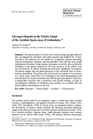
- 2. 48 Th.H. Ermak investigation was undertaken to examine the putative glycogen deposits in Styela clava and to re-evaluate the function of the pyloric gland. The identity of glycogen was elucidated by enzymatic digestion and the periodic acid Schiff (PAS) reaction. The cellular structure ofa pyloric tubule was also studied under starved conditions. Materials and Methods Specimens of S t y e l a clava were collected from Mission Bay, San Diego, and the Berkeley Marina, Berkeley, California. Parts of the intestinal wall immediately posterior to the stomach were fixed in 3 ~ glutaraldehyde in 0.1 M phosphate buffer (pH 7.3) with 0.7 M sucrose for 1.5 h at room temperature. Some specimens were then incubated at 35~ in 0.5 ~o alpha amylase (type III A, Sigma Chemical Co.; activity 50-100 units/mg) in 0.1 M phosphate buffer for 1 to 3 h. Prior to incubation, intestinal samples were rinsed in 0.1 M phosphate buffer to remove the sucrose in the glutaraldehyde fixative. Controls were incubated in heat-treated amylase in 0.I M phosphate buffer. Several animals were starved for 15 days before fixation. All specimens were postfixed in 1~o ice cold osmium tetroxide in 0.1 M phosphate buffer (pH 7.3) with 0.7 M sucrose for 1 h and embedded in Epon. Thick sections (1 ~tm) were stained with PAS (Leeson and Leeson, 1970) or toluidine blue. Silver-gold sections were stained with uranyl acetate and lead citrate and examined with an RCA-3G electron microscope operating at 100 kv. Results The pyloric gland of Styela clava is composed of numerous diffuse tubules which lie in the gut wall. The tubules are most concentrated in the intestine, the region of most intense nutrient absorption, and collect into a canal that leads into the stomach at its junction with the intestine. The intestinal wall (Fig. 1) consists of 3 layers: (1) an inner epithelium con- taining absorptive and secretory cells; (2) a middle layer of connective tissue, blood channels, blood cells, and pyloric tubules; and (3) an outer atrial epithelium, the lining of the body cavity. The pyloric tubules lie directly below the gut epi- thelium; none are located next to the atrial epithelium. Light microscopy of thick sections stained by the PAS technique shows large regions of intense reaction in the pyloric cells (Fig. 1). The atrial epithelium and some blood cells also show small regions of positive reaction, attributable possibly to glycogen or PAS positive mucous granules. Electron microscopy of the pyloric cells (Fig. 2) reveals large regions containing single (400 A) and aggregated particles of glycogen contained within a matrix of light, granular material. The aggregates are not arranged in typical alpha rosettes, and the single units appear compound (Fig. 3). The glycogen deposit, which excludes most cytoplasm and cytoplasmic organelles, is always surrounded by a layer of cytoplasm containing smooth endoplasmic reticulum and never borders Fig. 1. Light micrograph of intestinal wall, PAS staining, ae atrial epithelium; ct connective tissue; ie intestinal epithelium; pt pyloric tubules, x 250 Fig. 2. Electron micrograph of part of pyloric tubule showing large glycogen deposit (g). er endo- plasmic reticulum; M interdigitation; m mitochondrion; m y microvilli; tj tight junction, x 24,000
- 3. Glycogen in Ascidian Pyloric Gland 49 Fig. 3. Cytoplasmic matrix containing smooth endoplasmic reticulum (ser) in parallel with lateral plasma membranes (pm). g glycogen. • 48,000
- 4. 50 Th.H. Ermak Fig. 4. Light micrograph of pyloric tubule treated with alpha amylase for 1 h, toluidine blue staining. bc blood cells; l lumen; s space, x 640 Fig. 5. Electron micrograph ofpyloric cell treated with alpha amylase for 1 h. c connective tissue fibers; er endoplasmic reticulum; ly lysosome; m mitochondrion; n nucleus; s space, x 24,000 directly upon the plasma membrane. When the glycogen lies close to a lateral plasma membrane, the intervening cytoplasmic matrix may be as narrow as 0.1 to 0.2 gm, and the tubules of endoplasmic reticulum are arranged in parallel (Fig. 3). Mitochondria occur throughout the cytoplasm and frequently border directly on a glycogen deposit (Fig. 2). The cell also contains Golgi cisterns and lysosomes. Although no infoldings of the basal plasmalemma are observed, the pyloric
- 5. Glycogenin Ascidian Pyloric Gland 51 Fig. 6. Pyloric ceils from ascidian starved 15 days. g a Golgi apparatus; M interdigitation; m mito- chondrion; m v microvilli;c! cilia; tj tight junction, x 35,000 cells exhibit interdigitations of lateral plasma membranes (Figs. 2, 6). A tight junction (Mackie et al., 1974) occurs at the apical cell border (Figs. 1, 6); several gap-like junctions are seen between the tight junction and the basal border. The cell apex bears numerous microvilli 2 gm in length and at least one long cilium. Cilia in the tubules are sometimes so numerous that they fill the lumen. Treatment with alpha amylase eliminates the PAS-reaction leaving large unstained regions in the pyloric tubules but not in most other tissues. These spaces are particularly evident in toluidine blue stained 1 gm thick sections (Fig. 4). In thin sections, amylase removes the glycogen particles leaving large spaces (Fig. 5). A 1 h incubation period produces little alteration in cytoplasm, cellular membranes, and connective tissue fibers. Longer incubation, however,
- 6. 52 Th.H. Ermak disrupts microvilli and washes out cytoplasmic matrix. The reaction is inhibited by heat denaturation and sucrose in the incubation medium. Mucous granules in atrial epithelial cells (see below) are not digested by amylase. After 15 days starvation, glycogen deposits are lost from the pyloric epithelium (Fig. 6). The cells appear smaller in size; presumably at least a part of this decrease is due to the loss of glycogen. Elements of smooth and rough endoplasmic reti- culum fill most of the cytoplasm; however, smooth endoplasmic reticulum no longer occurs parallel to the plasma membrane (see Discussion). Atrial epithelial cells are low columnar cells containing several membrane- bounded mucous granules, presumably the PAS positive granules of light micro- scopy. They also have irregular apical and basal borders, and interdigitating baso-lateral plasma membranes. Several blood cells in the connective tissue also contain mucous granules and glycogen. Discussion The results of this investigation clarify several points concerning the function of the pyloric gland. Based upon ultrastructural evidence. Gaill (1974a) suggested that the gland might be excretory because of infoldings of the basal plasmalemma and the presence of microvilli and cilia. In Styela, the infoldings are actually from the lateral plasma membrane, a condition in common with the atrial epi- thelium and, in general, all transporting epithelia (Berridge and Oschman, 1972). The pyloric cells in Styela differ in morphology from true ascidian renal epithelial cells (Gaill, 1974b) and also plicated cells in the gut (Degail and Levi, 1964; Burighel and Milanesi, 1975) which exhibit distinct basal membrane infoldings containing numerous mitochondria. In the pyloric gland, mitochondria in the basal part of a cell are more intimately in contact with the glycogen deposit than with the plasmalemma. Fouque (1953) suggested that the pyloric cells are engaged in 're-working' substances absorbed by intestinal cells. The fine structure of the tubules supports this contention; the pyloric gland of Styela is most likely actively engaged in carbohydrate metabolism. Digestion of the large deposits by alpha amylase (as demonstrated by Biava, 1963, and Coimbra, 1967) but not of other PAS-positive sites in the intestinal wall indicates that the particles are glycogen. The deposits in Styela are much larger than those in Sidnyum (Gaill, 1974a). Since glycogen content typically varies with nutritional state, a difference between the two species might merely reflect variability in storage at the time of fixation. Alter- natively, different species of ascidians might store glycogen in the pyloric gland to different degrees. Among invertebrates, glycogen is usually concentrated in a specific region of the gut, such as the crustacean hepatopancreas (Vonk, 1960), the gastropod digestive gland (Chatterjee and Ghose, 1974), and the echinoderm intestine (Doezema and Phillips, 1970). In Styela, the pyloric gland is the major gut region to store glycogen. Small amounts of glycogen also occur in the ascidian stomach (Burighel and Milanesi, 1973), intestine (Gaill, 1974 a), and digestive diverticulum (Degail and Levi, t 964). Blood cells which contain glycogen are probably amoeb-
- 7. Glycogenin Ascidian PyloricGland 53 ocytes. In Perophora viridis, granular amoebocytes, which are presumed to be nutritive storage cells, contain masses of 200-300 A particles of glycogen (Overton, 1966). In an ascidian tadpole ocellus, the lens is a large glycogen deposit composed of beta granules (300-400 A in diameter) and surrounded by numerous mito- chondria (Eakin and Kuda, 1972). Refringent granules (Fouque, 1953) and solid concretions (Millar, 1949; 1953) have also been described in the pyloric gland. In light of the refractive qualities of glycogen, such as that in an ascidian tadpole lens (Eakin and Kuda, 1972), it is possible that the granules are actually glycogen. Those concretions illustrated by Millar (1953, Fig. 4, p. 33) in the pyloric gland of Ciona intestinalis correspond in size and shape to the glycogen deposits in Styela (compare to Fig. 1, this investigation). It is also possible that the vacuoles described in a pyloric cell (Millar, 1953) represent spaces where glycogen was lost in the preparation of the sections, since glycogen is frequently removed during routine histological procedures (Bloom and Fawcett, 1968, p. 591). The disposition of vacuoles in Ciona is like the distribution of spaces in Styela created by the digestion of glycogen by alpha amylase. Glycogen in a pyloric cell disappears with starvation. As in a mammalian hepatocyte (Cardell et al., 1973), beta and alpha particles of glycogen are associated with regions of smooth endoplasmic reticulum and mitochondria. Rough endo- plasmic reticulum, however, is not so well developed as that in the mammalian hepatocyte. In autoradiograms of liver cells exposed to tritiated glucose, label is localized over both glycogen particles and smooth endoplasmic reticulum (Coimbra and Leblond, 1966). Glucose is apparently added to growing glycogen particles, since peripheral D-glucosyl residues are renewed more rapidly than those in the core of a glycogen molecule (see Stetten and Stetten, 1960). The parallel arrangement of smooth endoplasmic reticulum between a glycogen deposit and a lateral plasma membrane is similar to subsurface endoplasmic reticula in mouse hepatocytes (Tandler, 1974). However, since this pattern disappears with the loss of glycogen during starvation, the tubules of smooth endoplasmic reticulum are probably not subsurface cisterns but only aligned in parallel because of the small amount of cytoplasm surrounding them. The fine structural similarities between ascidian pyloric cells and vertebrate hepatic cells as well as the distribution of pyloric tubules beneath the intestinal epithelium suggest that the pyloric gland might play a wider role in nutrient assimilation. In Styela, the entire intestinal lining is a renewing epithelium (Ermak, 1975). A groove of relatively undifferentiated cells runs along one side of the intestine. These germinal cells replace the absorptive and secretory cells on the side walls in about a month. Beneath the germinal cells, the pyloric tubules are few in number. Under the absorptive cells, however, the tubules are exceedingly numerous. All food products passing from the gut lumen to the blood channels pass through this meshwork of tubules. Like the vertebrate liver, which is inter- posed between the digestive tract and general circulation, the pyloric gland might act as a processing station transforming nutrients absorbed by the gut lining and passing them to blood cells which then distribute them to the rest of the body. Absorbed products of digestion, whether metabolized or transformed by the pyloric gland, would quickly be distributed throughout the body by way of
- 8. 54 Th.H. Ermak o p e n b l o o d channels (lined only by connective tissue), since the posterior end o f the heart leads directly to the gut. In ascidians, the heart is unusual in that it periodically reverses beat direction, one time p u m p i n g blood towards the pharynx, the next time towards the viscera. Like the vertebrate liver, the pyloric gland develops as a diverticulum o f the gut. Sections o f closely packed tubules in Styela are reminescent o f sections o f liver cords. A l t h o u g h the pyloric cells are involved in c a r b o h y d r a t e metabolism, lipid metabolism, which is also a function o f the vertebrate liver, is p r o b a b l y handled directly by the gut epithelium. Lipid droplets occur in great n u m b e r in the s t o m a c h lining (Burighel and Milanesi, 1973) but usually not in pyloric cells. In Styela, lipid reserves in absorptive cells o f the s t o m a c h and intestine fluctuate seasonally and are also depleted by starvation (Ermak, unpublished results). A l t h o u g h the pyloric gland does not have the ultrastructural features o f a renal o r g a n (see above), it p r o b a b l y has an hepatic-like excretory function. T h o r i u m dioxide which has been injected into the b o d y cavity o f Ciona intestinalis is p h a g o c y t o s e d by blood cells and eliminated by the intestine via the pyloric gland (Brown and Davies, 1971). In Styela plieata, the pyloric gland takes up vital dyes f r o m the blood (Fouque, 1953). Cilia in a pyloric tubule would, then, aid in transporting any secreted substances d o w n the pyloric duct to the intestine (see G o o d b o d y , 1974). Thus the ultrastructural, positional, and functional characteristics o f the pyloric gland are strikingly similar to those o f the vertebrate liver. Indeed, the likelihood that vertebrates evolved f r o m primitive ascidian stocks (Berrill, 1955) suggests that the pyloric gland is h o m o l o g o u s to the liver. If this is the case, the pyloric gland might be expected to possess other hepatic functions. References Berridge, M.J., Oschman, J.L.: Transporting epithelia. New York: Academic Press 1972 Berrill, N.J.: The origin of vertebrates. Oxford: Clarendon Press 1955 Biava, C.: The identification and structural forms of human particulate glycogen. Lab. Invest. 12, 1179 1197(1963) Bloom, W., Fawcett, D.W.: A textbook of histology. Philadelphia: W.B. Saunders Co. 1968 Brown, A.C., Davies, A.B.: The fate of thorium dioxide introduced into the body cavity of Ciona intestinalis. J. Invert. Path. 18, 276-279 (1971) Burighel, P., Milanesi, C.: Fine structure of the gastric epithelium of the ascidian Botryllus schlosseri. Vacuolated and zymogen cells. Z. Zellforsch. 145, 541-555 (1973) Burighel, P., Milanesi, C.: Fine structure of the gastric epithelium of the ascidian Botryllus schlosseri. Mucous, endocrine and plicated cells. Cell Tiss. Res. 158, 481-496 (1975) Cardell, R.R., Larner, J., Babcock, M.B.: Correlation between structure and glycogen content of livers from rats on a controlled feeding schedule. Anat. Rec. 177, 23-38 (1973) Chatterjee, B., Ghose, K.C.: Seasonal variation in stored glycogen and lipid in the digestive gland and genital organs of two freshwater prosobranchs. Proc. Mal. Soc. Lond. 40, 407-412 (1974) Coimbra, A.: Evaluation of the glycogenolytic effect of alpha amylase using radioautography and electron microscopy. J. Histochem. Cytochem. 14, 898-905 (1967) Coimbra, A., Leblond, C.P.: Sites of glycogen synthesis in rat liver cells as shown by EM radioauto- graphy after administration of glucose-H3. J. Cell Biol. 30, 151-176 (1966) Degail, L., Levi, C.: Etude au microscope 61ectronique de la glande digestive des Pyuridae (Ascidies). Cah. Biol. Mar. 5, 411-422 (1964) Doezema, P., Phillips Jr., J.H.: Glycogen storage and synthesis in the gut of the purple sea urchin, Strongylocentrotus purpuratus. Comp. Biochem. Phys. 34, 691-697 (1970)
- 9. Glycogen in Ascidian Pyloric Gland 55 Eakin, R.M., Kuda, A.: Glycogen in lens of tunicate tadpole (Chordata: Ascidiacea). J. exp. Zool. 180, 26%270 (1972) Ermak, T.H.: Cell proliferation in the digestive tract of Styela clara (Urochordata: Ascidiacea) as revealed by autoradiography with tritiated thymidine. J. exp. Zool. 194, 449-466 (1975) Fouque, G.: Contribution /l l'6tude de la glande pylorique des ascidiac6s. Ann. Inst. Oceanogr. (Paris) 28, 189-317 (1953) Gaill, F.: Etude histologique de la glande pylorique de Synoicum argus (Polyclinidae, Tuniciers). Arch. Zool. Exp. Gen. 114, 97-110 (1973) Gaill, F.: Aspect ultrastructural de la glande pylorique et de l'intestin post6rieur de Sidnyum argus (Polyclinidae, Tuniciers). Cah. Biol. Mar. 15, 337 341 (1974a) Gaill, F.: Observations ultrastructurales de l'epithelium r6nal de Molgula occulta (Ascidies, Tuniciers). C.R. Acad. Sci. (Paris) 279, 351-353 (1974b) Goodbody, I.: The physiology of ascidians. Adv. Mar. Biol. 12, 1-150 (1974) Leeson, C.R., Leeson, T.S.: Staining methods for sections of epon-embedded tissues for light micro- scopy. Canad. J. Zool. 48, 189-191 (1970) Mackie, G.O., Paul, D.H., Singla, C.M., Sleigh, M.A., Williams, D.E.: Branchial innervation and ciliary control in the ascidian Corella. Proc. roy. Soc. B 187, 1-35 (1974) Millar, R.H.: Concretions in the pyloric gland of Ciona intestinalis. Nature (Lond.) 164, 717-718 (1949) Millar, R.H.: Ciona, L.M.B.C. Memoirs. Liverpool: Colman 1953 Overton, J.: The fine structure of blood cells in the ascidian Perophora viridis. J. Morph. 119, 305-326 (1966) Stetten, D. Jr., Stetten, M.R.: Glycogen metabolism. Physiol. Rev. 40, 505-537 (1960) Tandler, B. : Subsurface cisterns in mouse hepatocytes. Anat. Rec. 179, 273-284 (1974) Vonk, H.J.: Digestion and metabolism. In: Physiology of crustacea (T.H. Waterman, ed.), Vol. 1, p. 291. New York: Academic Press 1960 Accepted August 6, 1976