
Recomendados
Mais conteúdo relacionado
Mais procurados
Mais procurados (16)
Semelhante a 52 ch11mitosis2008
Semelhante a 52 ch11mitosis2008 (20)
Mais de sbarkanic
Mais de sbarkanic (20)
Último
Último (20)
52 ch11mitosis2008
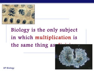
- 1. Biology is the only subject in which multiplication is the same thing as division… AP Biology 2007-2008
- 2. The Cell Cycle: Cell Growth, Cell Division AP Biology 2007-2008
- 3. Where it all began… You started as a cell smaller than a period at the end of a sentence… AP Biology
- 4. And now look at you… AP Biology How did you get from there to here?
- 5. Getting from there to here… Going from egg to baby…. the original fertilized egg has to divide… and divide… and divide… and divide… AP Biology
- 6. Why do cells divide? For reproduction asexual reproduction one-celled organisms For growth from fertilized egg to multi-celled organism For repair & renewal AP Biology replace cells that die from normal wear & tear or from injury amoeba
- 7. Making new cells Nucleus chromosomes DNA Cytoskeleton centrioles in animals AP Biology microtubule spindle fibers
- 8. Nucleus DNA Function chromosome protects DNA Structure histone protein nuclear envelope double membrane membrane fused in spots to create pores allows large macromolecules to pass through nuclear pores AP Biology What kind of molecules need to pass through? nuclear pore nucleolus nuclear envelope
- 9. AP Biology
- 10. Cytoskeleton Function structural support maintains shape of cell provides anchorage for organelles protein fibers microfilaments, intermediate filaments, microtubules motility cell locomotion cilia, flagella, etc. regulation organizes structures & activities of cell AP Biology
- 11. Cytoskeleton actin microtubule nuclei AP Biology
- 12. Centrioles Cell division in animal cells, pair of centrioles organize microtubules spindle fibers AP Biology guide chromosomes in mitosis
- 13. End of the Tour AP Biology
- 14. Getting the right stuff What is passed on to daughter cells? exact copy of genetic material = DNA mitosis organelles, cytoplasm, cell membrane, enzymes cytokinesis chromosomes (stained orange) in kangaroo rat epithelial cell AP Biology →notice cytoskeleton fibers
- 15. Overview of mitosis interphase prophase I.P.M.A.T. (pro-metaphase) cytokinesis AP Biology metaphase anaphase telophase
- 16. Interphase 90% of cell life cycle cell doing its “everyday job” produce RNA, synthesize proteins/enzymes prepares for duplication if triggered I’m working here! Time to divide & multiply! AP Biology
- 17. M Mitosis Cell cycle Cell has a “life cycle” cell is formed from a mitotic division cell grows & matures to divide again G1, S, G2, M epithelial cells, blood cells, stem cells AP Biology G2 Gap 2 S Synthesis cell grows & matures to never divide again liver cells G1→G0 brain / nerve cells muscle cells G1 Gap 1 G0 Resting
- 18. Interphase Divided into 3 phases: o al te n sig ivid d G1 = 1st Gap (Growth) cell doing its “everyday job” cell grows S = DNA Synthesis copies chromosomes G2 = 2nd Gap (Growth) prepares for division cell grows (more) produces organelles, proteins, membranes AP Biology G0
- 19. green = key features Interphase Nucleus well-defined DNA loosely packed in long chromatin fibers Prepares for mitosis replicates chromosome DNA & proteins AP Biology produces proteins & organelles
- 20. S phase: Copying / Replicating DNA Synthesis phase of Interphase dividing cell replicates DNA must separate DNA copies correctly to 2 daughter cells human cell duplicates ~3 meters DNA each daughter cell gets complete identical copy error rate = ~1 per 100 million bases 3 billion base pairs in mammalian genome ~30 errors per cell cycle mutations (to somatic (body) cells) AP Biology
- 21. ACTGGTCAGGCAATGTC Organizing DNA DNA DNA is organized in chromosomes double helix DNA molecule wrapped around histone proteins histones like thread on spools DNA-protein complex = chromatin chromatin organized into long thin fiber condensed further during mitosis double stranded chromosome AP Biology duplicated mitotic chromosome
- 22. Copying DNA & packaging it… After DNA duplication, chromatin condenses coiling & folding to make a smaller package mitotic chromosome DNA chromatin AP Biology
- 24. Mitotic Chromosome Duplicated chromosome 2 sister chromatids narrow at centromeres contain identical copies of original DNA homologous chromosomes homologous chromosomes single-stranded AP Biology sister chromatids double-stranded homologous = “same information”
- 25. Mitosis Dividing cell’s DNA between 2 daughter nuclei “dance of the chromosomes” 4 phases prophase metaphase anaphase telophase AP Biology
- 26. green = key features Prophase Chromatin condenses visible chromosomes chromatids Centrioles move to opposite poles of cell animal cell Protein fibers cross cell to form mitotic spindle microtubules actin, myosin coordinates movement of chromosomes Nucleolus disappears Nuclear membrane breaks down AP Biology
- 27. green = key features Transition to Metaphase Prometaphase spindle fibers attach to centromeres creating kinetochores microtubules attach at kinetochores connect centromeres to centrioles AP Biology chromosomes begin moving
- 28. green = key features Metaphase Chromosomes align along middle of cell metaphase plate meta = middle spindle fibers coordinate movement helps to ensure chromosomes separate properly so each new nucleus receives only 1 copy of each chromosome AP Biology
- 29. AP Biology
- 30. green = key features Anaphase Sister chromatids separate at kinetochores move to opposite poles pulled at centromeres pulled by motor proteins “walking”along microtubules actin, myosin increased production of ATP by mitochondria Poles move farther apart AP Biology polar microtubules lengthen
- 31. Separation of chromatids In anaphase, proteins holding together sister chromatids are inactivated separate to become individual chromosomes 1 chromosome 2 chromatids AP Biology double-stranded 2 chromosomes single-stranded
- 32. Chromosome movement Kinetochores use motor proteins that “walk” chromosome along attached microtubule AP Biology microtubule shortens by dismantling at kinetochore (chromosome) end
- 33. green = key features Telophase Chromosomes arrive at opposite poles daughter nuclei form nucleoli form chromosomes disperse no longer visible under light microscope Spindle fibers disperse Cytokinesis begins AP Biology cell division
- 34. Cytokinesis Animals constriction belt of actin microfilaments around equator of cell cleavage furrow forms splits cell in two like tightening a draw string AP Biology
- 35. Cytokinesis in Animals (play Cells Alive movies here) AP Thinkwell movies here) (playBiology
- 36. Mitosis in whitefish blastula AP Biology
- 37. Mitosis in animal cells AP Biology
- 38. Cytokinesis in Plants Plants cell plate forms vesicles line up at equator derived from Golgi vesicles fuse to form 2 cell membranes new cell wall laid down between membranes new cell wall fuses with existing cell wall AP Biology
- 39. Cytokinesis in plant cell AP Biology
- 40. Mitosis in plant cell AP Biology
- 41. onion root tip AP Biology
- 42. Evolution of mitosis Mitosis in chromosome: double-stranded replication of DNA DNA eukaryotes likely evolved from binary fission in bacteria single circular chromosome no membranebound organelles AP Biology Origin of replication elongation of cell ring of proteins cell pinches in two
- 43. Evolution of mitosis A possible progression of mechanisms intermediate between binary fission & mitosis seen in modern organisms prokaryotes (bacteria) protists dinoflagellates protists diatoms eukaryotes yeast eukaryotes animals AP Biology
- 44. Dinoflagellates algae “red tide” bioluminescence AP Biology
- 45. Diatoms microscopic algae marine freshwater AP Biology
- 47. Ghosts of Lectures Past (storage) AP Biology 2007-2008
- 48. Control of Cell Cycle AP Biology
- 49. Kinetochore Each chromatid has own kinetochore proteins AP Biology microtubules attach to kinetochore proteins
- 50. Chromosome structure chromatin loop scaffold protein n 30 rosettes of chromatin loops chromosome AP Biology m DNA nucleosome histone DNA double helix
- 51. M metaphase prophase Cell Division cycle C G2 Phases of a dividing cell’s life interphase AP Biology anaphase telophase S interphase (G1, S, G2 phases) mitosis (M) cytokinesis (C) G1 cell grows replicates chromosomes produces new organelles, enzymes, membranes… G1, S, G2 mitotic phase cell separates & divides chromosomes mitosis cell divides cytoplasm & organelles cytokinesis
- 52. Substitute Slides for Student Print version (for student note-taking) AP Biology 2007-2008
- 53. And now look at you… AP Biology How did you get from there to here?
Notas do Editor
- Unicellular organisms Cell division = reproduction Reproduces entire organism& increase population Multicellular organisms Cell division provides for growth & development in a multicellular organism that begins as a fertilized egg Also use cell division to repair & renew cells that die from normal wear & tear or accidents
- Centromeres are segments of DNA which have long series of tandem repeats = 100,000s of bases long. The sequence of the repeated bases is quite variable. It has proven difficult to sequence.
- Microtubules are NOT reeled in to centrioles like line on a fishing rod. The motor proteins walk along the microtubule like little hanging robots on a clothes line. In dividing animal cells, non-kinetochore microtubules are responsible for elongating the whole cell during anaphase, readying fro cytokinesis
- Division of cytoplasm happens quickly.
- Prokaryotes (bacteria) No nucleus; single circular chromosome. After DNA is replicated, it is partitioned in the cell. After cell elongation, FtsZ protein assembles into a ring and facilitates septation and cell division. Protists (dinoflagellates) Nucleus present and nuclear envelope remains intact during cell division. Chromosomes linear. Fibers called microtubules, composed of the protein tubulin, pass through tunnels in the nuclear membrane and set up an axis for separation of replicated chromosomes, and cell division. Protists (diatoms) A spindle of microtubules forms between two pairs of centrioles at opposite ends of the cell. The spindle passes through one tunnel in the intact nuclear envelope. Kinetochore microtubules form between kinetochores on the chromosomes and the spindle poles and pull the chromosomes to each pole. Eukaryotes (yeast) Nuclear envelope remains intact; spindle microtubules form inside the nucleus between spindle pole bodies. A single kinetochore microtubule attaches to each chromosome and pulls each to a pole. Eukaryotes (animals) Spindle microtubules begin to form between centrioles outside of nucleus. As these centrioles move to the poles, the nuclear envelope breaks down, and kinetochore microtubules attach kinetochores of chromosomes to spindle poles. Polar microtubules extend toward the center of the cell and overlap.