FFA OCT
•Transferir como PPTX, PDF•
16 gostaram•676 visualizações

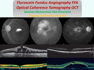
1. Fluorescein fundus angiography (FFA) involves injecting a dye and using filters to examine blood flow and leakage in the retina and choroid over time. 2. Optical coherence tomography (OCT) uses infrared light to create cross-sectional images of the retina, examining layer integrity and thickness. 3. Both FFA and OCT provide important diagnostic information but require careful descriptive analysis and avoiding premature diagnosis.

Recomendados
Mais conteúdo relacionado
Mais procurados
Mais procurados (20)
Semelhante a FFA OCT
Semelhante a FFA OCT (20)
Mais de Samhaa Mohammed
Mais de Samhaa Mohammed (20)
Último
Último (20)
FFA OCT
- 1. Fluroscein Fundus Angiography FFA Optical Coherence Tomography OCT Samhaa Mohammed Abd Elmoneim Zagazig Ophthalmic Hospital
- 2. FFA Fluorescence • Property of a substance to be stimulated by light of short wave length to emit light of longer wavelength.
- 3. Fluorescence • Blue excitation filter • Yellow-green barrier filter
- 4. Fluorescein ** (a water-soluble dye) is injected IV remains intravascular 85% bound to albumin & 15% free. ** The choriocapillaris are permeable to fluorescein molecules, while retinal blood vessels & capillaries are not. ** Excretion through kidney
- 5. • Fluorescein (water soluble) -----15% free. • Choriocapillaries ----- permeable. • Large choroidal vs, Capillaries & Retinal vs ----- impermeable.
- 6. Permeable • Choriocapillari es • Others: MA, NV Impermeable • Retinal vs • Large choroidal vs Impermeable • Capillaries
- 7. Facilities (reclining chair, resuscitation trolley & staff) Contraindication - Absolute (anaphylactic shock) - Relative (renal failure, asthma, pregnancy, cardiac dis.) Explanation (SE) & Formal consent Mydriasis Media opacity !!! - IV cannula - Compfortable sitting - Coloured , FAF photos - Injection (5ml of 10% Technique
- 9. Phases: • Prearterial. (10-13 sec) • Arterial. (12-15 sec after injection) • Arterial Venous Transit Time (AVT) • Venous (25-30 sec). • Late (recirculation).
- 10. Choroidal flush Arterial phase
- 11. Av phase
- 13. 4 3 5 6 2 1
- 14. Hyperfluorescence Autofluorescene (RPE, Drusen, early Stargardt) Pseudofluorescence (Filters issues) Hyperfluorescence ↑ Visualization of normal fluorescein (Transmission defect) Window defect (↑intensity) eg: RPE atrophy ↑ in fluorescein content Leakage (↑intensity, size, end ill defined) eg: NV, CME, CNV Pooling (↑intensity ,size, ends well defined) eg: CSR, PED (not size) Staining (dye retension ) eg: normal OD, fibrous tissue, drusen, exposed sclera)
- 15. Hypofluorescence Blockage (masking) Masking retinal fluorescence eg: preretinal blood Masking choroidal fluorescence Blood eg: intrarenal / subretinal hge, Pigment eg: choroidal naevus, CHRPE Inadequate perfusion (filling defect) Vascular occlusion eg: RVO, RAO, capillary drop out Loss of vascular bed eg: choroidemia, myopic degeneration
- 16. How to comment? Investigation is an art of description (donot jump to Dx)
- 17. 1. Filling timing (choroid, arteries, veins, recirculation) 2. Hyper/ Hypo flurescence lesion number site size(DD) shape edges intensity change of intensity. 3. Synchronicity 4. FAZ 5. Disc area
- 18. FAZ • Normal: hypoflurescent, oval or round. • Leakage: petalflower appearance, ink blot. • Ischaemic : enlarged, irregular capillary budding enhanced capillaries visualization
- 19. Hyperfluorescence early in choroidal phase ↑ intensity not size (window defect) RPE atrophy Hyper f starts well and ends ill defined, ↑intensity , size ( leakage) CNV
- 20. Hper f ↑size and intensity in early phases (pooling) smoke stalk , ink plot in CSR Hyper f ↑intensity not size (pooling) PED
- 21. Hyper f in late phase (staining) Scars of laser
- 22. Hper f ↑size , intensity ends ill defined CNV
- 23. Hyper f (flower petal appearance), ↑size, intensity (leakage)
- 24. Hypof (filling defect) BRAO Hypo f (filling defect) Capillary drop out Ischaemic CRVO
- 26. 1. Filling timing (choroid, arteries, veins, recirculation) 2. Hyper/ Hypo flurescence lesion number site size(DD) shape edges intensity change of intensity. 3. Synchronicity 4. FAZ 5. Disc area
- 27. Conclusion • Investigation is an art of description. • FFA reflects tissue integrity. • Donot jump to Dx, learn how to describe. • Describe then you easily find Dx
- 28. OCT • OCT (infrared light interferometry) is analogous of B scan (sound waves)
- 29. OCT Time domain Spectral domain Swept source Macular dis, retinoschesis Post segment OCT Glaucoma NFL OCT ACG, Corneal analysis Anterior segment OCT
- 30. GCL, INL, ONL Hyporeflective Layers IPL, OPL, NFL Hyperreflective Layers RPE, OS/IS, ELM Hypereflective Bands
- 31. OCT
- 32. OCT
- 34. OCT report • Scan pattern (3D, radial, line). • Vitreoretinal interface. • Epiretinal membrane. • Foveal contour. • Arrangement of retinal layers (regular, irregular, why?). • Thickness. • CME? • Neurosensory detachment. • RPE-choriocapillaris complex. • Associated retinoschisis. • Diagnosis.
- 37. OCT PED CME
- 38. OCT