SlideShare uma empresa Scribd logo
1 de 67
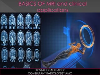
DR SAKHER-ALKHADERI
CONSULTANT RADIOLOGIST AMC
"Felix Bloch and Edward Purcell, both of whom were
awarded the Nobel Prize in 1952, discovered the
nuclear magnetic resonance (NMR) phenomenon
independently in 1946. In the period between 1950 and
1970, NMR was developed and used for chemical and
physical molecular analysis. In 1971 Raymond Damadian
showed that the nuclear magnetic relaxation times of
tissues and tumors differed, thus motivating
scientists to consider magnetic resonance for the
detection of diseases. Dr. Damadian and his team
spent the next seven years diligently designing and
creating the first MRI machine for medical imaging of
the human body."
MRI HISTORY
FONAR introduced the world's first commercial MRI (a whole-body MRI scanner)
in 1980, and went public in 1981.
The first MRI images produced by the machine .
1977
Modern MRI
TYPES OF MRI
The OPEN MRI's started to come into production . The initial intent was to provide a
tool to perform the MRI diagnostic testing for patients that were a) Claustrophobic
or 2) Too obese to fit into the Closed models.
Neutral Flexion
Dynamic MRI
Surface Coils.
Surface coils are designed to provide a very high RF sensitivity over a small reg
interest. These coils are often single or multi-turn loops which are placed direct
the anatomy of interest. The size of these coils can be optimized for the specifi
region of interest.
MRI : WHAT IS
THE EXACT
PRINCIPLE
RADIOFEQUENCY
TURN OFF
Camera
SO TO GET AN IMAGE WE NEED
THE SIGNAL IS DISPLAYED ON GREY SCALE
AND ACCORDING TO THE RELAXATION TIME WE GET
Dark signal on all sequences
SPECIAL MRI SEQUENCES
 Diffusion weighted imaging (DWI) is a form of MR
imaging based upon measuring the random
Brownian motion of water molecules within a
voxel of tissue. The relationship between histology
and diffusion is complex, however generally
densely cellular tissues or those with cellular
swelling exhibit lower diffusion coefficients, and
thus diffusion is particularly useful in tumour
characterisation and cerebral ischaemia.
 DW imaging has a major role in the following clinical
situations 3-5:
 early identification of ischemic stroke
 differentiation of acute from chronic stroke
 differentiation of acute stroke from other stroke mimics
 differentiation of epidermoid cyst from arachnoid cyst
 differentiation of abscess from necrotic tumors
 assessment of cortical lesions in CJD
 differentiation of herpes encephalitis from diffuse temporal
gliomas
 assessment of the extent of diffuse axonal injury
 grading of gliomas and meningiomas (need further study)
 assessment of active demyelination
EPIDERMOID CYST
Functional magnetic resonance
imaging or functional MRI (fMRI) is
a functional neuroimaging procedure
using MRItechnology that measures brain activity by
detecting associated changes in blood flow.[1][2] This
technique relies on the fact that cerebral blood flow
and neuronal activation are coupled. When an area of
the brain is in use, blood flow to that region also
increases.
The primary form of fMRI uses the blood-oxygen-level
dependent (BOLD) contrast,[3] discovered by Seiji
Ogawa. This is a type of specialized brain and body
scan used to map neural activity in the brain or spinal
cord of humans or other animals by imaging the
change in blood flow (hemodynamic response)
related to energy use by brain cells.
MRI
SPECTROSCOPY
The basic principle that enables MR
spectroscopy (MRS) is the electron
cloud around an atom that shields the
nucleus from the magnetic field to a
greater or lesser degree. This naturally
results in slightly resonant frequencies,
which in turn return a slightly different
signal.
MR spectroscopy
MR spectroscopy (MRS)
Hunter's angle
lactate peak: resonates at 1.3 ppm
lipids peak: resonate at 1.3 ppm
alanine peak: resonates at 1.48 ppm
N-acetylaspartate (NAA) peak: resonates at 2.0
glutamine-glutamate peak: resonate at 2.2-2.4 ppm
gamma-aminobutyric acid (GABA) peak: resonate at
2.2-2.4 ppm
citrate peak: resonates at 2.6 ppm
creatine peak: resonates at 3.0 ppm
choline peak: resonates at 3.2 ppm
myo-inositol peak: resonates at 3.5 ppm
 Magnetic resonance elastography (MRE) is a non-
invasive medical imaging technique that measures
the mechanical properties (stiffness) of soft tissues by
introducing shear waves and imaging their
propagation using MRI. Pathological tissues are often
stiffer than the surrounding normal tissue. For instance,
malignant breast tumors are much harder than
healthy fibro-glandular tissue. This characteristic has
been used by physicians for screening and diagnosis
of many diseases, through palpation. MRE calculates
the mechanical parameter as elicited by palpation,
in a non-invasive and objective way.
SUSEPTIBILTY WEIGHTED IMAGES ( SWI) FOR HEMORRHAGE
THE END

Mais conteúdo relacionado

Mais procurados

Distortion Artifacts in MRI and their correction
Distortion Artifacts in MRI and their correctionDistortion Artifacts in MRI and their correction
Distortion Artifacts in MRI and their correction
Miami Cancer Institute
 
Mri basics
Mri basicsMri basics
Mri basics
PS Deb
 

Mais procurados (20)

Fat suppression techniques in MRI
Fat suppression techniques in MRIFat suppression techniques in MRI
Fat suppression techniques in MRI
 
MRI PROCEDURE OF BRAIN
MRI PROCEDURE OF BRAIN MRI PROCEDURE OF BRAIN
MRI PROCEDURE OF BRAIN
 
MRI Brain
MRI BrainMRI Brain
MRI Brain
 
MRI artifacts
MRI artifactsMRI artifacts
MRI artifacts
 
Ct protocols of thorax
Ct protocols of thoraxCt protocols of thorax
Ct protocols of thorax
 
Ppt mri brain
Ppt mri brainPpt mri brain
Ppt mri brain
 
Basics of MRI Physics
Basics of MRI PhysicsBasics of MRI Physics
Basics of MRI Physics
 
CT SCAN spine
CT SCAN spineCT SCAN spine
CT SCAN spine
 
Mri hardware
Mri hardwareMri hardware
Mri hardware
 
Basics of mri
Basics of mriBasics of mri
Basics of mri
 
Basic Pulse Sequences In MRI
Basic Pulse Sequences In MRIBasic Pulse Sequences In MRI
Basic Pulse Sequences In MRI
 
Distortion Artifacts in MRI and their correction
Distortion Artifacts in MRI and their correctionDistortion Artifacts in MRI and their correction
Distortion Artifacts in MRI and their correction
 
Magnetic Resonance Spectroscopy
Magnetic Resonance SpectroscopyMagnetic Resonance Spectroscopy
Magnetic Resonance Spectroscopy
 
Radiologic anatomy of the cranial nerves
Radiologic anatomy of the cranial nervesRadiologic anatomy of the cranial nerves
Radiologic anatomy of the cranial nerves
 
The Physics of MRI
The Physics of MRIThe Physics of MRI
The Physics of MRI
 
CT artifact
CT artifact CT artifact
CT artifact
 
ULTRASOUND IMAGING PRINCIPLES
ULTRASOUND IMAGING PRINCIPLESULTRASOUND IMAGING PRINCIPLES
ULTRASOUND IMAGING PRINCIPLES
 
Mri basics
Mri basicsMri basics
Mri basics
 
Quantitative CT and MR Imaging of Liver
Quantitative CT and MR Imaging of Liver Quantitative CT and MR Imaging of Liver
Quantitative CT and MR Imaging of Liver
 
Ct brain presentation
Ct brain presentationCt brain presentation
Ct brain presentation
 

Destaque

Physical Principles Of Ultrasound
Physical Principles Of UltrasoundPhysical Principles Of Ultrasound
Physical Principles Of Ultrasound
u.surgery
 
Basics of CT & MRI
Basics of CT & MRIBasics of CT & MRI
Basics of CT & MRI
Vibhuti Kaul
 

Destaque (20)

Mri ppt
Mri pptMri ppt
Mri ppt
 
Physical Principles Of Ultrasound
Physical Principles Of UltrasoundPhysical Principles Of Ultrasound
Physical Principles Of Ultrasound
 
Ct Basics
Ct BasicsCt Basics
Ct Basics
 
CT Anatomy
CT AnatomyCT Anatomy
CT Anatomy
 
BASICS OF MRI
BASICS OF MRIBASICS OF MRI
BASICS OF MRI
 
Magnetic Resonance Imaging
Magnetic Resonance ImagingMagnetic Resonance Imaging
Magnetic Resonance Imaging
 
Basics Of MRI
Basics Of MRIBasics Of MRI
Basics Of MRI
 
MRI sequences
MRI sequencesMRI sequences
MRI sequences
 
MRI slides, second yr students 2008
MRI slides, second yr students 2008MRI slides, second yr students 2008
MRI slides, second yr students 2008
 
Basics of CT & MRI
Basics of CT & MRIBasics of CT & MRI
Basics of CT & MRI
 
Mri basic principle and sequences
Mri basic principle and sequencesMri basic principle and sequences
Mri basic principle and sequences
 
Mri physics PART 2
Mri physics PART 2Mri physics PART 2
Mri physics PART 2
 
MRI
MRIMRI
MRI
 
Mri
MriMri
Mri
 
Mri brain anatomy Dr Muhammad Bin Zulfiqar
Mri brain anatomy Dr Muhammad Bin ZulfiqarMri brain anatomy Dr Muhammad Bin Zulfiqar
Mri brain anatomy Dr Muhammad Bin Zulfiqar
 
Computer Tomography (CT Scan)
Computer Tomography (CT Scan)Computer Tomography (CT Scan)
Computer Tomography (CT Scan)
 
MRI Differences: Closed Bore, Open MRI & Wide Bore
MRI Differences: Closed Bore, Open MRI & Wide BoreMRI Differences: Closed Bore, Open MRI & Wide Bore
MRI Differences: Closed Bore, Open MRI & Wide Bore
 
PyMetris : A flexible Endoscope Tracker
PyMetris : A flexible Endoscope TrackerPyMetris : A flexible Endoscope Tracker
PyMetris : A flexible Endoscope Tracker
 
Mri Pulse Seqs
Mri Pulse SeqsMri Pulse Seqs
Mri Pulse Seqs
 
CT Scan Vs MRI Scan
CT Scan Vs MRI ScanCT Scan Vs MRI Scan
CT Scan Vs MRI Scan
 

Semelhante a Basics of MRI

Magnetic resonance spectroscopy
Magnetic resonance spectroscopyMagnetic resonance spectroscopy
Magnetic resonance spectroscopy
Anurag Singh
 
Neuroimaging in psychiatry
Neuroimaging in psychiatryNeuroimaging in psychiatry
Neuroimaging in psychiatry
Santanu Ghosh
 
Neuroimaging and its implications in psychiatry
Neuroimaging and its implications in psychiatryNeuroimaging and its implications in psychiatry
Neuroimaging and its implications in psychiatry
Rupinder Oberoi
 

Semelhante a Basics of MRI (20)

Brain imaging in psychiatry
Brain imaging in psychiatryBrain imaging in psychiatry
Brain imaging in psychiatry
 
Magnetic resonance imaging
Magnetic resonance imagingMagnetic resonance imaging
Magnetic resonance imaging
 
Introduction to Magnetic resonance imaging (mri)
Introduction to Magnetic resonance imaging (mri)Introduction to Magnetic resonance imaging (mri)
Introduction to Magnetic resonance imaging (mri)
 
Magnetic resonance imaging
Magnetic resonance imagingMagnetic resonance imaging
Magnetic resonance imaging
 
Mri by talha
Mri by talhaMri by talha
Mri by talha
 
Imaging of brain tumours
Imaging of brain tumoursImaging of brain tumours
Imaging of brain tumours
 
Neuroimaging Lecture
Neuroimaging LectureNeuroimaging Lecture
Neuroimaging Lecture
 
Magnetic resonance spectroscopy
Magnetic resonance spectroscopyMagnetic resonance spectroscopy
Magnetic resonance spectroscopy
 
Magnetic resonance
Magnetic resonanceMagnetic resonance
Magnetic resonance
 
Magnetic_Resonance_(MR)_spectroscopy-1.pptx
Magnetic_Resonance_(MR)_spectroscopy-1.pptxMagnetic_Resonance_(MR)_spectroscopy-1.pptx
Magnetic_Resonance_(MR)_spectroscopy-1.pptx
 
Magnetic resonance (mr) spectroscopy
Magnetic resonance (mr) spectroscopyMagnetic resonance (mr) spectroscopy
Magnetic resonance (mr) spectroscopy
 
Neuroimaging in psychiatry
Neuroimaging in psychiatryNeuroimaging in psychiatry
Neuroimaging in psychiatry
 
NMR PRESENTATION.pptx
NMR PRESENTATION.pptxNMR PRESENTATION.pptx
NMR PRESENTATION.pptx
 
MRI (Magnetic resonance imaging)
MRI (Magnetic resonance imaging)MRI (Magnetic resonance imaging)
MRI (Magnetic resonance imaging)
 
Advances in neuro-imaging
Advances in neuro-imaging Advances in neuro-imaging
Advances in neuro-imaging
 
Clinical correlation and interpretation of Brain MRI by dr.Sagor
Clinical correlation and interpretation of Brain MRI by dr.SagorClinical correlation and interpretation of Brain MRI by dr.Sagor
Clinical correlation and interpretation of Brain MRI by dr.Sagor
 
Neruoimaging final
Neruoimaging finalNeruoimaging final
Neruoimaging final
 
Neurography Supplement to Issue 50
Neurography Supplement to Issue 50Neurography Supplement to Issue 50
Neurography Supplement to Issue 50
 
MRI Presentation Final
MRI Presentation FinalMRI Presentation Final
MRI Presentation Final
 
Neuroimaging and its implications in psychiatry
Neuroimaging and its implications in psychiatryNeuroimaging and its implications in psychiatry
Neuroimaging and its implications in psychiatry
 

Mais de Sakher Alkhaderi

Imaging of Focal Lung Lesions
Imaging of Focal Lung Lesions Imaging of Focal Lung Lesions
Imaging of Focal Lung Lesions
Sakher Alkhaderi
 

Mais de Sakher Alkhaderi (18)

Imaging of Acute Diverticulitis
Imaging of Acute Diverticulitis  Imaging of Acute Diverticulitis
Imaging of Acute Diverticulitis
 
Imaging of Acute Appendicitis
Imaging of Acute Appendicitis Imaging of Acute Appendicitis
Imaging of Acute Appendicitis
 
CT Imaging of Bowel Wall Thickening
CT Imaging  of Bowel Wall Thickening CT Imaging  of Bowel Wall Thickening
CT Imaging of Bowel Wall Thickening
 
Imaging of Acute Abdomen
Imaging of Acute Abdomen Imaging of Acute Abdomen
Imaging of Acute Abdomen
 
Imaging Of Peritoneal Pathology
Imaging Of Peritoneal Pathology  Imaging Of Peritoneal Pathology
Imaging Of Peritoneal Pathology
 
CT ABDOMEN ANATOMY
 CT ABDOMEN ANATOMY CT ABDOMEN ANATOMY
CT ABDOMEN ANATOMY
 
HRCT Low attenuation pattern
HRCT Low attenuation pattern HRCT Low attenuation pattern
HRCT Low attenuation pattern
 
HRCT High attenuation pattern
HRCT High attenuation pattern HRCT High attenuation pattern
HRCT High attenuation pattern
 
Imaging of Aortic Dissection
Imaging of Aortic DissectionImaging of Aortic Dissection
Imaging of Aortic Dissection
 
HRCT Nodular pattern
HRCT Nodular pattern HRCT Nodular pattern
HRCT Nodular pattern
 
HRCT Reticular pattern
HRCT Reticular pattern HRCT Reticular pattern
HRCT Reticular pattern
 
Imaging of Focal Lung Lesions
Imaging of Focal Lung Lesions Imaging of Focal Lung Lesions
Imaging of Focal Lung Lesions
 
CT OF THE MEDIASTINUM
CT OF THE MEDIASTINUMCT OF THE MEDIASTINUM
CT OF THE MEDIASTINUM
 
CT CHEST ANATOMY
CT CHEST ANATOMYCT CHEST ANATOMY
CT CHEST ANATOMY
 
Brain Tumors
Brain TumorsBrain Tumors
Brain Tumors
 
CT Imaging of Cerebral Ischemia and Infarction
CT Imaging of Cerebral Ischemia and Infarction CT Imaging of Cerebral Ischemia and Infarction
CT Imaging of Cerebral Ischemia and Infarction
 
Cerebral Hemorrhage and Traumatic Brain Injury
Cerebral Hemorrhage and Traumatic Brain InjuryCerebral Hemorrhage and Traumatic Brain Injury
Cerebral Hemorrhage and Traumatic Brain Injury
 
Brain CT Anatomy and Basic Interpretation Part II
Brain CT Anatomy and Basic Interpretation Part IIBrain CT Anatomy and Basic Interpretation Part II
Brain CT Anatomy and Basic Interpretation Part II
 

Último

Call Girls in Gagan Vihar (delhi) call me [🔝 9953056974 🔝] escort service 24X7
Call Girls in Gagan Vihar (delhi) call me [🔝  9953056974 🔝] escort service 24X7Call Girls in Gagan Vihar (delhi) call me [🔝  9953056974 🔝] escort service 24X7
Call Girls in Gagan Vihar (delhi) call me [🔝 9953056974 🔝] escort service 24X7
9953056974 Low Rate Call Girls In Saket, Delhi NCR
 
College Call Girls in Haridwar 9667172968 Short 4000 Night 10000 Best call gi...
College Call Girls in Haridwar 9667172968 Short 4000 Night 10000 Best call gi...College Call Girls in Haridwar 9667172968 Short 4000 Night 10000 Best call gi...
College Call Girls in Haridwar 9667172968 Short 4000 Night 10000 Best call gi...
perfect solution
 
Call Girls Aurangabad Just Call 8250077686 Top Class Call Girl Service Available
Call Girls Aurangabad Just Call 8250077686 Top Class Call Girl Service AvailableCall Girls Aurangabad Just Call 8250077686 Top Class Call Girl Service Available
Call Girls Aurangabad Just Call 8250077686 Top Class Call Girl Service Available
Dipal Arora
 

Último (20)

Call Girls Guntur Just Call 8250077686 Top Class Call Girl Service Available
Call Girls Guntur  Just Call 8250077686 Top Class Call Girl Service AvailableCall Girls Guntur  Just Call 8250077686 Top Class Call Girl Service Available
Call Girls Guntur Just Call 8250077686 Top Class Call Girl Service Available
 
Call Girls Siliguri Just Call 8250077686 Top Class Call Girl Service Available
Call Girls Siliguri Just Call 8250077686 Top Class Call Girl Service AvailableCall Girls Siliguri Just Call 8250077686 Top Class Call Girl Service Available
Call Girls Siliguri Just Call 8250077686 Top Class Call Girl Service Available
 
Call Girls in Gagan Vihar (delhi) call me [🔝 9953056974 🔝] escort service 24X7
Call Girls in Gagan Vihar (delhi) call me [🔝  9953056974 🔝] escort service 24X7Call Girls in Gagan Vihar (delhi) call me [🔝  9953056974 🔝] escort service 24X7
Call Girls in Gagan Vihar (delhi) call me [🔝 9953056974 🔝] escort service 24X7
 
Premium Call Girls In Jaipur {8445551418} ❤️VVIP SEEMA Call Girl in Jaipur Ra...
Premium Call Girls In Jaipur {8445551418} ❤️VVIP SEEMA Call Girl in Jaipur Ra...Premium Call Girls In Jaipur {8445551418} ❤️VVIP SEEMA Call Girl in Jaipur Ra...
Premium Call Girls In Jaipur {8445551418} ❤️VVIP SEEMA Call Girl in Jaipur Ra...
 
♛VVIP Hyderabad Call Girls Chintalkunta🖕7001035870🖕Riya Kappor Top Call Girl ...
♛VVIP Hyderabad Call Girls Chintalkunta🖕7001035870🖕Riya Kappor Top Call Girl ...♛VVIP Hyderabad Call Girls Chintalkunta🖕7001035870🖕Riya Kappor Top Call Girl ...
♛VVIP Hyderabad Call Girls Chintalkunta🖕7001035870🖕Riya Kappor Top Call Girl ...
 
Premium Bangalore Call Girls Jigani Dail 6378878445 Escort Service For Hot Ma...
Premium Bangalore Call Girls Jigani Dail 6378878445 Escort Service For Hot Ma...Premium Bangalore Call Girls Jigani Dail 6378878445 Escort Service For Hot Ma...
Premium Bangalore Call Girls Jigani Dail 6378878445 Escort Service For Hot Ma...
 
Call Girls in Delhi Triveni Complex Escort Service(🔝))/WhatsApp 97111⇛47426
Call Girls in Delhi Triveni Complex Escort Service(🔝))/WhatsApp 97111⇛47426Call Girls in Delhi Triveni Complex Escort Service(🔝))/WhatsApp 97111⇛47426
Call Girls in Delhi Triveni Complex Escort Service(🔝))/WhatsApp 97111⇛47426
 
Call Girls Coimbatore Just Call 9907093804 Top Class Call Girl Service Available
Call Girls Coimbatore Just Call 9907093804 Top Class Call Girl Service AvailableCall Girls Coimbatore Just Call 9907093804 Top Class Call Girl Service Available
Call Girls Coimbatore Just Call 9907093804 Top Class Call Girl Service Available
 
College Call Girls in Haridwar 9667172968 Short 4000 Night 10000 Best call gi...
College Call Girls in Haridwar 9667172968 Short 4000 Night 10000 Best call gi...College Call Girls in Haridwar 9667172968 Short 4000 Night 10000 Best call gi...
College Call Girls in Haridwar 9667172968 Short 4000 Night 10000 Best call gi...
 
Call Girls Aurangabad Just Call 8250077686 Top Class Call Girl Service Available
Call Girls Aurangabad Just Call 8250077686 Top Class Call Girl Service AvailableCall Girls Aurangabad Just Call 8250077686 Top Class Call Girl Service Available
Call Girls Aurangabad Just Call 8250077686 Top Class Call Girl Service Available
 
Call Girls Ooty Just Call 8250077686 Top Class Call Girl Service Available
Call Girls Ooty Just Call 8250077686 Top Class Call Girl Service AvailableCall Girls Ooty Just Call 8250077686 Top Class Call Girl Service Available
Call Girls Ooty Just Call 8250077686 Top Class Call Girl Service Available
 
Call Girls Gwalior Just Call 8617370543 Top Class Call Girl Service Available
Call Girls Gwalior Just Call 8617370543 Top Class Call Girl Service AvailableCall Girls Gwalior Just Call 8617370543 Top Class Call Girl Service Available
Call Girls Gwalior Just Call 8617370543 Top Class Call Girl Service Available
 
All Time Service Available Call Girls Marine Drive 📳 9820252231 For 18+ VIP C...
All Time Service Available Call Girls Marine Drive 📳 9820252231 For 18+ VIP C...All Time Service Available Call Girls Marine Drive 📳 9820252231 For 18+ VIP C...
All Time Service Available Call Girls Marine Drive 📳 9820252231 For 18+ VIP C...
 
Call Girls Varanasi Just Call 8250077686 Top Class Call Girl Service Available
Call Girls Varanasi Just Call 8250077686 Top Class Call Girl Service AvailableCall Girls Varanasi Just Call 8250077686 Top Class Call Girl Service Available
Call Girls Varanasi Just Call 8250077686 Top Class Call Girl Service Available
 
Best Rate (Patna ) Call Girls Patna ⟟ 8617370543 ⟟ High Class Call Girl In 5 ...
Best Rate (Patna ) Call Girls Patna ⟟ 8617370543 ⟟ High Class Call Girl In 5 ...Best Rate (Patna ) Call Girls Patna ⟟ 8617370543 ⟟ High Class Call Girl In 5 ...
Best Rate (Patna ) Call Girls Patna ⟟ 8617370543 ⟟ High Class Call Girl In 5 ...
 
Russian Call Girls Service Jaipur {8445551418} ❤️PALLAVI VIP Jaipur Call Gir...
Russian Call Girls Service  Jaipur {8445551418} ❤️PALLAVI VIP Jaipur Call Gir...Russian Call Girls Service  Jaipur {8445551418} ❤️PALLAVI VIP Jaipur Call Gir...
Russian Call Girls Service Jaipur {8445551418} ❤️PALLAVI VIP Jaipur Call Gir...
 
Call Girls Bareilly Just Call 8250077686 Top Class Call Girl Service Available
Call Girls Bareilly Just Call 8250077686 Top Class Call Girl Service AvailableCall Girls Bareilly Just Call 8250077686 Top Class Call Girl Service Available
Call Girls Bareilly Just Call 8250077686 Top Class Call Girl Service Available
 
(Low Rate RASHMI ) Rate Of Call Girls Jaipur ❣ 8445551418 ❣ Elite Models & Ce...
(Low Rate RASHMI ) Rate Of Call Girls Jaipur ❣ 8445551418 ❣ Elite Models & Ce...(Low Rate RASHMI ) Rate Of Call Girls Jaipur ❣ 8445551418 ❣ Elite Models & Ce...
(Low Rate RASHMI ) Rate Of Call Girls Jaipur ❣ 8445551418 ❣ Elite Models & Ce...
 
Call Girls Visakhapatnam Just Call 9907093804 Top Class Call Girl Service Ava...
Call Girls Visakhapatnam Just Call 9907093804 Top Class Call Girl Service Ava...Call Girls Visakhapatnam Just Call 9907093804 Top Class Call Girl Service Ava...
Call Girls Visakhapatnam Just Call 9907093804 Top Class Call Girl Service Ava...
 
Call Girls Dehradun Just Call 9907093804 Top Class Call Girl Service Available
Call Girls Dehradun Just Call 9907093804 Top Class Call Girl Service AvailableCall Girls Dehradun Just Call 9907093804 Top Class Call Girl Service Available
Call Girls Dehradun Just Call 9907093804 Top Class Call Girl Service Available
 

Basics of MRI

  • 2. "Felix Bloch and Edward Purcell, both of whom were awarded the Nobel Prize in 1952, discovered the nuclear magnetic resonance (NMR) phenomenon independently in 1946. In the period between 1950 and 1970, NMR was developed and used for chemical and physical molecular analysis. In 1971 Raymond Damadian showed that the nuclear magnetic relaxation times of tissues and tumors differed, thus motivating scientists to consider magnetic resonance for the detection of diseases. Dr. Damadian and his team spent the next seven years diligently designing and creating the first MRI machine for medical imaging of the human body." MRI HISTORY FONAR introduced the world's first commercial MRI (a whole-body MRI scanner) in 1980, and went public in 1981.
  • 3.
  • 4. The first MRI images produced by the machine .
  • 7.
  • 8. The OPEN MRI's started to come into production . The initial intent was to provide a tool to perform the MRI diagnostic testing for patients that were a) Claustrophobic or 2) Too obese to fit into the Closed models.
  • 9.
  • 10.
  • 12.
  • 13.
  • 14. Surface Coils. Surface coils are designed to provide a very high RF sensitivity over a small reg interest. These coils are often single or multi-turn loops which are placed direct the anatomy of interest. The size of these coils can be optimized for the specifi region of interest.
  • 15.
  • 16.
  • 17.
  • 18.
  • 19.
  • 20. MRI : WHAT IS THE EXACT PRINCIPLE
  • 21.
  • 22.
  • 23.
  • 24.
  • 25.
  • 27.
  • 30. SO TO GET AN IMAGE WE NEED
  • 31. THE SIGNAL IS DISPLAYED ON GREY SCALE
  • 32. AND ACCORDING TO THE RELAXATION TIME WE GET
  • 33.
  • 34.
  • 35.
  • 36.
  • 37.
  • 38.
  • 39.
  • 40. Dark signal on all sequences
  • 41.
  • 42.
  • 44.
  • 45.
  • 46.
  • 47.
  • 48.
  • 49.
  • 50.  Diffusion weighted imaging (DWI) is a form of MR imaging based upon measuring the random Brownian motion of water molecules within a voxel of tissue. The relationship between histology and diffusion is complex, however generally densely cellular tissues or those with cellular swelling exhibit lower diffusion coefficients, and thus diffusion is particularly useful in tumour characterisation and cerebral ischaemia.
  • 51.  DW imaging has a major role in the following clinical situations 3-5:  early identification of ischemic stroke  differentiation of acute from chronic stroke  differentiation of acute stroke from other stroke mimics  differentiation of epidermoid cyst from arachnoid cyst  differentiation of abscess from necrotic tumors  assessment of cortical lesions in CJD  differentiation of herpes encephalitis from diffuse temporal gliomas  assessment of the extent of diffuse axonal injury  grading of gliomas and meningiomas (need further study)  assessment of active demyelination
  • 52.
  • 54.
  • 55.
  • 56.
  • 57. Functional magnetic resonance imaging or functional MRI (fMRI) is a functional neuroimaging procedure using MRItechnology that measures brain activity by detecting associated changes in blood flow.[1][2] This technique relies on the fact that cerebral blood flow and neuronal activation are coupled. When an area of the brain is in use, blood flow to that region also increases. The primary form of fMRI uses the blood-oxygen-level dependent (BOLD) contrast,[3] discovered by Seiji Ogawa. This is a type of specialized brain and body scan used to map neural activity in the brain or spinal cord of humans or other animals by imaging the change in blood flow (hemodynamic response) related to energy use by brain cells.
  • 58.
  • 59. MRI SPECTROSCOPY The basic principle that enables MR spectroscopy (MRS) is the electron cloud around an atom that shields the nucleus from the magnetic field to a greater or lesser degree. This naturally results in slightly resonant frequencies, which in turn return a slightly different signal.
  • 60. MR spectroscopy MR spectroscopy (MRS) Hunter's angle lactate peak: resonates at 1.3 ppm lipids peak: resonate at 1.3 ppm alanine peak: resonates at 1.48 ppm N-acetylaspartate (NAA) peak: resonates at 2.0 glutamine-glutamate peak: resonate at 2.2-2.4 ppm gamma-aminobutyric acid (GABA) peak: resonate at 2.2-2.4 ppm citrate peak: resonates at 2.6 ppm creatine peak: resonates at 3.0 ppm choline peak: resonates at 3.2 ppm myo-inositol peak: resonates at 3.5 ppm
  • 61.
  • 62.
  • 63.
  • 64.  Magnetic resonance elastography (MRE) is a non- invasive medical imaging technique that measures the mechanical properties (stiffness) of soft tissues by introducing shear waves and imaging their propagation using MRI. Pathological tissues are often stiffer than the surrounding normal tissue. For instance, malignant breast tumors are much harder than healthy fibro-glandular tissue. This characteristic has been used by physicians for screening and diagnosis of many diseases, through palpation. MRE calculates the mechanical parameter as elicited by palpation, in a non-invasive and objective way.
  • 65.
  • 66. SUSEPTIBILTY WEIGHTED IMAGES ( SWI) FOR HEMORRHAGE

Notas do Editor

  1. On july,3,1977 the first human scan by Dr damadian