

Recomendados
Mais conteúdo relacionado
Mais procurados
Mais procurados (20)
Semelhante a Asthma copd
Semelhante a Asthma copd (20)
Mais de Muhammad Saim
Mais de Muhammad Saim (20)
Asthma copd
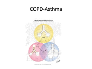
- 1. COPD-Asthma
- 3. Risk Factors for COPD Nutrition Infections Socio-economic status Aging Populations
- 5. Percent Change in Age-Adjusted Death Rates, U.S., 1965-1998 Proportion of 1965 Rate 3.0 Coronary Stroke Other CVD COPD All Other 2.5 Heart Causes Disease 2.0 1.5 1.0 0.5 –59% –64% –35% +163% –7% 0 1965 - 1998 1965 - 1998 1965 - 1998 1965 - 1998 1965 - 1998 Source: NHLBI/NIH/DHHS
- 6. Prevalence of allergies and asthma in Pakistan M.Y. Noori, S.M. Hasnain, and M.A. Waqar. World Allergy Organization Journal & November 2007 • The frequency of wheezing was found to be 15.2%, • while the diagnosed cases of asthma were 9.5%. • The frequency of allergic rhinitis was found to be 34.3%. • The frequency of those having allergic rhinitis as well as wheezing episodes was 8%. • There was no statistically significant difference between asthmatics and non-asthmatics by sex (P-value:0.402). • Socioeconomic status was found to affect significantly (p value 0.001) as the prevalence of diagnosed asthma cases was 6.17% in high socioeconomic class,13.11% in the middle-class and 2.4% in the low socioeconomic class. • Family history of atopy was also found to be significantly higher in asthmatics.
- 7. NOCTURNAL ASTHMA IN SCHOOL CHILDREN OF SOUTH PUNJAB,PAKISTAN Ghulam Mustafa, Pervez Akber Khan, Imran Iqbal J Ayub Med Coll Abbottabad 2008;20(3) • The parents reported nocturnal asthma in 177 (6%) of their children with an equal prevalence in boys and girls,
- 8. Anatomy
- 9. Pathogenesis of Cigarette smoke COPD Biomass particles Particulates Host factors Amplifying mechanisms LUNG INFLAMMATION Anti-oxidants Anti-proteinases Oxidative stress Proteinases Repair mechanisms COPD PATHOLOGY Source : Peter J. Barnes,
- 10. Differences in Inflammation and its Consequences: Asthma and COPD ASTHMA COPD Allergens Cigarette smoke Y Y Y Ep cells Mast cell Alv macrophage Ep cells CD4+ cell Eosinophil CD8+ cell Neutrophil (Th2) (Tc1) Bronchoconstriction Small airway narrowing AHR Alveolar destruction Airflow Limitation Reversible Irreversible Source : Peter J. Barnes,
- 11. COPD airway
- 12. Asthma airway
- 13. Changes in Large Airways of COPD Patients Mucus hypersecretion Neutrophils in sputum Squamous metaplasia of epithelium No basement membrane thickening Goblet cell hyperplasia ↑ Macrophages ↑ CD8+ lymphocytes Mucus gland hyperplasia Little increase in airway smooth muscle Source : Peter J. Barnes, MD
- 14. Air Trapping in COPD Normal Mild/moderate Severe Inspiration COPD COPD small airway alveolar attachments loss of elasticity loss of alveolar attachments Expiration closure ↓ Health Dyspnea Air trapping status ↓ Exercise capacity Hyperinflation Source : Peter J. Barnes,
- 15. Changes in Small Airways in COPD Patients Inflammatory exudate in lumen Disrupted alveolar attachments Thickened wall with inflammatory cells - macrophages, CD8+ cells, fibroblasts Peribronchial fibrosis Lymphoid follicle Source : Peter J. Barnes, MD
- 16. Changes in the Lung Parenchyma in COPD Patients Alveolar wall destruction Loss of elasticity Destruction of pulmonary capillary bed ↑ Inflammatory cells macrophages, CD8+ lymphocytes Source : Peter J. Barnes, MD
- 17. Inflammatory Cells Involved in COPD Cigarette smoke (and other irritants) Epithelial Alveolar macrophage cells Chemotactic factors CD8+ Fibroblast lymphocyte Neutrophil Monocyte Neutrophil elastase PROTEASES Cathepsins MMPs Fibrosis Alveolar wall destruction Mucus hypersecretion (Obstructive (Emphysema) bronchiolitis) Source : Peter J. Barnes, MD
- 18. Oxidative Stress in COPD Macrophage Neutrophil Anti-proteases SLPI α 1-AT NF-κ B Proteolysis IL-8 TNF-α ↓ HDAC2 O2-, H202 Neutrophil OH., ONOO- recruitment ↑Inflammation Steroid resistance Isoprostanes Plasma leak Bronchoconstriction ↑ Mucus secretion Source : Peter J. Barnes, MD
- 19. Pulmonary Hypertension in COPD Chronic hypoxia Pulmonary vasoconstriction Muscularization Pulmonary hypertension Intimal hyperplasia Fibrosis Cor pulmonale Obliteration Edema Death Source : Peter J. Barnes, MD
- 20. Asthma
- 22. Emphysema
- 24. PEF meters
- 25. Spirometry: Normal and Patients with COPD
- 27. Lung Volumes and Capacities
- 28. PFTs ASTHMA COPD FEV1 Decreased in active asthma Decreased-stage of disease FVC Decreased Decreased FEV1/FVC Decreased decreased TLC Normal or increased Normal or increased FRC Normal or increased Normal or increased RV Normal or Increased Normal or increased DLCO Normal or Increased Decreased in Emphysema
- 29. Therapy at Each Stage of COPD I: Mild II: Moderate III: Severe IV: Very Severe FEV 1 /FVC < 70% FEV 1 /FVC < 70% FEV 1 < 30% FEV 1 /FVC < 70% predicted FEV 1 /FVC < 70% 30% < FEV 1 < or FEV 1 < 50% 50% < FEV 1 < 80% 50% predicted predicted plus FEV 1 > 80% predicted chronic respiratory Active reduction of risk factor(s); influenza vaccination predicted failure Add short-acting bronchodilator (when needed) Add regular treatment with one or more long-acting bronchodilators (when needed); Add rehabilitation Add inhaled glucocorticosteroids if repeated exacerbations Add long term oxygen if chronic respiratory failure. Consider surgical treatments
Notas do Editor
- Pathogenesis of COPD, illustrating the central role of inflammation
- Changes in large airways of COPD patients. The epithelium often shows squamous metaplasia and there is goblet cell and submucosal gland hyperplasia, resulting in mucus hypersecretion. The airway wall is infiltrated with macrophages and CD8+ lymphocytes, whereas neutrophils predominate in the airway lumen and around submucosal glands. Airway smooth muscle and basement membrane are minimally increased compared to the findings in asthma.
- Air trapping in COPD. During expiration small airways narrow but closure is prevented by the elasticity of alveolar attachments. In COPD patients there is a loss of elasticity with greater narrowing in small airways, which may close completely when there is loss of alveolar attachments as a result of emphysema. This results in air trapping and hyperinflation, leading to dyspnea and reduced exercise capacity.
- Changes in small airways in COPD patients. The airway wall is thickened and infiltrated with inflammatory cells, predominately macrophages and CD8+ lymphocytes, with increased numbers of fibroblasts. In severe COPD there are also lymphoid follicles. The lumen is often filled with an inflammatory exudate and mucus. There is peribronchial fibrosis and airway smooth muscle may be increased, resulting in narrowing of the airway.
- Changes in the lung parenchyma in COPD patients. There is loss of elasticity and alveolar wall destruction, and accumulation of inflammatory cells, predominantly macrophages and CD8+ lymphocytes. The destructive changes reduce the pulmonary capillary bed. The left panel shows a scanning electron micrograph of a patient with emphysema demonstrating the enlargement of alveoli and destruction of the alveolar walls.
- Inflammatory cells involved in COPD. Cigarette smoke activates macrophages and epithelial cells to release chemotactic factors that recruit neutrophils, monocytes and CD8+ T-lymphocytes from the circulation. They also release factors that activate fibroblasts leading to small airway obstruction (obstructive bronchiolitis). Proteases released from neutrophils and macrophages may cause mucus hypersecretion and emphysema.
- Oxidative stress in COPD has several detrimental consequences, including activation of the transcription factor nuclear factor-κB (NF-κB), reduction in antiproteases, plasma leakage and mucus hypersecretion. In addition it reduces histone deacetylase-2, resulting in amplified inflammation and reduced anti-inflammatory response to corticosteroids.
- This provides a summary of the recommended treatment at each stage of COPD.