Mammary glands
•Transferir como PPTX, PDF•
59 gostaram•55,651 visualizações

Mammary glands breasts

Recomendados
Mais conteúdo relacionado
Mais procurados
Mais procurados (20)
Semelhante a Mammary glands
Semelhante a Mammary glands (20)
Mais de Nepalese army institute of health sciences
Mais de Nepalese army institute of health sciences (20)
Último
Último (20)
Mammary glands
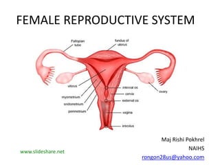
- 1. FEMALE REPRODUCTIVE SYSTEM Maj Rishi Pokhrel NAIHS rongon28us@yahoo.com www.slideshare.net
- 3. OVARY
- 4. 4 MAMMARY GLAND – Modified sweat gland in sup fascia – No connective tissue covering. – Accessory female reproductive org
- 5. Mammary Gland EXTENT • Vertical : 2-6 ribs in mid-clavicular line • Hori : lat border sternum – mid axillary line at level of 4th rib • Extends into axilla – Axillary tail of SPENCE – Foramen of LANGER SHAPE • hemispherical, conical, pendulous, etc
- 6. MAMMARY GLAND – Superficial & deep surface – Superficial surface • Skin, nipple & areola • Under skin, superficial fascia has nerves/vessels • Nipple and areola - No subcutaneous fat and hair. 6
- 7. Nipple • 4th ICS, 4 inch from midline • 15 – 20 lactiferous ducts open • Presence of circular muscle, longitudinal muscle • Rich nerve supply
- 8. 8 Areola – Circular pigmented area, pink or brown. – Periphery : sebaceous glands – Enlarged as Tubercles of MONTGOMERY during pregnancy – Lubrication of nipple and areola – Lactiferous sinus
- 9. 9 Deep surface Retro mammary space – Separates mam gland from Pectoralis major fascia – Contains areolar tissue – Helps in mobility of breast – Space for breast implants
- 10. 10 DEEP RELATIONS – MAMMARY BED • Covered by deep fascia – Pectoralis major : • Medial 2/3, – Serratus Anterior : • Upper two digitations • Lat 1/3, – EO aponeurosis • Inferomedially – separate it from rectus sheath
- 11. 11 STRUCTURE • Glandular portion with parenchyma • Connective tissue i.e stroma Fibrous tissue Fatty tissue Suspensory lig of cooper
- 12. Glandular portion – 15 -20 lobes each with multiple lobules containing acini or alveoli – lactiferous duct- commence toward nipple from each lobe – lactiferous sinus opens into tip of nipple – Lobes radially arranged, hence incision radially given – Glandular tissue increase during pregnancy and lactation 12
- 14. 14 Mammary Gland: Structure Alveoli opening into ductSuspensory ligament running from skin to P Major
- 15. 15 BLOOD SUPPLY 1. Internal thoracic artery (subclavian) – perforating br – 2,3,4 ICS 2. Br from Axillary : – Sup thoracic Art – Thoraco acromial – pectoral br – Lat thoracic art – Subscapular art 3. Intercostal art – – 2,3,4th ICS lat br – 2nd IC Art largest br – • supply upper breast, Nipple and areola)
- 16. 16 Mammary Gland : Blood Supply Branches of Axillary 1. Sup thoracic Art 2. Thoraco-acromial pectoral br 3. Lat thoracic art 4. Subscapular art
- 17. 17 Communication via Post IC vein, Azygous and Internal vert plexus which in turn communicate with transverse and sagittal sinus spreads malignancy to abdominal organs, brain, vertebrae, ribs and skull VENOUS DRAINAGE – Superficial and deep veins – Circulus venosus (part of superficial vein): sub areolar plexus of vein – Superficial and deep vein drain into • Int mammary V • Axillary V • Post IC vein – which drain into Azygous vein
- 18. 18 Venous drainage of mammary gland
- 19. 19 LYMPHATIC DRAINAGE – Axillary (five sub group) – Internal mammary LN • along Internal mammary V – Supraclavicular – Posterior IC Lymph nodes
- 20. 20 Summary Lymphatic drainage – 75 % Parenchyma = Axillary LN – 20 % Parenchyma = Internal mammary LN – 5 % parenchyma = Post IC Nodes along Post IC vein
- 21. 21 • Investigations – Mammography • Soft tissue radiographs of breast. • Cyst (well defined smooth opacity) and carcinomas (irregular density, distortion of breast tissue, calcification) – FNAC (fine needle aspiration cytology) • Used for cell diagnosis APPLIED ANATOMY
- 22. 22 • AXILLARY TAIL – Well developed axillary tail mistaken for enlarged lymph nodes/Lipoma • Nipple – Cracked nipple • in later pregnancy and lactation. • Nipple to be washed, and lubricated with lanolin – Discharges • management depends upon presence of lump
- 23. 23 – Infections and inflammations – cause mastitis with abscess – Cysts – Tumors • Benign – Lipoma, fibro adenoma • Malignant – carcinoma “more in nulliparous and bearing child protective” – Spread by local, lymphatic and blood stream. – LN involvement shows metastatic potential. – Advanced disease – involve supraclavicular
- 24. 24 – Malignant tumours cont,d • Presentation – – Hard lump with retracted nipple – Peau d’ orange (orange like skin) – involvement of skin of breast due to cutaneous lymphatic oedema – Advanced – ulceration, fixation to chest wall, metastatsis to viscera, bone • Treatment – Mastectomy – Radiotherapy – Harmone therapy – chemotherapy
- 25. 25 Breast Cancer • Breast cancer – Peau d orange – nipple retraction, – skin dimpling – Metastasis : • skull and brain (Batsons plexus of veins) A - Dimpling of skin B - Retracted nipple C - Peau d orange A – due to pull by lig of cooper B - due to retraction of milk ducts C – due to lymphatic obstruction
- 26. 26
- 27. 27 KRUKENBERGS TUMOUR Secondary deposits in ovaries due to spread from Ca breast : • Lymph inferomedial part • communicate with rectus sheath – • pierce Linea alba – forms Sub peritoneal plexus – drain into subdiaphragmatic LN – • pass through Falciform lig – • reach hepatic node – – Cause obstructive jaundice • Tumor cells drop from sub peritoneal plexus into general cavity – • reach surface of ovary and enter through Ostia left by ovulating Graafian follicle – KRUKENBERGS (secondary deposits on surface of ovary)
- 29. 29 Congenital anomalies – Polythelia • Supernumery nipples over breast – Athelia • No nipple over breast (mainly accessory breast) – Polymastia • Accessory breast along milk ridge – Amastia • No breast development – Amazia • Nipple developed, no breast development
- 30. 30