SlideShare uma empresa Scribd logo
1 de 78
COMPARTMENT SYNDROME
PRESENTER: DR RAMIN MAHARJAN
FIRST YEAR RESIDENT, PAHS
DEPARTMENT OF ORTHOPAEDICS
DATE: 2078/03/03
CONTENTS
• Relevant anatomy
• Compartment syndrome
• Definition
• Epidemiology
• Causes
• Evaluation of patient with Acute compartment syndrome
• Treatment
• Fasciotomy
• Compartment of thigh, leg, forearm, foot and hand
• Chronic exertional compartment syndrome
RELEVANT ANATOMY
FASCIA
• The muscle groups of the human
limbs are divided into sections,
or compartments, formed by
strong, unyielding fascial
membranes.
• Compartments are present in
various part of the body.
Compartment syndrome
• It is an elevation of the interstitial pressure in a closed osteofascial
compartment that results in microvascular compromise.
• Commonly are involved, especially the anterior and deep posterior
compartments of the leg and the volar compartment of the forearm.
• It can develop in the buttock, thigh, shoulder, hand, foot, arm, and
lumbar paraspinous muscles.
• It is an emergency condition.
Epidemiology
• Compartment syndrome is most
commonly seen in Younger age group.
It is seen in 7.3 per 100,000 males and
0.7 per 100,000 females.
Tibial shaft fracture is the most common
associated with a 1 to 10 percent incidence
Classification of Compartment syndrome
• Depending on the cause of the increased pressure and the duration of
symptoms.
• Acute compartment syndrome
• Chronic exertional compartment syndrome
Etiology
Increase Compartment Volume
• Fracture
• Soft tissue injury
• Crush syndrome
• Revascularization
• Exercise
• Bleeding
diathesis/anticoagula
nts
• Fluid infusion
(including
arthroscopy)
• Arterial puncture
• Osteotomy
• Snake bite
• Nephrotic syndrome
• Leukemic infiltration
• Viral myositis
• Acute hematogenous
osteomyelitis
• Ruptured
ganglia/cysts
Reduce Compartment Volume
• Burns
• Tight closure of fascial defects
• Repair of muscle hernia
• Tight casts, dressings, tourniquet or splints
• Entrapment under collapsed weights
• Excessive traction to fractured limbs
• Intramedullary nailing
• Military antishock garments
Etiology • Most common cause: fracture (69% of cases)
Underlying condition % of cases Underlying condition % of cases
Tibial diaphyseal fracture 36 Tibial plateau fracture 3.0
Soft tissue injury 23.2 Hand fracture(s) 2.5
Distal radius fracture 9.8 Tibial pilon fractures 2.5
Crush syndrome 7.9 Foot fracture(s)
Diaphyseal fracture
forearm
7.9 Ankle fracture 0.6
Femoral diaphyseal
fracture
3.0 Elbow fracture dislocation 0.6
Vicious cycle of Volkmann’s Ischemia
• Normal tissue pressure
• 0-4 mmHg
8-10 with exertion
• Absolute intra-compartment pressure
• 30 mmHg -Mubarak
• 45 mmHg -Matsen
Effect of Ischemia
Muscle
• 3-4 hrs: reversible changes
• 6 hrs: variable changes
• 8 hrs: irreversible changes
Nerves
• 1 hour: loses nerve
conduction
• 4 hours: neuropraxia
• 8 hours: irreversible changes
Evaluation of Patient with ACS
• Acute compartment syndrome is a clinical diagnosis and needs prompt
treatment.
Evaluation of Patient with ACS
• History and clinical examinations
• Measurement of intra-compartment pressure
• Creatinine phosphokinase, RFT, Urine myoglobin
History
• 5 P’s
• Pain out of proportion
• Paresthesia
• Paresis
• Pulselessness
• Pallor
• Pain on passive stretching
Physical examinations
• Observe skin for lesions, swelling or color change
• Palpate over the compartment, observing temperature, tension,
tenderness
• Check pulses
• Evaluate two-point discrimination and sensation
• Evaluate motor function
• Due to the potential for rapid progression of compartment syndrome,
clinicians should perform serial exams.
Measurement of intracompartmental pressure
• is not required
• but can aid in diagnosis if uncertainty exists.
• Different devices can be used to detect intracompartmental pressure
• The normal pressure within the compartment is between 0 mmHg to 8
mmHg.
• An intra-compartmental pressure greater than 30 mmHg indicates
compartment syndrome and a need for fasciotomy.
• When intra-compartmental pressure increases to within 10 mmHg to
30 mmHg of the patient's diastolic blood pressure, this indicates
inadequate perfusion and relative ischemia of the involved extremity.
• The perfusion pressure of a compartment, also known as the
compartment delta pressure:
• clinicians often utilize delta pressure less than or equal to 30 mmHg as
indicative of the need for fasciotomy.
Delta Pressure = Diastolic Pressure - Measured Intracompartmental Pressure
Synthes (West
Chester, PA) hand-
held
compartment
pressure monitor.
Noninvasive methods
• Ultrasonic device
• Measures submicrometric displacement of fascia caused by volume
expansion.
• Near-Infrared Spectroscopy (NIRS)
• Tracking of variations in the oxygenation of muscle tissue.
• Laser Doppler Flowmetry
• Flexible fibre optic wire is introduced into the muscle compartment
and signals from this wire are recorded on a computer.
The Reported Sensitivities and Specificities of
the Clinical Symptoms and Signs of ACS
Symptom or Sign Sensitivity (%) Specificity (%) PPV (%) NPV (%)
Pain 19 97 14 98
Pain on passive stretch 19 97 14 98
Paresis/motor changes 13 97 11 98
Paresthesia/sensory changes 13 98 15 98
Swelling 54 76 70 63
ICP monitoring 94 98 93 99
Precaution- Pain may be absent
• Patient with epidural analgesia
• Patients who are intubated or sedated
• Patient with head injuries, under influence of ethanol or drugs
Management
• Prompt diagnosis and treatment
• Provide supplemental oxygen.
• Remove any restrictive casts, dressings or bandages to relieve
pressure.
• Keep the extremity at the level of the heart to prevent hypo-perfusion.
• Prevent hypotension and provide blood pressure support in patients
with hypotension.
• If ICP greater than or equal to 30 mmHg or delta pressure less
than or equal to 30mmHg, fasciotomy should be done. For
hypotensive delta pressure <20mmHg is indication.
Fasciotomy
• The ideal time frame for fasciotomy is within six hours of injury.
• Not recommended after 36 hours following injury.
• If necrosis occurs before fasciotomy is performed, there is a high
likelihood of infection which may require amputation.
• Patients must be closely monitored for complications which include
infection, acute renal failure, and rhabdomyolysis.
Fasciotomy Principles
• Make early diagnosis
• Long extensile incisions
• Release all fascial compartments
• Preserve neurovascular structures
• Debride necrotic tissues
• Coverage within 7-10 days
Prognosis
• depends mainly on how quickly the condition is diagnosed and treated.
• Long-term studies on survivors do reveal residual pain, Volkmann's
contracture, mild neurological deficits and marked cosmetic defects in
the affected extremity.
Fasciotomy Recovery of limb function
Within 6 hours Almost 100%
After 6 hours Residual nerve damage
Within 12 hours 2/3rd of patients have normal
In very delayed May require amputation
Delayed Fasciotomy
• Infection rate of 46% and amputation rate of 21 % after a delay of 12
hours
• 4.5 % complications for early fasciotomies and 54 % for delayed ones
• Recommedations
• If Compartment syndrome has existed > 8-10 hrs, supportive treatment of
acute renal failure should be considered.
• Skin is left intact and late reconstructions may be planned.
Sheridan, Matsen JBJS
1976
Delayed Fasciotomy
• 5 patients with fasciotomies in lower limbs
• Average delay 56hr (35 -95 hrs)
• 1 patient died of septicemia and multiorgan failure, the others
required
Recommendations:
• In delayed cases, routine fasciotomy may not be successful
Finkelstein et al. J Trauma 1996
Postoperative care
• After the Fasciotomy, a bulky compression dressing and a splint are
applied.
• Wound is not closed at initial surgery
• Second look debridement with consideration for coverage after 2-3
days
• Limb should not be at risk for further swelling
• Patient should be adequately stabilized
• Usually requires skin graft
• Flap coverage needed if nerves, vessels or bone exposed
Goal is to obtain definitive coverage within 7-10 days
Wound closure
Wound closure
Split thickness skin graft
Rehabilitation Care
• Physical therapy to regain function and strength and prevent
contractures and stiffness.
• Wound care and monitoring for any ischemia, infection, gangrene.
• Antibiotics
• Analgesics
• The patient will need to learn how to use an ambulatory device
like crutches until healing is complete.
• An occupational therapy consult is recommended to help teach the
patient how to perform daily living activities.
Complications related to fasciotomy
• Altered sensation within the margins of the wound (77%)
• Dry, scaly skin (40%)
• Pruritus (33%)
• Discoloured wounds (30%)
• Swollen limbs (25%)
• Tethered scars (26%)
• Recurrent ulceration (13%)
• Muscle herniation (13%)
• Pain related to the wound (10%)
• Tethered tendons (7%)
• Fitzderald, McQueen Br J plastic Surgery, 2000
Complications of compartment syndrome
• Pain
• Contractures
• Rhabdomyolysis
• Nerve damage and associated numbness and/or weakness
• Infection
• Renal failure
• Death
Volkmann ‘s ischaemic contracture
Differential Diagnosis
• Deep vein thrombosis
• Cellulitis
• Gas gangrene
• Phlegmasia cerulean dolens
• Rhabdomyolysis
• Peripheral vascular injuries
Compartment
of Leg
COMPARTMENTS NERVES SIGNS
Anterior Deep peroneal(anterior
tibial) nerve and vessel
Numbness=1st web space
Weakness=ankle/toe extension
Lateral Superficial peroneal nerve Pain on=passive foot inversion
Numbness=dorsum of foot
Weakness=eversion
Superficial posterior Sural nerve Pain on =passive ankle extension
Numbness=dorsolateral foot
Weakness=plantar flexion
Deep posterior Posterior tibial nerve Pain on=passive ankle/toe
extension/foot eversion
Numbness=sole of foot
Weakness=toe/ankle flexion, foot inversion
Single incision Fasciotomy
Single-incision fasciotomy
landmarks.
a = anterior tibial crest,
b = lateral knee joint line,
c = fibular head,
d = lateral malleolus, and
e = gastrocnemius muscle body.
Full-thickness skin flaps with
visualization of anterior and
lateral compartments.
a = peroneus longus tendon
under lateral compartment
fascia,
b =anterior intermuscular
septum, and
c = lateral intermuscular septum.
Fascial incisions to decompress
superficial posterior, lateral, and
then anterior compartments.
a = lateral compartment fascia,
b = anterior tibialis tendon, c =
anterior compartment incision,
d = lateral compartment incision,
e = superficial posterior
compartment incision,
f = superficial posterior
compartment fascia, and
g = gastrocnemius muscle body.
Identifying the lateral
intermuscular septum.
a = lateral compartment fascia,
b = peroneus longus tendon, c =
peroneus brevis muscle body,
d = lateral intermuscular septum,
and
e = gastrocnemius muscle body.
Entrance to deep posterior compartment.
a = posterior border of fibula and site of
longitudinal incision,
b = lateral intermuscular septum,
c = flexor hallucis longus muscle body,
d = peroneus longus muscle body, and
e = fascia overlying lateral compartment
Double incision Fasciotomy
Landmarks for anterolateral
skin incision in dual-incision
fasciotomy.
a = anterior tibial crest,
b = anterolateral skin incision,
c = fibular head,
d = lateral knee joint line, and
e = tibiotalar joint.
Fascial incisions to
decompress lateral and
anterior compartments.
a = anteroinferior border of
patella,
b = fascial incision in anterior
compartment,
c = tibialis anterior tendon,
d = peroneus longus tendon,
e = fascial incision in lateral
compartment, and
f = anterior intermuscular
septum
Landmarks for medial skin
incision in dual-incision
fasciotomy.
a = medial knee joint line, b =
anterior tibial crest,
c = medial malleolus,
d = posterior border of tibia, and
e = incision mark
Approach to superficial and
deep posterior
compartments through
medial incision.
a = gastrocnemius muscle
body under superficial
posterior compartment
fascia,
b = Achilles tendon, and
c = medial malleolus.
Decompression of deep posterior
compartment.
a = soleus muscle attachment to
posterior part of tibia, b = superficial
posterior compartment fascia over
gastrocnemius muscle body,
c = soleus muscle as seen through
longitudinal incision that provides
access into deep posterior
compartment, and
d = tibia.
Release of soleus bridge.
a = inferomedial border of patella,
b = soleus muscle body origin after
release from posteromedial aspect of
tibia,
c = fascia over gastrocnemius muscle, d
= posteromedial border of tibia, and
e = incision to access deep posterior
compartment
Compartment
of Thigh
Thigh compartment contents
COMPARTMENTS NERVES SIGNS
Anterior Saphaneous Pain on passive knee flexion
Numbness on medial leg/foot
Weakness – knee extension
Posterior Sciatic Pain on=passive knee extension
Sensory changes rare
Weakness – knee flexion
Medial Obturator Pain on passive hip abduction
Sensory changes rare
Weakness – hip adduction
Compartment
of Forearm
Henry incision
Ulnar incision
Dorsal incision
Incision for release of the volar forearm compartment syndrome.
Hand
• 10 separate osteofascial compartments
• Doral interossei -4
• Palmar interossei-3
• Thenar and hypothenar-2
• Adductor policis – 1
• Non specific aching of the hand
• Disproportionate pain
• Loss of digit motion and continued swelling
• Pain on MCP extension and PIP flexion
• Compartment Contents
Medial Intrinsic muscles of the great toe
Lateral Flexor digiti minimi & Abductor digiti minimi
Central
- Superficial Flexor digitorum brevis
-Deep (calcaneal) Quadratus plantae
Adductor hallucis Adductor hallucis
Interosseous (×4) Interosseous muscles
Digital nerves
The Foot
Foot – compartment syndrome
• Careful examination with any swelling
• Clinical suspicion with certain mechanism of
injury:
• Lisfrac fracture dislocation
• Calcaneus fracture
Chronic exertional compartment syndrome
• Reversible ischemia secondary to a noncompliant osteofascial
compartment that is unresponsive to the expansion of muscle volume
that occurs with exercise.
• Muscle volume can increase upto 20% of its resting size
• Muscle hypertrophy
• Common in young adult recreational runners, elite athletes, and
military recruits.
Clinical findings
• Presents with
• exercise induced pain and
• a feeling of tightness that begins after 20-30 minutes of running.
• It resolves within 15 to 30 minutes of cessation of exercise.
• Paresthesia
• Physical examination
• Tenderness over the musculature of involved compartment and
• muscle herniation through fascial defect may be palpated.
Diagnosis – Pedowitz criteria
• The most commonly accepted values are the presence of one or more
of the following criteria:
(1) preexercise, resting pressure of 15 mm Hg or more;
(2) pressure of 30 mm Hg or more 1 minute after exercise; and
(3) pressure of 20 mm Hg or more 5 minutes after exercise.
Treatment
• Conservative measures
• Relative rest
• Anti-inflammatory medications
• Stretching and strengthening of the involved muscles
• Orthotics
Operative treatment
fasciotomy
References
• Campbell’s Operative Orthopaedics 4th Edition
• J. Maheshwari 5th Edition
• Apley’s 9th Edition
• UpToDate Version 3.0
• Torlincasi AM, Lopez RA, Waseem M. Acute Compartment Syndrome.
[Updated 2021 Feb 10]. In: StatPearls [Internet]. Treasure Island (FL):
StatPearls Publishing; 2021 Jan-. Available from:
https://www.ncbi.nlm.nih.gov/books/NBK448124/
• Thank you

Mais conteúdo relacionado

Mais procurados

Hoffa's Fracture: Diagnosis, management & New Classification System by BAGARI...
Hoffa's Fracture: Diagnosis, management & New Classification System by BAGARI...Hoffa's Fracture: Diagnosis, management & New Classification System by BAGARI...
Hoffa's Fracture: Diagnosis, management & New Classification System by BAGARI...Vaibhav Bagaria
 
39. tibial plafond (pilon) fractures
39. tibial plafond (pilon) fractures39. tibial plafond (pilon) fractures
39. tibial plafond (pilon) fracturesMuhammad Abdelghani
 
Damage control orthopaedics
Damage control orthopaedicsDamage control orthopaedics
Damage control orthopaedicsRohit Vikas
 
Osteotomies around hip by dr rohit kumar
Osteotomies around hip by dr rohit kumarOsteotomies around hip by dr rohit kumar
Osteotomies around hip by dr rohit kumarDr Rohit Kumar
 
Septic arthritis sequelae
Septic arthritis sequelaeSeptic arthritis sequelae
Septic arthritis sequelaeorthoprince
 
Stages of Bone healing and madalities to enhance bone healing
Stages of Bone healing and madalities to enhance bone healing Stages of Bone healing and madalities to enhance bone healing
Stages of Bone healing and madalities to enhance bone healing Surya Vijay Singh
 
Masquelet technique ppt
Masquelet technique pptMasquelet technique ppt
Masquelet technique pptApoorv Garg
 
Mangled extremity and its Management
  Mangled extremity and its Management  Mangled extremity and its Management
Mangled extremity and its ManagementSiddhartha Naru
 
Aseptic loosening total hip arthroplasty
Aseptic loosening total hip arthroplastyAseptic loosening total hip arthroplasty
Aseptic loosening total hip arthroplastyImran Ali
 
Septic arthritis sequelae.
Septic arthritis sequelae.Septic arthritis sequelae.
Septic arthritis sequelae.sabique mp
 
DDH BY DR AJAY SHAH
DDH BY DR AJAY SHAHDDH BY DR AJAY SHAH
DDH BY DR AJAY SHAHAjay Shah
 
Proximal tibia fractures(Plateau, spine ,Tubercle and Epiphyseal )
Proximal tibia fractures(Plateau, spine ,Tubercle and Epiphyseal ) Proximal tibia fractures(Plateau, spine ,Tubercle and Epiphyseal )
Proximal tibia fractures(Plateau, spine ,Tubercle and Epiphyseal ) Prasanthmuddada
 

Mais procurados (20)

Hoffa's Fracture: Diagnosis, management & New Classification System by BAGARI...
Hoffa's Fracture: Diagnosis, management & New Classification System by BAGARI...Hoffa's Fracture: Diagnosis, management & New Classification System by BAGARI...
Hoffa's Fracture: Diagnosis, management & New Classification System by BAGARI...
 
39. tibial plafond (pilon) fractures
39. tibial plafond (pilon) fractures39. tibial plafond (pilon) fractures
39. tibial plafond (pilon) fractures
 
Non union neck of femur
Non union neck of femurNon union neck of femur
Non union neck of femur
 
Damage control orthopaedics
Damage control orthopaedicsDamage control orthopaedics
Damage control orthopaedics
 
Osteotomies around hip by dr rohit kumar
Osteotomies around hip by dr rohit kumarOsteotomies around hip by dr rohit kumar
Osteotomies around hip by dr rohit kumar
 
Ortho Journal Club 11 by Dr Saumya Agarwal
Ortho Journal Club 11 by Dr Saumya AgarwalOrtho Journal Club 11 by Dr Saumya Agarwal
Ortho Journal Club 11 by Dr Saumya Agarwal
 
Fracture IT Femur
Fracture IT FemurFracture IT Femur
Fracture IT Femur
 
Septic arthritis sequelae
Septic arthritis sequelaeSeptic arthritis sequelae
Septic arthritis sequelae
 
Non union
Non unionNon union
Non union
 
Stages of Bone healing and madalities to enhance bone healing
Stages of Bone healing and madalities to enhance bone healing Stages of Bone healing and madalities to enhance bone healing
Stages of Bone healing and madalities to enhance bone healing
 
Masquelet technique ppt
Masquelet technique pptMasquelet technique ppt
Masquelet technique ppt
 
Mangled extremity and its Management
  Mangled extremity and its Management  Mangled extremity and its Management
Mangled extremity and its Management
 
Aseptic loosening total hip arthroplasty
Aseptic loosening total hip arthroplastyAseptic loosening total hip arthroplasty
Aseptic loosening total hip arthroplasty
 
Septic arthritis sequelae.
Septic arthritis sequelae.Septic arthritis sequelae.
Septic arthritis sequelae.
 
Mipo
Mipo Mipo
Mipo
 
Discoid meniscus
Discoid meniscusDiscoid meniscus
Discoid meniscus
 
DDH BY DR AJAY SHAH
DDH BY DR AJAY SHAHDDH BY DR AJAY SHAH
DDH BY DR AJAY SHAH
 
Floating Knee
Floating KneeFloating Knee
Floating Knee
 
Seminar k nail
Seminar k nailSeminar k nail
Seminar k nail
 
Proximal tibia fractures(Plateau, spine ,Tubercle and Epiphyseal )
Proximal tibia fractures(Plateau, spine ,Tubercle and Epiphyseal ) Proximal tibia fractures(Plateau, spine ,Tubercle and Epiphyseal )
Proximal tibia fractures(Plateau, spine ,Tubercle and Epiphyseal )
 

Semelhante a Compartment syndrome

COMPARTMENT SYNDROME
COMPARTMENT SYNDROME  COMPARTMENT SYNDROME
COMPARTMENT SYNDROME Salman Syed
 
Pathology and management of compartment syndrome in orthopedics 1
Pathology and management of compartment syndrome in orthopedics 1Pathology and management of compartment syndrome in orthopedics 1
Pathology and management of compartment syndrome in orthopedics 1EnejoJoseph
 
Compartment syndromes
Compartment syndromesCompartment syndromes
Compartment syndromesOladele Situ
 
Compartment Syndrome by dr bedru mo.pptx
Compartment Syndrome by dr bedru mo.pptxCompartment Syndrome by dr bedru mo.pptx
Compartment Syndrome by dr bedru mo.pptxBedrumohammed2
 
Compartment syndrome
Compartment syndromeCompartment syndrome
Compartment syndromeAvid Listener
 
Clinical features,presentation,clinical and surgical management of TMJ disloc...
Clinical features,presentation,clinical and surgical management of TMJ disloc...Clinical features,presentation,clinical and surgical management of TMJ disloc...
Clinical features,presentation,clinical and surgical management of TMJ disloc...EUROUNDISA
 
EMERGENCY ORTHOPAEDI trauma.pptx
EMERGENCY ORTHOPAEDI trauma.pptxEMERGENCY ORTHOPAEDI trauma.pptx
EMERGENCY ORTHOPAEDI trauma.pptxngurah123
 
Calcified Tenditinitis of The Shoulder.pptx
Calcified Tenditinitis of The Shoulder.pptxCalcified Tenditinitis of The Shoulder.pptx
Calcified Tenditinitis of The Shoulder.pptxKavivarma Raj Rajendran
 
Unit 1_ Orthopedic Nursing^J Educational Platform copy.pptx
Unit 1_ Orthopedic Nursing^J Educational Platform copy.pptxUnit 1_ Orthopedic Nursing^J Educational Platform copy.pptx
Unit 1_ Orthopedic Nursing^J Educational Platform copy.pptxRawalRafiqLeghari
 
Carpal tunell syndrme
Carpal tunell syndrmeCarpal tunell syndrme
Carpal tunell syndrmeRam Mohan
 
Carpel tunnel syndrome
Carpel tunnel syndromeCarpel tunnel syndrome
Carpel tunnel syndromeShruti Shirke
 
Complication of traumatology theme lecture
Complication of traumatology theme lectureComplication of traumatology theme lecture
Complication of traumatology theme lectureGMCA Block 4.4 @ KFU
 

Semelhante a Compartment syndrome (20)

COMPARTMENT SYNDROME.pptx
COMPARTMENT SYNDROME.pptxCOMPARTMENT SYNDROME.pptx
COMPARTMENT SYNDROME.pptx
 
Compartment_Syndrome - COK ver 02112022.ppt
Compartment_Syndrome - COK ver 02112022.pptCompartment_Syndrome - COK ver 02112022.ppt
Compartment_Syndrome - COK ver 02112022.ppt
 
COMPARTMENT SYNDROME
COMPARTMENT SYNDROME  COMPARTMENT SYNDROME
COMPARTMENT SYNDROME
 
Pathology and management of compartment syndrome in orthopedics 1
Pathology and management of compartment syndrome in orthopedics 1Pathology and management of compartment syndrome in orthopedics 1
Pathology and management of compartment syndrome in orthopedics 1
 
Compartment syndrome
Compartment syndromeCompartment syndrome
Compartment syndrome
 
Compartment syndromes
Compartment syndromesCompartment syndromes
Compartment syndromes
 
Compartment Syndrome by dr bedru mo.pptx
Compartment Syndrome by dr bedru mo.pptxCompartment Syndrome by dr bedru mo.pptx
Compartment Syndrome by dr bedru mo.pptx
 
Compartment syndrome
Compartment syndromeCompartment syndrome
Compartment syndrome
 
Clinical features,presentation,clinical and surgical management of TMJ disloc...
Clinical features,presentation,clinical and surgical management of TMJ disloc...Clinical features,presentation,clinical and surgical management of TMJ disloc...
Clinical features,presentation,clinical and surgical management of TMJ disloc...
 
EMERGENCY ORTHOPAEDI trauma.pptx
EMERGENCY ORTHOPAEDI trauma.pptxEMERGENCY ORTHOPAEDI trauma.pptx
EMERGENCY ORTHOPAEDI trauma.pptx
 
Calcified Tenditinitis of The Shoulder.pptx
Calcified Tenditinitis of The Shoulder.pptxCalcified Tenditinitis of The Shoulder.pptx
Calcified Tenditinitis of The Shoulder.pptx
 
Unit 1_ Orthopedic Nursing^J Educational Platform copy.pptx
Unit 1_ Orthopedic Nursing^J Educational Platform copy.pptxUnit 1_ Orthopedic Nursing^J Educational Platform copy.pptx
Unit 1_ Orthopedic Nursing^J Educational Platform copy.pptx
 
Amputation
AmputationAmputation
Amputation
 
Spinal stenosis
Spinal stenosisSpinal stenosis
Spinal stenosis
 
Carpal tunell syndrme
Carpal tunell syndrmeCarpal tunell syndrme
Carpal tunell syndrme
 
Carpel tunnel syndrome
Carpel tunnel syndromeCarpel tunnel syndrome
Carpel tunnel syndrome
 
Complication of traumatology theme lecture
Complication of traumatology theme lectureComplication of traumatology theme lecture
Complication of traumatology theme lecture
 
compartment syndrome
compartment syndromecompartment syndrome
compartment syndrome
 
Carpal tunnel
Carpal tunnelCarpal tunnel
Carpal tunnel
 
Carpal tunnel
Carpal tunnelCarpal tunnel
Carpal tunnel
 

Último

The basics of sentences session 2pptx copy.pptx
The basics of sentences session 2pptx copy.pptxThe basics of sentences session 2pptx copy.pptx
The basics of sentences session 2pptx copy.pptxheathfieldcps1
 
Sociology 101 Demonstration of Learning Exhibit
Sociology 101 Demonstration of Learning ExhibitSociology 101 Demonstration of Learning Exhibit
Sociology 101 Demonstration of Learning Exhibitjbellavia9
 
ICT Role in 21st Century Education & its Challenges.pptx
ICT Role in 21st Century Education & its Challenges.pptxICT Role in 21st Century Education & its Challenges.pptx
ICT Role in 21st Century Education & its Challenges.pptxAreebaZafar22
 
This PowerPoint helps students to consider the concept of infinity.
This PowerPoint helps students to consider the concept of infinity.This PowerPoint helps students to consider the concept of infinity.
This PowerPoint helps students to consider the concept of infinity.christianmathematics
 
Class 11th Physics NEET formula sheet pdf
Class 11th Physics NEET formula sheet pdfClass 11th Physics NEET formula sheet pdf
Class 11th Physics NEET formula sheet pdfAyushMahapatra5
 
How to Give a Domain for a Field in Odoo 17
How to Give a Domain for a Field in Odoo 17How to Give a Domain for a Field in Odoo 17
How to Give a Domain for a Field in Odoo 17Celine George
 
Russian Escort Service in Delhi 11k Hotel Foreigner Russian Call Girls in Delhi
Russian Escort Service in Delhi 11k Hotel Foreigner Russian Call Girls in DelhiRussian Escort Service in Delhi 11k Hotel Foreigner Russian Call Girls in Delhi
Russian Escort Service in Delhi 11k Hotel Foreigner Russian Call Girls in Delhikauryashika82
 
General Principles of Intellectual Property: Concepts of Intellectual Proper...
General Principles of Intellectual Property: Concepts of Intellectual  Proper...General Principles of Intellectual Property: Concepts of Intellectual  Proper...
General Principles of Intellectual Property: Concepts of Intellectual Proper...Poonam Aher Patil
 
Z Score,T Score, Percential Rank and Box Plot Graph
Z Score,T Score, Percential Rank and Box Plot GraphZ Score,T Score, Percential Rank and Box Plot Graph
Z Score,T Score, Percential Rank and Box Plot GraphThiyagu K
 
ICT role in 21st century education and it's challenges.
ICT role in 21st century education and it's challenges.ICT role in 21st century education and it's challenges.
ICT role in 21st century education and it's challenges.MaryamAhmad92
 
TỔNG ÔN TẬP THI VÀO LỚP 10 MÔN TIẾNG ANH NĂM HỌC 2023 - 2024 CÓ ĐÁP ÁN (NGỮ Â...
TỔNG ÔN TẬP THI VÀO LỚP 10 MÔN TIẾNG ANH NĂM HỌC 2023 - 2024 CÓ ĐÁP ÁN (NGỮ Â...TỔNG ÔN TẬP THI VÀO LỚP 10 MÔN TIẾNG ANH NĂM HỌC 2023 - 2024 CÓ ĐÁP ÁN (NGỮ Â...
TỔNG ÔN TẬP THI VÀO LỚP 10 MÔN TIẾNG ANH NĂM HỌC 2023 - 2024 CÓ ĐÁP ÁN (NGỮ Â...Nguyen Thanh Tu Collection
 
Ecological Succession. ( ECOSYSTEM, B. Pharmacy, 1st Year, Sem-II, Environmen...
Ecological Succession. ( ECOSYSTEM, B. Pharmacy, 1st Year, Sem-II, Environmen...Ecological Succession. ( ECOSYSTEM, B. Pharmacy, 1st Year, Sem-II, Environmen...
Ecological Succession. ( ECOSYSTEM, B. Pharmacy, 1st Year, Sem-II, Environmen...Shubhangi Sonawane
 
Presentation by Andreas Schleicher Tackling the School Absenteeism Crisis 30 ...
Presentation by Andreas Schleicher Tackling the School Absenteeism Crisis 30 ...Presentation by Andreas Schleicher Tackling the School Absenteeism Crisis 30 ...
Presentation by Andreas Schleicher Tackling the School Absenteeism Crisis 30 ...EduSkills OECD
 
1029 - Danh muc Sach Giao Khoa 10 . pdf
1029 -  Danh muc Sach Giao Khoa 10 . pdf1029 -  Danh muc Sach Giao Khoa 10 . pdf
1029 - Danh muc Sach Giao Khoa 10 . pdfQucHHunhnh
 
Basic Civil Engineering first year Notes- Chapter 4 Building.pptx
Basic Civil Engineering first year Notes- Chapter 4 Building.pptxBasic Civil Engineering first year Notes- Chapter 4 Building.pptx
Basic Civil Engineering first year Notes- Chapter 4 Building.pptxDenish Jangid
 
Explore beautiful and ugly buildings. Mathematics helps us create beautiful d...
Explore beautiful and ugly buildings. Mathematics helps us create beautiful d...Explore beautiful and ugly buildings. Mathematics helps us create beautiful d...
Explore beautiful and ugly buildings. Mathematics helps us create beautiful d...christianmathematics
 
Energy Resources. ( B. Pharmacy, 1st Year, Sem-II) Natural Resources
Energy Resources. ( B. Pharmacy, 1st Year, Sem-II) Natural ResourcesEnergy Resources. ( B. Pharmacy, 1st Year, Sem-II) Natural Resources
Energy Resources. ( B. Pharmacy, 1st Year, Sem-II) Natural ResourcesShubhangi Sonawane
 
Advanced Views - Calendar View in Odoo 17
Advanced Views - Calendar View in Odoo 17Advanced Views - Calendar View in Odoo 17
Advanced Views - Calendar View in Odoo 17Celine George
 

Último (20)

The basics of sentences session 2pptx copy.pptx
The basics of sentences session 2pptx copy.pptxThe basics of sentences session 2pptx copy.pptx
The basics of sentences session 2pptx copy.pptx
 
INDIA QUIZ 2024 RLAC DELHI UNIVERSITY.pptx
INDIA QUIZ 2024 RLAC DELHI UNIVERSITY.pptxINDIA QUIZ 2024 RLAC DELHI UNIVERSITY.pptx
INDIA QUIZ 2024 RLAC DELHI UNIVERSITY.pptx
 
Sociology 101 Demonstration of Learning Exhibit
Sociology 101 Demonstration of Learning ExhibitSociology 101 Demonstration of Learning Exhibit
Sociology 101 Demonstration of Learning Exhibit
 
ICT Role in 21st Century Education & its Challenges.pptx
ICT Role in 21st Century Education & its Challenges.pptxICT Role in 21st Century Education & its Challenges.pptx
ICT Role in 21st Century Education & its Challenges.pptx
 
This PowerPoint helps students to consider the concept of infinity.
This PowerPoint helps students to consider the concept of infinity.This PowerPoint helps students to consider the concept of infinity.
This PowerPoint helps students to consider the concept of infinity.
 
Class 11th Physics NEET formula sheet pdf
Class 11th Physics NEET formula sheet pdfClass 11th Physics NEET formula sheet pdf
Class 11th Physics NEET formula sheet pdf
 
Asian American Pacific Islander Month DDSD 2024.pptx
Asian American Pacific Islander Month DDSD 2024.pptxAsian American Pacific Islander Month DDSD 2024.pptx
Asian American Pacific Islander Month DDSD 2024.pptx
 
How to Give a Domain for a Field in Odoo 17
How to Give a Domain for a Field in Odoo 17How to Give a Domain for a Field in Odoo 17
How to Give a Domain for a Field in Odoo 17
 
Russian Escort Service in Delhi 11k Hotel Foreigner Russian Call Girls in Delhi
Russian Escort Service in Delhi 11k Hotel Foreigner Russian Call Girls in DelhiRussian Escort Service in Delhi 11k Hotel Foreigner Russian Call Girls in Delhi
Russian Escort Service in Delhi 11k Hotel Foreigner Russian Call Girls in Delhi
 
General Principles of Intellectual Property: Concepts of Intellectual Proper...
General Principles of Intellectual Property: Concepts of Intellectual  Proper...General Principles of Intellectual Property: Concepts of Intellectual  Proper...
General Principles of Intellectual Property: Concepts of Intellectual Proper...
 
Z Score,T Score, Percential Rank and Box Plot Graph
Z Score,T Score, Percential Rank and Box Plot GraphZ Score,T Score, Percential Rank and Box Plot Graph
Z Score,T Score, Percential Rank and Box Plot Graph
 
ICT role in 21st century education and it's challenges.
ICT role in 21st century education and it's challenges.ICT role in 21st century education and it's challenges.
ICT role in 21st century education and it's challenges.
 
TỔNG ÔN TẬP THI VÀO LỚP 10 MÔN TIẾNG ANH NĂM HỌC 2023 - 2024 CÓ ĐÁP ÁN (NGỮ Â...
TỔNG ÔN TẬP THI VÀO LỚP 10 MÔN TIẾNG ANH NĂM HỌC 2023 - 2024 CÓ ĐÁP ÁN (NGỮ Â...TỔNG ÔN TẬP THI VÀO LỚP 10 MÔN TIẾNG ANH NĂM HỌC 2023 - 2024 CÓ ĐÁP ÁN (NGỮ Â...
TỔNG ÔN TẬP THI VÀO LỚP 10 MÔN TIẾNG ANH NĂM HỌC 2023 - 2024 CÓ ĐÁP ÁN (NGỮ Â...
 
Ecological Succession. ( ECOSYSTEM, B. Pharmacy, 1st Year, Sem-II, Environmen...
Ecological Succession. ( ECOSYSTEM, B. Pharmacy, 1st Year, Sem-II, Environmen...Ecological Succession. ( ECOSYSTEM, B. Pharmacy, 1st Year, Sem-II, Environmen...
Ecological Succession. ( ECOSYSTEM, B. Pharmacy, 1st Year, Sem-II, Environmen...
 
Presentation by Andreas Schleicher Tackling the School Absenteeism Crisis 30 ...
Presentation by Andreas Schleicher Tackling the School Absenteeism Crisis 30 ...Presentation by Andreas Schleicher Tackling the School Absenteeism Crisis 30 ...
Presentation by Andreas Schleicher Tackling the School Absenteeism Crisis 30 ...
 
1029 - Danh muc Sach Giao Khoa 10 . pdf
1029 -  Danh muc Sach Giao Khoa 10 . pdf1029 -  Danh muc Sach Giao Khoa 10 . pdf
1029 - Danh muc Sach Giao Khoa 10 . pdf
 
Basic Civil Engineering first year Notes- Chapter 4 Building.pptx
Basic Civil Engineering first year Notes- Chapter 4 Building.pptxBasic Civil Engineering first year Notes- Chapter 4 Building.pptx
Basic Civil Engineering first year Notes- Chapter 4 Building.pptx
 
Explore beautiful and ugly buildings. Mathematics helps us create beautiful d...
Explore beautiful and ugly buildings. Mathematics helps us create beautiful d...Explore beautiful and ugly buildings. Mathematics helps us create beautiful d...
Explore beautiful and ugly buildings. Mathematics helps us create beautiful d...
 
Energy Resources. ( B. Pharmacy, 1st Year, Sem-II) Natural Resources
Energy Resources. ( B. Pharmacy, 1st Year, Sem-II) Natural ResourcesEnergy Resources. ( B. Pharmacy, 1st Year, Sem-II) Natural Resources
Energy Resources. ( B. Pharmacy, 1st Year, Sem-II) Natural Resources
 
Advanced Views - Calendar View in Odoo 17
Advanced Views - Calendar View in Odoo 17Advanced Views - Calendar View in Odoo 17
Advanced Views - Calendar View in Odoo 17
 

Compartment syndrome

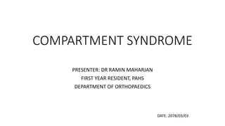
  • 1. COMPARTMENT SYNDROME PRESENTER: DR RAMIN MAHARJAN FIRST YEAR RESIDENT, PAHS DEPARTMENT OF ORTHOPAEDICS DATE: 2078/03/03
  • 2. CONTENTS • Relevant anatomy • Compartment syndrome • Definition • Epidemiology • Causes • Evaluation of patient with Acute compartment syndrome • Treatment • Fasciotomy • Compartment of thigh, leg, forearm, foot and hand • Chronic exertional compartment syndrome
  • 3. RELEVANT ANATOMY FASCIA • The muscle groups of the human limbs are divided into sections, or compartments, formed by strong, unyielding fascial membranes. • Compartments are present in various part of the body.
  • 4. Compartment syndrome • It is an elevation of the interstitial pressure in a closed osteofascial compartment that results in microvascular compromise. • Commonly are involved, especially the anterior and deep posterior compartments of the leg and the volar compartment of the forearm. • It can develop in the buttock, thigh, shoulder, hand, foot, arm, and lumbar paraspinous muscles. • It is an emergency condition.
  • 5. Epidemiology • Compartment syndrome is most commonly seen in Younger age group. It is seen in 7.3 per 100,000 males and 0.7 per 100,000 females. Tibial shaft fracture is the most common associated with a 1 to 10 percent incidence
  • 6. Classification of Compartment syndrome • Depending on the cause of the increased pressure and the duration of symptoms. • Acute compartment syndrome • Chronic exertional compartment syndrome
  • 7. Etiology Increase Compartment Volume • Fracture • Soft tissue injury • Crush syndrome • Revascularization • Exercise • Bleeding diathesis/anticoagula nts • Fluid infusion (including arthroscopy) • Arterial puncture • Osteotomy • Snake bite • Nephrotic syndrome • Leukemic infiltration • Viral myositis • Acute hematogenous osteomyelitis • Ruptured ganglia/cysts Reduce Compartment Volume • Burns • Tight closure of fascial defects • Repair of muscle hernia • Tight casts, dressings, tourniquet or splints • Entrapment under collapsed weights • Excessive traction to fractured limbs • Intramedullary nailing • Military antishock garments
  • 8. Etiology • Most common cause: fracture (69% of cases) Underlying condition % of cases Underlying condition % of cases Tibial diaphyseal fracture 36 Tibial plateau fracture 3.0 Soft tissue injury 23.2 Hand fracture(s) 2.5 Distal radius fracture 9.8 Tibial pilon fractures 2.5 Crush syndrome 7.9 Foot fracture(s) Diaphyseal fracture forearm 7.9 Ankle fracture 0.6 Femoral diaphyseal fracture 3.0 Elbow fracture dislocation 0.6
  • 9.
  • 10. Vicious cycle of Volkmann’s Ischemia
  • 11. • Normal tissue pressure • 0-4 mmHg 8-10 with exertion • Absolute intra-compartment pressure • 30 mmHg -Mubarak • 45 mmHg -Matsen
  • 12. Effect of Ischemia Muscle • 3-4 hrs: reversible changes • 6 hrs: variable changes • 8 hrs: irreversible changes Nerves • 1 hour: loses nerve conduction • 4 hours: neuropraxia • 8 hours: irreversible changes
  • 13. Evaluation of Patient with ACS • Acute compartment syndrome is a clinical diagnosis and needs prompt treatment.
  • 14. Evaluation of Patient with ACS • History and clinical examinations • Measurement of intra-compartment pressure • Creatinine phosphokinase, RFT, Urine myoglobin
  • 15. History • 5 P’s • Pain out of proportion • Paresthesia • Paresis • Pulselessness • Pallor • Pain on passive stretching
  • 16. Physical examinations • Observe skin for lesions, swelling or color change • Palpate over the compartment, observing temperature, tension, tenderness • Check pulses • Evaluate two-point discrimination and sensation • Evaluate motor function • Due to the potential for rapid progression of compartment syndrome, clinicians should perform serial exams.
  • 17. Measurement of intracompartmental pressure • is not required • but can aid in diagnosis if uncertainty exists. • Different devices can be used to detect intracompartmental pressure • The normal pressure within the compartment is between 0 mmHg to 8 mmHg. • An intra-compartmental pressure greater than 30 mmHg indicates compartment syndrome and a need for fasciotomy.
  • 18. • When intra-compartmental pressure increases to within 10 mmHg to 30 mmHg of the patient's diastolic blood pressure, this indicates inadequate perfusion and relative ischemia of the involved extremity. • The perfusion pressure of a compartment, also known as the compartment delta pressure: • clinicians often utilize delta pressure less than or equal to 30 mmHg as indicative of the need for fasciotomy. Delta Pressure = Diastolic Pressure - Measured Intracompartmental Pressure
  • 19.
  • 20.
  • 21.
  • 22. Synthes (West Chester, PA) hand- held compartment pressure monitor.
  • 23.
  • 24. Noninvasive methods • Ultrasonic device • Measures submicrometric displacement of fascia caused by volume expansion. • Near-Infrared Spectroscopy (NIRS) • Tracking of variations in the oxygenation of muscle tissue. • Laser Doppler Flowmetry • Flexible fibre optic wire is introduced into the muscle compartment and signals from this wire are recorded on a computer.
  • 25. The Reported Sensitivities and Specificities of the Clinical Symptoms and Signs of ACS Symptom or Sign Sensitivity (%) Specificity (%) PPV (%) NPV (%) Pain 19 97 14 98 Pain on passive stretch 19 97 14 98 Paresis/motor changes 13 97 11 98 Paresthesia/sensory changes 13 98 15 98 Swelling 54 76 70 63 ICP monitoring 94 98 93 99
  • 26. Precaution- Pain may be absent • Patient with epidural analgesia • Patients who are intubated or sedated • Patient with head injuries, under influence of ethanol or drugs
  • 27. Management • Prompt diagnosis and treatment • Provide supplemental oxygen. • Remove any restrictive casts, dressings or bandages to relieve pressure. • Keep the extremity at the level of the heart to prevent hypo-perfusion. • Prevent hypotension and provide blood pressure support in patients with hypotension.
  • 28. • If ICP greater than or equal to 30 mmHg or delta pressure less than or equal to 30mmHg, fasciotomy should be done. For hypotensive delta pressure <20mmHg is indication.
  • 29.
  • 30.
  • 31. Fasciotomy • The ideal time frame for fasciotomy is within six hours of injury. • Not recommended after 36 hours following injury. • If necrosis occurs before fasciotomy is performed, there is a high likelihood of infection which may require amputation. • Patients must be closely monitored for complications which include infection, acute renal failure, and rhabdomyolysis.
  • 32. Fasciotomy Principles • Make early diagnosis • Long extensile incisions • Release all fascial compartments • Preserve neurovascular structures • Debride necrotic tissues • Coverage within 7-10 days
  • 33. Prognosis • depends mainly on how quickly the condition is diagnosed and treated. • Long-term studies on survivors do reveal residual pain, Volkmann's contracture, mild neurological deficits and marked cosmetic defects in the affected extremity. Fasciotomy Recovery of limb function Within 6 hours Almost 100% After 6 hours Residual nerve damage Within 12 hours 2/3rd of patients have normal In very delayed May require amputation
  • 34. Delayed Fasciotomy • Infection rate of 46% and amputation rate of 21 % after a delay of 12 hours • 4.5 % complications for early fasciotomies and 54 % for delayed ones • Recommedations • If Compartment syndrome has existed > 8-10 hrs, supportive treatment of acute renal failure should be considered. • Skin is left intact and late reconstructions may be planned. Sheridan, Matsen JBJS 1976
  • 35. Delayed Fasciotomy • 5 patients with fasciotomies in lower limbs • Average delay 56hr (35 -95 hrs) • 1 patient died of septicemia and multiorgan failure, the others required Recommendations: • In delayed cases, routine fasciotomy may not be successful Finkelstein et al. J Trauma 1996
  • 36. Postoperative care • After the Fasciotomy, a bulky compression dressing and a splint are applied. • Wound is not closed at initial surgery • Second look debridement with consideration for coverage after 2-3 days • Limb should not be at risk for further swelling • Patient should be adequately stabilized • Usually requires skin graft • Flap coverage needed if nerves, vessels or bone exposed Goal is to obtain definitive coverage within 7-10 days
  • 39. Rehabilitation Care • Physical therapy to regain function and strength and prevent contractures and stiffness. • Wound care and monitoring for any ischemia, infection, gangrene. • Antibiotics • Analgesics • The patient will need to learn how to use an ambulatory device like crutches until healing is complete. • An occupational therapy consult is recommended to help teach the patient how to perform daily living activities.
  • 40. Complications related to fasciotomy • Altered sensation within the margins of the wound (77%) • Dry, scaly skin (40%) • Pruritus (33%) • Discoloured wounds (30%) • Swollen limbs (25%) • Tethered scars (26%) • Recurrent ulceration (13%) • Muscle herniation (13%) • Pain related to the wound (10%) • Tethered tendons (7%) • Fitzderald, McQueen Br J plastic Surgery, 2000
  • 41. Complications of compartment syndrome • Pain • Contractures • Rhabdomyolysis • Nerve damage and associated numbness and/or weakness • Infection • Renal failure • Death
  • 42. Volkmann ‘s ischaemic contracture
  • 43. Differential Diagnosis • Deep vein thrombosis • Cellulitis • Gas gangrene • Phlegmasia cerulean dolens • Rhabdomyolysis • Peripheral vascular injuries
  • 45. COMPARTMENTS NERVES SIGNS Anterior Deep peroneal(anterior tibial) nerve and vessel Numbness=1st web space Weakness=ankle/toe extension Lateral Superficial peroneal nerve Pain on=passive foot inversion Numbness=dorsum of foot Weakness=eversion Superficial posterior Sural nerve Pain on =passive ankle extension Numbness=dorsolateral foot Weakness=plantar flexion Deep posterior Posterior tibial nerve Pain on=passive ankle/toe extension/foot eversion Numbness=sole of foot Weakness=toe/ankle flexion, foot inversion
  • 47. Single-incision fasciotomy landmarks. a = anterior tibial crest, b = lateral knee joint line, c = fibular head, d = lateral malleolus, and e = gastrocnemius muscle body.
  • 48. Full-thickness skin flaps with visualization of anterior and lateral compartments. a = peroneus longus tendon under lateral compartment fascia, b =anterior intermuscular septum, and c = lateral intermuscular septum.
  • 49. Fascial incisions to decompress superficial posterior, lateral, and then anterior compartments. a = lateral compartment fascia, b = anterior tibialis tendon, c = anterior compartment incision, d = lateral compartment incision, e = superficial posterior compartment incision, f = superficial posterior compartment fascia, and g = gastrocnemius muscle body.
  • 50. Identifying the lateral intermuscular septum. a = lateral compartment fascia, b = peroneus longus tendon, c = peroneus brevis muscle body, d = lateral intermuscular septum, and e = gastrocnemius muscle body.
  • 51. Entrance to deep posterior compartment. a = posterior border of fibula and site of longitudinal incision, b = lateral intermuscular septum, c = flexor hallucis longus muscle body, d = peroneus longus muscle body, and e = fascia overlying lateral compartment
  • 53. Landmarks for anterolateral skin incision in dual-incision fasciotomy. a = anterior tibial crest, b = anterolateral skin incision, c = fibular head, d = lateral knee joint line, and e = tibiotalar joint.
  • 54. Fascial incisions to decompress lateral and anterior compartments. a = anteroinferior border of patella, b = fascial incision in anterior compartment, c = tibialis anterior tendon, d = peroneus longus tendon, e = fascial incision in lateral compartment, and f = anterior intermuscular septum
  • 55. Landmarks for medial skin incision in dual-incision fasciotomy. a = medial knee joint line, b = anterior tibial crest, c = medial malleolus, d = posterior border of tibia, and e = incision mark
  • 56. Approach to superficial and deep posterior compartments through medial incision. a = gastrocnemius muscle body under superficial posterior compartment fascia, b = Achilles tendon, and c = medial malleolus.
  • 57. Decompression of deep posterior compartment. a = soleus muscle attachment to posterior part of tibia, b = superficial posterior compartment fascia over gastrocnemius muscle body, c = soleus muscle as seen through longitudinal incision that provides access into deep posterior compartment, and d = tibia.
  • 58. Release of soleus bridge. a = inferomedial border of patella, b = soleus muscle body origin after release from posteromedial aspect of tibia, c = fascia over gastrocnemius muscle, d = posteromedial border of tibia, and e = incision to access deep posterior compartment
  • 60. Thigh compartment contents COMPARTMENTS NERVES SIGNS Anterior Saphaneous Pain on passive knee flexion Numbness on medial leg/foot Weakness – knee extension Posterior Sciatic Pain on=passive knee extension Sensory changes rare Weakness – knee flexion Medial Obturator Pain on passive hip abduction Sensory changes rare Weakness – hip adduction
  • 61.
  • 64. Incision for release of the volar forearm compartment syndrome.
  • 65. Hand • 10 separate osteofascial compartments • Doral interossei -4 • Palmar interossei-3 • Thenar and hypothenar-2 • Adductor policis – 1 • Non specific aching of the hand • Disproportionate pain • Loss of digit motion and continued swelling • Pain on MCP extension and PIP flexion
  • 66.
  • 67.
  • 68. • Compartment Contents Medial Intrinsic muscles of the great toe Lateral Flexor digiti minimi & Abductor digiti minimi Central - Superficial Flexor digitorum brevis -Deep (calcaneal) Quadratus plantae Adductor hallucis Adductor hallucis Interosseous (×4) Interosseous muscles Digital nerves The Foot
  • 69. Foot – compartment syndrome • Careful examination with any swelling • Clinical suspicion with certain mechanism of injury: • Lisfrac fracture dislocation • Calcaneus fracture
  • 70.
  • 71. Chronic exertional compartment syndrome • Reversible ischemia secondary to a noncompliant osteofascial compartment that is unresponsive to the expansion of muscle volume that occurs with exercise. • Muscle volume can increase upto 20% of its resting size • Muscle hypertrophy • Common in young adult recreational runners, elite athletes, and military recruits.
  • 72. Clinical findings • Presents with • exercise induced pain and • a feeling of tightness that begins after 20-30 minutes of running. • It resolves within 15 to 30 minutes of cessation of exercise. • Paresthesia • Physical examination • Tenderness over the musculature of involved compartment and • muscle herniation through fascial defect may be palpated.
  • 73. Diagnosis – Pedowitz criteria • The most commonly accepted values are the presence of one or more of the following criteria: (1) preexercise, resting pressure of 15 mm Hg or more; (2) pressure of 30 mm Hg or more 1 minute after exercise; and (3) pressure of 20 mm Hg or more 5 minutes after exercise.
  • 74. Treatment • Conservative measures • Relative rest • Anti-inflammatory medications • Stretching and strengthening of the involved muscles • Orthotics Operative treatment fasciotomy
  • 75.
  • 76.
  • 77. References • Campbell’s Operative Orthopaedics 4th Edition • J. Maheshwari 5th Edition • Apley’s 9th Edition • UpToDate Version 3.0 • Torlincasi AM, Lopez RA, Waseem M. Acute Compartment Syndrome. [Updated 2021 Feb 10]. In: StatPearls [Internet]. Treasure Island (FL): StatPearls Publishing; 2021 Jan-. Available from: https://www.ncbi.nlm.nih.gov/books/NBK448124/

Notas do Editor

  1. Compartment present in the buttock, thigh, shoulder, hand, foot, arm, and lumbar paraspinous muscles. Fascia is a thin, inelastic sheet of connective tissue that surrounds muscle compartments and limits the capacity for rapid expansion.
  2. Compartments with relatively noncompliant fascial or osseous structures most commonly are involved, especially the anterior and deep posterior compartments of the leg and the volar compartment of the forearm. Compartment syndrome can develop anywhere skeletal muscle is surrounded by substantial fascia, however, such as in the buttock, thigh, shoulder, hand, foot, arm, and lumbar paraspinous muscles.
  3. Active age group is mostly involved. Youth has been found to be the most important risk factor for developing ACS, possibly due to the relatively high muscle bulk in a fixed compartment and involve in high energy trauma, and thus a reduced capacity for swelling in these patients. Older patients often have reduced muscle bulk secondary to sarcopenia, with an associated increased perfusion pressure due to hypertension, which could potentially explain the protective effects of increasing age.
  4. Acute compartment syndrome typically occurs within a few hours of inciting trauma. However, it can present up to 48 hours after. Chronic exertional compartment syndrome (CECS) is recurrence of increased pressure, most often in the anterior or deep posterior compartment of the leg. Exercise can increase muscle volume by 20%, causing an increase in pressure in a noncompliant compartment. Exertional compartment syndrome of the lower extremity is most common in long-distance runners and military recruits pushed past normal limits of functional tolerance. It also has been reported to occur elsewhere, including the forearms in weightlifters, rowers, welders, and others who place large demands on their upper extremities. Chronic compartment syndrome occurs most probably due to increased muscle mass in a closed fascial space occurring during exercise or exertion. Muscle volume can increase up to 20% of its resting size during exercise.
  5. any condition that restricts the intracompartmental space or increases the fluid volume in the intracompartmental space.
  6. The effect of ischemia on muscle and nerve are temporal; prolonged delay results in greater loss of function. Muscles retain the electrical response up to 3 hours. They can tolerate ischemia up to 4 hours and irreversible damage occurs at 8 hours. Nerves can conduct impulses up to 1 hours. They can survive up to 4 hours (neuropraxia), and at 8 hours irreversible damage occurs. Although these are experimental figures often quoted, in reality muscle necrosis occurs within the first 3 hours for unknown reasons
  7. How to evaluate a patient for acute compartment syndrome
  8. Elevations in creatine phosphokinase (CPK) may suggest muscle breakdown from ischemia, damage, or rhabdomyolysis.If rhabdomyolysis is being considered, renal function tests, urine myoglobin, and urinalysis should be done. outcome?
  9. Acute compartment syndrome typically occurs within a few hours of inciting trauma. However, it can present up to 48 hours after. Pain is typically severe, out of proportion to the injury. Early on, pain may only be present with passive stretching. However, this symptom may be absent in advanced acute compartment syndrome. In the initial stages, pain may be characterized as a burning sensation or as a deep ache of the involved compartment. Paresthesia, hypoesthesia, or poorly localized deep muscular pain may also be present. Classically, the presentation of acute compartment syndrome has been remembered by "The Five P's": pain, pulselessness, paresthesia, paralysis, and pallor. However, aside from paresthesia, which may occur earlier in the course of the condition, these are typically late findings. Beware that the presence or absence of a palpable arterial pulse may not accurately indicate relative tissue pressure or predict the risk for compartment syndrome. In some patients, a pulse is still present, even in a severely compromised extremity. 
  10. Physical exam should focus on the neurovascular territory of the involved compartment: The earliest objective physical finding is the tense, or ''wood-like" feeling of the involved compartment.  Although the clinical features discussed above can help identify compartment syndrome, they have limited sensitivity and specificity. Other factors, such as compartment pressures, can help make the diagnosis.[18] [19]
  11. often measured with a manometer, a device that detects intracompartmental pressure by measuring the resistance that is present when a saline solution is injected into the compartment. Another method employs a slit catheter, whereby a catheter is placed within the compartment, and the pressure measured with an arterial line transducer. The slit catheter method is more accurate and allows for continuous monitoring. Its use is also recommended to measure all the surrounding compartments.
  12. Compartment pressures are often measured with a manometer, a device that detects intracompartmental pressure by measuring the resistance that is present when a saline solution is injected into the compartment. Another method employs a slit catheter, whereby a catheter is placed within the compartment, and the pressure measured with an arterial line transducer. The slit catheter method is more accurate and allows for continuous monitoring. Its use is also recommended to measure all the surrounding compartments. Slit technique required a polyethylene tubing filled with air and no air bubbles present withing the tubing, connected to a pressure transducer
  13. any other circumstance that alters patient’s ability to accurately sense and communicate pain levels.
  14. Acute compartment syndrome is an emergency condition. Less time should be spent on confirmation of the diagnosis, as delayed treatment may result in loss of limb. Remove circumferential cast and padding if any(35% one side; 65% both side ;padding 15%) Splitting of cast reduces pressure by 30% that increases to 65% if the cast is split and spread. Complete splitting and padding the cast adds 10% to relieve pressure, and complete removal adds another 15%. There could be total of 85–90% reduction if cast is fully removed. Limb should not be elevated as this reduces the mean pressure but not the compartment pressure.
  15. For patients who do not meet diagnostic criteria for acute compartment syndrome but who are at high risk based on history and physical exam findings, or for patients with intracompartmental pressures between 15 to 20 mmHg, serial intracompartmental pressure measurements are recommended. Patients with ICPs between 20-30 mmHg should be admitted and the surgical team should be consulted. For patients with intracompartmental pressures greater than 30 mmHg or delta pressures less than 30 mmHg, surgical fasciotomy should be done.
  16. When tissue pressure remains elevated for that amount of time, irreversible damage may occur, and fasciotomy may not be beneficial in this situation. If infection occurs, debridement is necessary to prevent the systemic spread or other complications.
  17. Recurrent compartment syndrome has been known to occur in athletes due to scarring. There are some individuals who may die from acute compartment syndrome. Often these cases are caused by infection, which ultimately leads to sepsis and multiorgan failure. than outcomes for the posterior compartment of the leg worse since it is difficult to perform inadequate decompression of the posterior compartment.
  18. POSTOPERATIVE CARE  At 48 to 72 hours, the patient is returned to the operating room for debridement of any necrotic material. Intravenous fluorescein and a Wood light can be helpful in evaluating muscle viability. If there is no evidence of muscle necrosis, the skin is loosely closed. If closure is not accomplished, the debridement is repeated after another 48- to 72-hour interval, after which skin closure or skin grafting can be done.
  19. Contractures results from insufficient arterial perfusion and venous stasis followed by ischaemic degeneration of muscle; irreversible muscle necrosis begins after 4-6 hrs Resulting progressive muscle necrosis; Muscle degeneration is most affected at the middle third of muscle belly being most severe closer to bone, less involvement toward proximal and distal surfaces Necrosis of muscle with secondary fibrosis that may develop followed by calcification in its final stage Ulnar artery – AIN branch – FDP, FDS, pronator teres and FPL FDP and FDS contracted and scared lead to wrist flexion and clawing of fingers
  20. The authors noted that a pressure of 105 mm Hg immediately after 5 minutes of sustained walking at 4 miles per hour with a 5% incline and the patient wearing a 15-kg backpack had better diagnostic accuracy than the Pedowitz criteria above.
  21. relative rest (limiting activity to a level that avoids all but minimal symptoms)