Cloning vectors based on m13 and lambda bacteriophage
•
4 gostaram•2,278 visualizações

m13 and lamba phage based vectors, problems associated with using m13 and lambda phages as vectors

Recomendados
Mais conteúdo relacionado
Mais procurados
Mais procurados (20)
Semelhante a Cloning vectors based on m13 and lambda bacteriophage
Semelhante a Cloning vectors based on m13 and lambda bacteriophage (20)
Último
Último (20)
Cloning vectors based on m13 and lambda bacteriophage
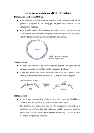
- 1. Cloning vectors based on M13 bacteriophage Difficulties in constructing M13 vector Main limitation is limited scope of alteration in M13 genes as most of the genome is composed of ten closely packed genes, each essential for the replication of the phage. There is only a single 507-nucleotide intergenic sequence into which new DNA could be inserted without disrupting one of these genes, and this region includes the replication origin which must itself remain intact. M13mp1 vector M13mp1 was constructed by introducing introduce the lacZ′ gene into the intergenic sequence. M13mp1 forms blue plaques on X-gal agar. It does not possess any unique restriction sites in the lacZ′ gene. It does, however, contain the hexanucleotide GGATTC near the start of the gene. M13mp2 vector M13mp2 was constructed by a single nucleotide change in GGATTC to GAATTC which is present in M13mp1at the start of lacZ′ gene. This alteration was carried out using in vitro mutagenesis. M13mp2 has a slightly altered lacZ′ gene (the sixth codon now specifies asparagine instead of aspartic acid), but the b-galactosidase enzyme produced by cells infected with M13mp2 is still perfectly functional.
- 2. M13mp7 vector Additional restriction sites were introduced into the lacZ′ by inserting a short oligonucleotide, called a polylinker. A polylinker consists of a series of restriction sites and has EcoRI sticky ends. This polylinker was inserted into the EcoRI site of M13mp2, to give M13mp7. M13mp7 has four possible cloning sites (EcoRI, BamHI, SalI, and PstI). The polylinker is designed so that it does not totally disrupt the lacZ′ gene: a reading frame is maintained throughout the polylinker, and a functional, though altered, β-galactosidase enzyme is still produced. M13mp8 vector M13mp8 has the same series of restriction sites as the plasmid pUC8. One advantage of M13mp8 is its ability to take DNA fragments with two different sticky ends. Cloning vectors based on λ bacteriophage Problems associated with using λ bacteriophage as vector: Two problems had to be solved before λ -based cloning vectors could be developed: The λ DNA molecule can be increased in size by only about 5%. If the total size of the molecule is more than 52 kb, then it cannot be packaged into the λ head structure and infective phage particles are not formed. This severely limits the size of a DNA fragment that can be inserted into an unmodified λ vector. The λ genome is so large that it has more than one recognition sequence for virtually every restriction endonuclease. Restriction cannot be used to cleave
- 3. the normal λ molecule in a way that will allow insertion of new DNA, because the molecule would be cut into several small fragments that would be very unlikely to re-form a viable λ genome on religation. By overcoming these difficulties a wide variety of λ cloning vectors have been developed, their primary use being to clone large pieces of DNA, from 5 to 25 kb, much too big to be handled by plasmid or M13 vectors. Overcoming problems associated with using λ bacteriophage as vector: 1) Segments of the λ genome can be deleted without impairing viability A large segment in the central region of the λ DNA molecule can be removed without affecting the ability of the phage to infect E. coli cells. Removal of all or part of this non-essential region, between positions 20 and 35 decreases the size of the resulting λ molecule by up to 15 kb. This means that as much as 18 kb of new DNA can now be added before the cut-off (52 kb) point for packaging is reached. This “non-essential” region contains most of the genes involved in integration and excision of the λ prophage from the E. coli chromosome. A deleted λ genome is therefore non-lysogenic and can follow only the lytic infection cycle. This is desirable for a cloning vector as it means induction is not needed before plaques are formed. 2) Natural selection can be used to isolate modified λ that lack certain restriction sites Even a deleted λ genome, with the non-essential region removed, has multiple recognition sites for most restriction endonucleases. If just one or two sites need to be removed, then the technique of in vitro mutagenesis can be used. For example, an EcoRI site, GAATTC, could be changed to GGATTC, which is not recognized by the enzyme.However, in vitro mutagenesis was in its infancy when the first λ vectors were under development, and even today would not be an efficient means of changing more than a few sites in a single molecule. Instead, natural selection was used to provide strains of λ that lack the unwanted restriction sites. Natural selection achieved by using an E. coli strain that produces EcoRI, as a host. Most λ DNA molecules that invade the cell are
- 4. destroyed by this restriction endonuclease, but a few survive and produce plaques. These are mutant phages, from which one or more EcoRI sites have been lost spontaneously. Several cycles of infection will eventually result in λ molecules that lack all or most of the EcoRI sites. λ Insertion and replacement vectors The first two classes of vector to be produced were λ insertion and λ replacement (or substitution) vectors. λ Insertion vectors With an insertion vector, a large segment of the non-essential region has been deleted, and the two arms ligated together. An insertion vector possesses at least one unique restriction site into which new DNA can be inserted. The size of the DNA fragment that an individual vector can carry depends on the extent to which the non-essential region has been deleted.
- 5. Figure: Cloning with lambda phage vectors Two popular insertion vectors are: λ gt10 vector It can carry up to 8 kb of new DNA, inserted into a unique EcoRI site located in the cI gene. Insertional inactivation of this gene means that recombinants are distinguished as clear rather than turbid plaques. λ ZAPII vector It carries lacZ′ gene and 6 restriction sites within a polylinker. DNA fragment of size up to 10 kb can be inserted into any of 6 restriction sites within the polylinker.
- 6. Insertion of DNA fragment inactivates the lacZ′ gene carried by the vector. Recombinants give clear rather than blue plaques on X-gal agar. Replacement vectors A λ replacement vector has two recognition sites for the restriction endonuclease used for cloning. These sites flank a segment of DNA that is replaced by the DNA to be cloned. Often the replaceable fragment (or “stuffer fragment” in cloning jargon) carries additional restriction sites that can be used to cut it up into small pieces, so that its own re-insertion during a cloning experiment is very unlikely. Replacement vectors are generally designed to carry larger pieces of DNA than insertion vectors can handle. Recombinant selection is often on the basis of size, with non-recombinant vectors being too small to be packaged into λ phage heads. An example of a replacement vectors is: λ EMBL4 It can carry up to 20 kb of inserted DNA by replacing a segment flanked by pairs of EcoRI, BamHI, and SalI sites. Any of these three restriction endonucleases can be used to remove the stuffer fragment, so DNA fragments with a variety of sticky ends can be cloned. Recombinant selection with λ EMBL4 can be on the basis of size, or can utilize the Spi phenotype.
- 7. REFERENCE: Gene Cloning and DNA Analysis, T.A. Brown, 6th edition