Tumours of external and middle ear
•Transferir como PPT, PDF•
68 gostaram•23,005 visualizações

Tumors of external and middle ear

Recomendados
Mais conteúdo relacionado
Mais procurados
Mais procurados (20)
Destaque
Destaque (20)
Semelhante a Tumours of external and middle ear
Semelhante a Tumours of external and middle ear (20)
Mais de Ramesh Parajuli
Mais de Ramesh Parajuli (15)
Último
Último (20)
Tumours of external and middle ear
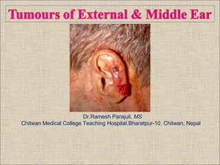
- 1. Dr.Ramesh Parajuli, MS Chitwan Medical College Teaching Hospital,Bharatpur-10, Chitwan, Nepal
- 2. Contents • Introduction • Tumours of auricle • Benign tumours of external auditory canal (EAC) & middle ear cleft • Malignant tumours of external auditory canal (EAC) & middle ear cleft
- 3. Introduction • Less common • Little known • Some surgeons - never encounter • Difficult to treat • Outcome: unsatisfactory
- 5. Basal cell & Squamous cell carcinoma Epidemiology: • Mean age 70 yr • Lower in darker-skinned ethnic group • Basal cell (BCC)>Squamous (SCC)>Melanoma (MM) • 85-95% of all BCC & SCC occur in head & neck region • 12% of these tumours in auricle • SCC: External ear & upper face • BCC: Midface & auricle
- 6. Pathophysiology/Risk factors 1. Sun exposure: ultraviolet radiation (SCC> BCC) 2. Actinic keratosis: small, scaly lesions, common (Actinic keratosis60% SCC) 3. Keratoacanthoma: benign conditionspontaneous resolution or SCC 4. Exposure to radiation 5. Immunosupression: 5-16 fold
- 7. 7. Xeroderma pigmentosa: Autosomal recessive, DNA repair mechanism, SCC and BCC at young age 8. Trauma 9. Frostbite 10. Psoriasis 11.Aflatoxin B 12. Nevoid basal cell syndrome (Gorlin’s syndrome): Autosomal dominant, multiple pigmented BCC once reach puberty 13. Chronic otitis externa
- 8. Pathology Basal Cell Carcinoma (BCC) Variants of BCC: Nodular: most common, least aggressive, bleeds easily Ulcerative: “Rodent ulcer” Pigmented: Superficial: Morpheaform/sclerosing: Most aggressive, highest rate of recurrence Basaloid squamous
- 9. Squamous cell carcinoma (SCC) • Plaque to nodule or ulcer • Variants of SCC, less common than BCC • Keratin pearl • Variants of SCC: Nodular Ulcerative Pigmented: confused with melanoma Spindle shaped subtype: radiated skin Verrucous- locally destructive Basaloid squamous Adenoid squamous
- 10. Diagnosis • Clinical evaluation • Biopsy: • Lesion with change in colour, size, shape, friable or ulcerated Punch biopsy- preserves architecture of the lesion for histologic analysis, depth of lesion, excision with precise margin. • Imaging studies (CT, MRI): rarely needed
- 11. Staging of Tumour (AJCC)
- 12. Stage Primary tumour Regional lymph node Distant metastasis Stage 0 Tis N0 M 0 Stage I T1 N0 M 0 Stage II T 2 N 0 M 0 T 3 N 0 M 0 Stage III T 4 N 0 M 0 Any T N 1 M 0 Stage IV Any T Any N M 1
- 13. Limitations of AJCC staging in malignancy of ear Thin skin of ear early involvement of deeper structure T4 T4- doesn’t have similar prognosis or require radical treatment like in other sites Staging based on sizeless practical (unique anatomy of ear) Even small tumours eg. preauricular, concha or tragusextensive surgery Doesn’t account for different histologies: BCC may act differently than SCC
- 14. Treatment Depends on • Location & size • General condition of patient • Expertise of doctor • Desire of patient
- 15. Surgical Excision • Most common form of treatment • Appropriate margin of resection difficult • BCC: – 8 mm for <3 cm – 1.5 cm for >3cm (Bumstead et al., 1981) • 2-3 mm for <1cm • 3-5mm for 1-2 cm • 7-10mm-morpheaform histology (Scotto et al., 1983) • SCC: 1-2 cm surgical margin (Bumstead et al., 1981)
- 16. Mohs’ Surgery Dr. Frederick Mohs in 1941 • Serial horizontal sectioning of tumor & surrounding tissue with immediate microscopic analysisconfirm the margins clear of tumour • Fixed in vivo with zinc chloride • Several advantages over traditional wide local excision Horizontal section of entire margin analysis of small islands of tumor cells Avoids unnecessary excision of normal tissue Less recurrences following Mohs’ surgery
- 17. • Recommended method for Malignancy arising in vital area Recurrent or previously treated area Aggressive histopathology Larger carcinoma >1cm Poorly defined margin
- 18. • Mohs’ surgery: overall cure rates for auricular carcinoma- 98% for BCC and 92% for SCC (Mohs F, 1998 and Niparko et al., 1990)
- 19. Other surgical techniques: For small non-agressive tumours with clearly defined margin • Curettage and electrodesiccation • Cryosurgery: -40°c, cure rate exceeds 95% Cryogun used to spray liquid nitrogen
- 20. 5 year recurrence rate for primary BCC (Rowe et al., 1989)
- 21. 5 year recurrence rates for previously treated BCC (Rowe et al., 1989)
- 22. Radiation Therapy • Advantages Avoids tissue defects, unfit patients, refuse surgery & tumour involving adjacent areas • Disadvantages Multiple treatments, more costly, risk of radiation induced tumour • Regimen vary from center to center • Common regimen – 20 Gy, 2# very small tumor – 30-40 Gy for 2-4 cm volume – 60-65 Gy for large tumors • Cure rate comparable to surgery for <1 cm
- 23. • Non surgical options – Topical 5-Fluoruracil (5-20% concentration) – Intralesional interferon-alpha – Use of photodynamic therapy • Treatment of recurrent disease- more difficult Aggressive tumours, margins less precise and disrupted anatomy due to prior treatmentreconstruction more difficult • Treatment of metastatic neck disease – Overall recurrence rate for BCC- <5% and SCC- 10 to 15% – Elective neck dissection - Controversy
- 28. Malignant melanoma Epidemiology: • Incidence increasing • Accounts for majority of deaths from skin malignancies • 7-13% of all head & neck malignancy, 1% of all melanoma • 65-79 yr • Lower in black & dark skinned people • Lower in woman- more attention paid on appearance & more regular physician visits
- 29. Pathophysiology • Sun exposure: Acute severe & cumulative • Actinic precursor • Congenital naevi • Familial dysplastic naevus syndrome • Xeroderma pigmentosa • Immunosuppression Dysplastic nevus
- 30. Types of Melanoma • Cutaneous melanoma Acral lentiginous Superfical spreading melanoma- most common Lentigo maligna melanoma Nodular melanoma Amelanotic melanoma • Mucosal melanoma Acral lentiginous Nodular melanoma
- 31. Diagnosis • Keen eyes, diligent physical examination • Majority arises from pre-existing naevi • A B C D (Asymmetry, Border, Color, Diameter) • Excisional biopsy • HPE: Variable • Immunohistochemistry: S-100, Vimentin, HMB- 45 • Role of imaging
- 34. Treatment Surgery • Tumour thickness- correlates with survival • 1cm for 1mm thickness & 2cm for larger • Mohs’ Surgery: Controversy • Overall cure rate: 68% • Recurrence between 1-3 yr, follow up every 1-2 month • Role of radiation • Traditionally considered to be radioresistant • For local control of paritally resected melanoma
- 35. Treatment of neck metastasis • 17% nodal disease at presentation • Auricular melanoma risk 42% • Parotid & upper jugular digastric • Radical or modified neck dissection for melanoma- No studies to demonstrate differences • Post-operative RT • Elective neck dissection: Controversy • Treatment of advanced disease: 10% 10 yr survival
- 36. Other tumours • Merkel cell carcinoma: One of the most aggressive High recurrence and metastases rate Neuroendocrine tumour Pleuripotent basal cells Rapidly growing, firm, nodular Wide local excision Radiosensitive
- 37. • Malignant fibrous histiocytoma • Dermatofibrosarcoma • Angiosarcoma • Metastatic disease from other sites
- 39. Contd… • All three primordial layers temporal bone spectrum of neoplasms • Benign & malignant • Neither AJCC nor the UICCsystem for classifying temporal bone tumours • Difference in growth rate & sites of origin varied presentations
- 40. Classification of tumours of the temporal bone:
- 41. Benign tumours of EAC and middle ear cleft Exostosis • Benign • Deep part of bony canal, adjacent to TM • Smooth, sessile, multiple, inner part of meatus & bilateral • Relationship with cold water exposure • Asymptomatic • >80% stenosis: Symptomatic
- 42. • Diagnosis – Otoscopy – CT • Treatment – Conservative – Excision with meatoplasty – Postaural approach
- 43. Osteoma • Smooth, rounded & pedunculated • Single • Unilateral • Arise from lateral part of bony EAC • Asymptomatic • Diagnosis • Management
- 44. Other benign tumours: • Keratoacanthoma – Relatively common – Arise from hair follicle – Elevated mass, fleshy colored or pinkish – Linked to chemical carcinogens, sun exposure, trauma & viral – Premalignant – Complete excision • Papilloma: squamous papilloma and inverted papilloma • Pleomorphic adenoma • Choriostoma: commonly salivary gland tissue, not a true neoplasm, no aggressive potential, surveillance
- 45. • Haemangioma • Langerhans’ cell histiocytosis • Nerve sheath neoplasm • Paraganglioma • Hamartoma Haemangioma Hamartoma
- 46. • Meningioma • Chordoma • Haemangiopericytoma • Fibrous dysplasia: Fibrous tissue, bony spicules- undergoing resorption and formation Islands of cartilage replacing bone marrow Monostotic and polyostotic Mc Cune Albright syndrome ‘Ground glass’ appearance Surgical excision
- 47. Malignant Tumours of Temporal Bone • Squamous cell carcinoma • Basal cell carcinoma • Adenocarcinoma • Acinic cell carcinoma • Adenoid cystic carcinoma • Melanoma • Osteosarcoma • Ewing’s sarcoma • Chondrosarcoma • Rhabdomyosarcoma • Metastases to temporal bone • Lymphoma • Malignant neuroma • Malignant paraganglioma • CNS malignancy • Endolymphatic sac tumour
- 48. Pathophysiology/Risk factors: • Risk factors - not well defined • Chronic otitis media: Strongly suggests but not invariably associated • Human papilloma virus: HPV 16 and 18 (Jin et al., 1997) • Radium dial workers • External beam radiation • Ultraviolent radiation and other forms of radiation: no strong link?
- 49. • Incidence: old age EAC • Squamous cell carcinoma: most common • Hidradenocarcinoma • BCC, melanoma & mucoepidermoid carcinoma Middle ear • SCC (80%), adenocarcinoma, BCC & adenoid cystic carcinoma Well differentiated SCC of EAC
- 50. Behavior of SCC of external auditory canal & middle ear • Escape from the EAC and middle ear cleft several directions • Superiorly • Posteriorly • Anteriorly • Inferiorly • Medially • Laterally
- 53. MRI coronal view of the temporal bone showing mass from the left mastoid area extending out to involve external ear canal and skin
- 54. MRI axial view of the temporal bone showing tumour eroding left lateral semicircular canal
- 55. MRI axial view showing the left temporal lobe involvement and cerebellar compression
- 56. Signs & Symptoms • Change in pattern: may be only clue • Growth • Pain • Discharge • Deteriorating hearing • Facial paralysis, hemi facial spasm • Lower cranial nerve palsy • Trismus • Lymphadenopathy Differential diagnosis: Otitis Externa, COM An exophytic mass within EAC
- 57. (Gustafson ML and Pensak ML, 2002)
- 58. Investigations • Multimodality imaging • CT + MRI – Coronal: EAC, Outer Attic Wall, Tegmen tympani – Axial: Posterior & medial invasion of petrous pyramid, carotid canal, lymph node detection, prevertebral spread • HPE: Multiple biopsy • FNAC (CT guided) • Angiography – Major vessel invasion • Audiometry: Every patient
- 59. Staging (Ariaga et al., 1990)
- 60. (Pensak et al., 1996)
- 61. Treatment • Surgical treatment followed by radiotherapy • Extent of surgery controversial • En block Vs Piecemeal • Radical mastoidectomy • Problem of local recurrence • Extensive surgery Complete temporal bone resection
- 62. Sleeve Resection • Tumours confined to skin & soft tissue of cartilaginous portion of EAC • T 1 • Red line • Incision – medial and lateral • Involved skin & underlying cartilage resectedwide meatoplasty • Split-thickness skin graft
- 63. Lateral Temporal Bone Resection • Tumours involving bony & cartilaginous part (not involved annulus & tympanic cavity) • T 2 • Entire external auditory canal + TM + Malleus + Incus resected ‘en-bloc’ (Blue Line) • Facial nerve dissected from stylomastoid foramen to pes anserinus • Superficial parotidectomy • Eustachian tube - plugged
- 66. En-bloc lateral temporal bone resection with radical neck dissection
- 67. Modified Lateral Temporal Bone Resection • Tumor extension to tympanic cavity or mastoid air cell • T3 • Facial nerve sacrificed • Posterior petrosectomy- bone removal posteriorly back to the transverse sinus & posterior fossa dura • Tegmen tympani removed • Perilabyrinthine, retrofacial cells opened down to jugular bulb • More extensive defect: temporalis and sternocleidomastoid muscle flaps rotated inobliterate the defect
- 68. Subtotal Temporal Bone Resection • Extensively encroach tympanic cavity • T3 • Entire temporal bone lateral to petrous carotid artery en bloc • If necessary: Portion of dura, sigmoid sinus, parotid gland, ramus of mandible, subtotal parotidectomy • Facial nerve transected
- 69. Total Temporal Bone Resection • Sacrifice of carotid artery • T 4 Light green line • Most important decision is not to operate at all (Gustafson et al., 2003) • Invasion of cavernous sinus, ICA, infratemporal fossa, paraspinous muscle-surgically incurable
- 70. Radiation therapy • Radiotherapy for curative treatment – limited success • 5-year cure rate RT alone 28.7%, RT+ Surgery 59.6% (Zhang et al.,1999) • Higher doses- toxicity to brainstem • Adjuvant therapy & palliation • Side effects and complications- osteoradionecrosis, facial nerve pasly & brain necrosis • No randomized study-effect of radiation on survival & recurrence
- 71. Treatment of metastasis • Regional & distant metastasis from temporal bone malignancy-low • Nodal diseases at the time of presentation- 9% to 18% (Pensak ML, 2003) • Upper jugulodiagastric & parotid nodes- most commonly involved • Neck dissection - no improvement in survival • Superficial parotidectomy in every patient with more than superficial disease & neck dissection reserved for known adenopathy
- 72. Other epithelial malignancy: • BCC • Melanoma Glandular malignant lesion • Adenoid cystic carcinoma- most common glandular malignancy in EAC • Ceruminous adenocarcinoma • Mucoepidermoid carcinoma Sarcomas: Fibrosarcoma, osteogenic sarcoma, Ewing’s sarcoma, Kaposi’s sarcoma & chondrosarcoma Meatastasis to temporal bone: breast, lung, kidney, stomach, brochus & prostate Secondary tumor of temporal bone Spreading from adjaent areas: Parotid, nasopharyngeal, auricular & meningioma
- 74. Tumours in childhood Rhabdomyosarcoma (RMS) • Rhabdo-rod shaped, myo- muscle • Most common soft tissue sarcoma in children • Ear 3rd most common after nasopharynx & orbit • Meningeal, parameningeal & orbital • Majority - before 12 years (average age - 4.4 years) • Types: 1. Embryonal–nearly all of the head & neck rhabdomyosarcoma 2. Alveolar – worst prognosis 3. Pleomorphic 4. Botryoid – best survival
- 75. RMS contd… Clinical features: (Prat J, 1997) 1. Mass in ear region 56% 2. Aural polyp 54% 3. Ear discharge 40% 4. Bleeding from ear 30% 5. Ear pain 22% 6. Hearing loss 14% 7. Facial paralysis 14% Diagnosis • Biopsy • PCR • Bone marrow • CT • MRI
- 76. International Rhabdomyosarcoma Study (IRS) Based on tumour resectability: • Group I - Tumour completely removed • Group II - Microscopic residual tumour, involved regional nodes, or both • Group III - Gross residual tumour • Group IV - Distant metastatic disease
- 77. Treatment • Multimodality & multidisciplinary • Triple therapy • Surgery: biopsy to confirm the diagnosis • Chemotherapy: Vincristine, Dactinomycin, Cyclophosphamide, Ifosfamide • Radiotherapy: Intensity Modulated Radiotherapy (IMRT)