23204943
•
4 gostaram•582 visualizações

The document describes 3 radiology case studies involving the gastrointestinal system: 1) A case of scleroderma diagnosed based on an x-ray showing dilation of the small bowel with closely spaced folds. 2) A case of appendicitis diagnosed on CT showing an enlarged appendix with periappendiceal inflammation and an appendicolith. 3) A case of a gastric leiomyoma diagnosed on barium study and CT showing a rounded filling defect in the stomach with central ulceration arising from the gastric wall.

Recomendados
Mais conteúdo relacionado
Mais procurados
Mais procurados (20)
Destaque
Destaque (20)
Semelhante a 23204943
Semelhante a 23204943 (20)
Mais de radgirl
Último
Último (20)
23204943
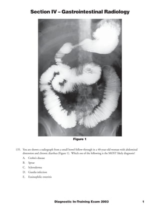
- 1. Section IV – Gastrointestinal Radiology Figure 1 135. You are shown a radiograph from a small bowel follow-through in a 40-year-old woman with abdominal distension and chronic diarrhea (Figure 1). Which one of the following is the MOST likely diagnosis? A. Crohn’s disease B. Sprue C. Scleroderma D. Giardia infection E. Eosinophilic enteritis Diagnostic In-Training Exam 2003 1
- 2. Section IV – Gastrointestinal Radiology Question #135 Findings: The radiograph demonstrates dilation of the small bowel with closely spaced normal thickness folds. Rationales: A) Incorrect. Crohn’s disease involves the small bowel in 75% of patients at the time of presentation. Affected segments show fold thickening and ulceration. Sinus tracts and fistulae are also noted. Surrounding fibrofatty proliferation may produce a mass effect or displace adjacent bowel. None of these findings, however, are noted here. B) Incorrect. Sprue or gluten sensitive enteropathy produces villous atrophy and radiographically shows reversal of the normal fold pattern with loss of normal jejunal folds and an increased number of folds per inch in the ilium. Transient intussusceptions, hypomotility, and flocculation of barium can also be seen. However, none of these findings are present on this film. C) Correct. Scleroderma causes smooth muscle atrophy and fibrosis. Radiographically the small bowel appears dilated with closely spaced but normal caliber valvulae conniventes as shown in this radiograph. Wide mouth diverticula are frequently noted on the mesenteric side of the bowel, although they are not seen here. Hypomotility is common and along with the dilation can produce bacterial overgrowth syndrome and the symptoms noted here. The small bowel is the second most commonly affected portion of the GI tract in scleroderma. D) Incorrect. Giardia lamblia is a common intestinal parasite. The radiographic findings in this protozoal infection are most common in the duodenum and proximal jejunum and consist of thickened folds associated with bowel irritability, hypermotility, and increased secretion. E) Incorrect. Eosinophilic enteritis is a benign infiltration of the bowel wall with eosinophils. The etiology is unclear but the disease responds rapidly to steroids. Radiographically fold thickening that can be nodular is noted most prominently in the proximal small bowel. Gastric antral involvement is also common. 2 American College of Radiology
- 3. Section IV – Gastrointestinal Radiology Figure 2A 136. You are shown three images from a contrast enhanced CT in a 33-year-old man with a 4 day history of abdominal pain (Figures 2A, 2B and 2C). Which one of the following is the MOST likely diagnosis? A. Mesenteric adenitis B. Appendicitis C. Crohn’s disease D. Carcinoid E. Epiploic appendagitis Diagnostic In-Training Exam 2003 3
- 4. Section IV – Gastrointestinal Radiology Figure 2B Figure 2C 4 American College of Radiology
- 5. Section IV – Gastrointestinal Radiology Question #136 Findings: The CT image demonstrates an enlarged and partially air filled appendix with an appendicolith noted at its juncture with the cecum. There is extensive periappendiceal and pericecal inflammation. Rationales: A) Incorrect. Mesenteric adenitis is a benign inflammatory process usually involving the mesenteric lymph nodes in the right lower quadrant. On CT, it appears as a cluster of enlarged nodes. Occasionally ileal or cecal wall thickening is noted. The extensive perienteric and periappendiceal inflammation shown here would not be seen. B) Correct. Appendicitis is usually 2ry to luminal obstruction. In this case, a discreet high-density appendicolith is noted at the juncture of the cecum and appendix. The appendix is enlarged and there is significant inflammatory stranding about the appendix with wall thickening noted involving the cecal tip. The findings are characteristic of appendicitis. C) Incorrect. Crohn’s disease is an idiopathic inflammatory condition noted predominantly in the small and large bowel. On CT wall thickening is noted in the involved bowel with fibrofatty proliferation often also seen. Isolated involvement of the appendix would be extremely unusual. D) Incorrect. Carcinoid is a slow growing tumor derived from enterochromaffin cells. Approximately 50 % of carcinoids are found in the appendix. The lesions typically appear as a small mural mass on CT. Nodal metastasis in the mesentery can show extensive surrounding desmoplasia and retraction. Although appendiceal carcinoid can present as an appendicitis 2ry to luminal obstruction, this would not be the most likely diagnosis. E) Incorrect. Epiploic Appendagitis is a rare inflammatory condition resulting from either appendageal torsion or spontaneous venous thrombosis of the draining vein. CT findings of epiploic appendagitis include a small paracolic fat lesion with adjacent inflammatory stranding. A central high attenuating dot and or thickening in the adjacent bowel and peritoneum is also noted. Diagnostic In-Training Exam 2003 5
- 6. Section IV – Gastrointestinal Radiology Figure 3A Figure 3B 137. You are shown a radiograph of the gastric fundus from a biphasic upper gastrointestinal exam (UGI) (Figure 3A) along with a CT scan (Figure 3B) through the upper abdomen in a 62-year-old man with melena. Which one of the following is the MOST likely diagnosis? A. Lymphoma B. Adenocarcinoma C. Brunner’s gland hamartoma D. Carcinoid E. Leiomyoma 6 American College of Radiology
- 7. Section IV – Gastrointestinal Radiology Question #137 Findings: The radiograph from the UGI and CT demonstrates a smooth bordered rounded filling defect in the gastric fundus with a central ulceration. The lesion appears to arise from the gastric wall with an abrupt margin with the remainder of the wall. Rationales: A) Incorrect. The stomach is the most common site of lymphomatous involvement in the GI tract, but gastric lymphoma makes up only 3% of all gastric malignancy. Radiographically lymphoma can appear as an infiltrative, ulcerative, or nodular mass that often mimics the appearance of adenocarcinoma. The antrum and body are most commonly involved. The smoothly and very discreetly marginated mass seen on the radiograph and CT in this case would be unusual for lymphoma. B) Incorrect. Adenocarcinoma is the most common gastric malignancy making up 95% of cancers. They can present as an infiltrative, either polypoid or ulcerative mass. A linitis plastica appearance is also noted. It would be unusual however for an adenocarcinoma to present as the rounded submucosal process shown in this case. C) Incorrect. Brunner’s glands occur in the duodenum and secrete alkaline mucus. Hyperplastic changes of the glands may produce a small mass like lesion. These glands do not occur in the gastric fundus nor do Brunner’s gland hamartomas become this large D) Incorrect. Less than 5% of gastrointestinal carcinoid tumors are located in the stomach. Radiographically they appear as small (1-4 cm) submucosal masses. Central ulceration may occur. E) Correct. Leiomyomas make up 90% of mesenchymal gastric tumors. They typically appear as a submucosal mass with smooth and sharply circumscribed margins. The majority has an endogastric growth pattern with about 20% showing an exogastric or endo-exogastric configuration. Although lesions are usually less than 3 cm, lesions as large as 25 cm have been reported. Central ulceration is not uncommon, being noted in 50-70% of leiomyomas greater than 2 cm in size. Diagnostic In-Training Exam 2003 7
- 8. Section IV – Gastrointestinal Radiology Figure 4 138. You are shown a single image from an abdominal CT in a 67-year-old woman with abdominal pain (Figure 4). Which one of the following is the MOST likely diagnosis? A. Gastroduodenal artery pseudoaneurysm B. Pseudocyst C. Pancreatic adenocarcinoma D. Superior mesenteric vein thrombosis E. Peripancreatic adenopathy 8 American College of Radiology
- 9. Section IV – Gastrointestinal Radiology Question #138 Findings: The image demonstrates thrombosis of the superior mesenteric vein. Rationales: A) Incorrect. The gastroduodenal artery is located on the ventral surface of the head of the pancreas. GDA pseudoaneurysm can occur as sequelae of pancreatitis and present as an enhancing mass in the head of the pancreas. Although the appearance would be possible with a thrombosed pseudoaneurysm, its location would not be consistent with the GDA. B) Incorrect. Pancreatic pseudocysts form as a sequelae of pancreatitis. They are usually located in or adjacent to the pancreas although less commonly they can be seen in locations that are more distant. C) Incorrect. Pancreatic adenocarcinoma is the most common pancreatic malignancy and typically presents as an ill-defined hypo enhancing pancreatic mass. D) Correct. The superior mesenteric vein is located adjacent and medial to the pancreatic head, usually to the right, and slightly anterior to the superior mesenteric artery. Thrombosis of the SMV is associated with a variety of factors including infection, hypercoagulable states, and recent surgery. Hyper enhancment of the vein wall, while not uncommon, or residual flow about the thrombus is noted. E) Incorrect. Low-density adenopathy can be seen in a variety of diseases including mycobacterial infection, Whipple’s disease, and testicular neoplasms. This location and appearance however would be unusual and would not explain the appearance of the SMV. Diagnostic In-Training Exam 2003 9
- 10. Section IV – Gastrointestinal Radiology Figure 5A 139. You are shown two images from a CT scan in a 30-year-old woman being worked up for a liver mass. Figure 5A was obtained during the portal venous phase. Figure 5B is a 4 minute delayed image. Which one of the following is the MOST likely diagnosis? A. Hypervascular metastasis B. Hepatic hemangioma C. Hepatic adenoma D. Fibronodular hyperplasia E. Fibrolamellar carcinoma 10 American College of Radiology
- 11. Section IV – Gastrointestinal Radiology Figure 5B Diagnostic In-Training Exam 2003 11
- 12. Section IV – Gastrointestinal Radiology Question #139 Findings: The images show a large well-circumscribed lesion in the right lobe of the liver that demonstrates nodular peripheral discontinuous enhancement that is equal to that of the vessels. The delayed image shows gradual peripheral fill-in. Rationales: A) Incorrect. Although most metastases are hypovascular relative to normal liver, several are hypervascular particularly in the early arterial phase. These include renal cell cancer, islet cell tumors, carcinoid as well as occasionally sarcomas, melanoma, adrenal tumors and breast cancer. Most become iso or hypo attenuating on more delayed scanning. None would demonstrate the discontinuous nodular peripheral enhancement shown here with persistent fill-in over time. B) Correct. Hemangioma is the most common benign hepatic tumor. On enhanced CT or MRI, they typically show a discontinuous nodular peripheral enhancement that is equal to that of the vessels and gradually fills in over time as shown here. C) Incorrect. Hepatic adenomas are benign tumors that occur most commonly in women. They are associated with oral contraceptive use. They typically show early homogenous enhancement with rapid fading to isoattenuation. D) Incorrect. FNH is the second most common benign hepatic tumor and is more common in women. Radiographically it appears as a well-circumscribed lesion with homogenous early enhancement. Delayed images typically show fading to isoattenuation with normal liver. A central scar is frequently present. E) Incorrect. Fibrolamellar carcinoma is a subtype of hepatocellular carcinoma occurring most commonly in younger patients. It has a better prognosis than HCC and is not associated with underlying cirrhosis. On CT, it typically appears as a large lesion with heterogeneous enhancement and often a central calcified scar. 12 American College of Radiology