

Recomendados
Mais conteúdo relacionado
Mais procurados
Mais procurados (20)
Destaque
Destaque (20)
Semelhante a 23204913
Semelhante a 23204913 (20)
Mais de radgirl
Último
Último (20)
23204913
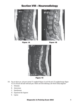
- 1. Section VIII – Neuroradiology Figure 1A Figure 1B Figure 1C 321. You are shown pre- and post-contrast T1-weighted (Figures 1A and 1B) and a T2-weighted image (Figure 1C) in a 28-year-old male with back pain. Which one of the following is the MOST likely diagnosis? A. Metastasis B. Astrocytoma C. Ependymoma D. Sequestered disc fragment E. Lipoma Diagnostic In-Training Exam 2002 1
- 2. Section VIII – Neuroradiology Question #321 Findings: There is a well-defined intradural mass at the filum terminale. The mass is isointense to the spinal cord on T1 and T2 sequences and shows contrast enhancement. Rationales: A) Incorrect. Most metastases to the spine are extradural. They involve the vertebral bodies or epidural space. Intradural metastases are uncommon and usually result from primary central nervous system neoplasm. Intradural metastases most commonly demonstrate thickening and enhancement of the nerve roots of the filum terminale. Lumbar puncture shows malignant cells in 90% of cases. B) Incorrect. Astrocytoma is the second most common primary spinal cord neoplasm. However, it occurs within the cord, more commonly in the cervical spine. They are generally infiltrative tumors that show contrast enhancement. C) Correct. Myxopapillary ependymoma is subtype of ependymal tumor which occurs only in the filum terminale. Ependymoma is the most common primary spinal cord neoplasm. It presents with increased T2 signal, enlargement of the cord, and contrast enhancement. Astrocytoma is the primary differential diagnosis for cord lesions. Schwannoma and meningioma could be considered as a differential for filum lesions. D) Incorrect. Sequestered disc fragments are a common extradural mass. They may show peripheral contrast enhancement due to surrounding granulation tissue. They are usually associated with degenerative disc disease at a nearby level. Most disc herniations remain in contact with the disc space rather than becoming sequestered. Disc fragments are rarely found in an intradural location at surgery. This case is unlikely to represent a disc fragment as the lesion is intradural and shows homogenous contrast enhancement. E) Incorrect. Lipoma is a common incidental finding. It is characterized by high T1 signal on pre-contrast images due to the presence of fat. This diagnosis may be confirmed with fat-suppressed MR sequences. 2 American College of Radiology
- 3. Section VIII – Neuroradiology Figure 2A Figure 2B 322. You are shown right common (Figure 2A) and external (Figure 2B) carotid artery angiograms from a 40-year- old female with headaches. Which one of the following is the MOST likely diagnosis? A. Arteriovenous malformation B. Meningioma C. Cerebral aneurysm D. Hemangiopericytoma E. Glioblastoma multiforme Diagnostic In-Training Exam 2002 3
- 4. Section VIII – Neuroradiology Question #322 Findings: Cerebral angiography shows a dural based mass. This mass shows dense contrast enhancement on selective external carotid artery injection. The mass is poorly seen on common carotid artery injection due to preferential flow of contrast into the internal carotid. This demonstrates the value of selective injections. Rationales: A) Incorrect. The most important feature of pial (parenchymal) AVMs is the presence of an early draining vein. A nidus of abnormal vessels as well as enlarged feeding arteries may be seen. Branches of the internal carotid arteries or vertebrobasilar system supply pial AVMs. Dural AVMs are less common and also are characterized by early venous drainage. B) Correct. Meningiomas most commonly are supplied by branches of the external carotid artery. They show dense, persistent contrast enhancement and have a dural attachment. Meningiomas are more common in women and are usually benign with slow growth. C) Incorrect. An aneurysm is a focal enlargement of an artery. Most cerebral aneurysms are saccular projections found at arterial branch points. The most common locations are the terminal internal carotid arteries, the middle cerebral artery bifurcations, and the anterior communicating artery. Most cerebral aneurysms are found around the Circle of Willis. D) Incorrect. Hemangiopericytoma is a rare neoplasm that may mimic meningioma. It may be large and dural based but frequently shows osseous destruction rather than the hyperostosis seen with some meningiomas. Hemangiopericytomas are quite vascular and bizarre tumoral vessels may be seen on angiography. These are seen as prominent flow voids on MR. E) Incorrect. Glioblastoma multiforme is the most common primary intra-axial neoplasm of adults. These tumors may show staining on angiography but are usually supplied by internal carotid branches. 4 American College of Radiology
- 5. Section VIII – Neuroradiology Figure 3 323. You are shown an axial CT image (Figure 3) of the orbits from a 15-month-old boy. Which one of the following is the MOST likely diagnosis? A. Langerhan’s cell histiocytosis B. Osteosarcoma C. Osteomyelitis D. Fibrous dysplasia E. Neuroblastoma Diagnostic In-Training Exam 2002 5
- 6. Section VIII – Neuroradiology Question #323 Findings: Computed tomography shows spiculated bone formation involving both orbits. Rationales: A) Incorrect. Langerhans cell histiocytosis (Eosinophilic granuloma). Histiocytosis may present with skull lesions in children. However, the lesions are lytic without bone production. B) Incorrect. Spiculated bone formation may be seen with osteosarcoma. However, this symmetric involvement of the orbits is not seen. Also, the peak incidence is older children and young adults. Osteosarcoma most frequently occurs at the ends of rapidly growing bones, such as at the knee and shoulder joints. C) Incorrect. Osteomyelitis may occur in this age group, but usually is a lytic lesion. Although bone formation may occur in chronic osteomyelitis, the symmetric involvement of the orbits makes this unlikely. D) Incorrect. Fibrous dysplasia may cause relatively symmetric facial lesions in children. However, the bone involvement typically has a “ground glass” appearance rather than the malignant spiculation seen here. E) Correct. Neuroblastoma is frequently metastatic at the time of presentation, with bone being a common site of metastasis. The peak incidence of neuroblastoma is in the first two years of life. Up to 20% of patients with neuroblastoma have orbital involvement. Some patients (including this case) have orbital masses due to osseous neuroblastoma metastasis as their presenting symptom. 6 American College of Radiology
- 7. Section VIII – Neuroradiology Figure 4 324. You are shown a coronal CT image (Figure 4) of a 60-year-old male with nasal congestion. Which one of the following is the MOST likely diagnosis? A. Squamous cell carcinoma B. Sinonasal polyposis C. Chronic bacterial sinusitis D. Rhabdomyosarcoma E. Melanoma Diagnostic In-Training Exam 2002 7
- 8. Section VIII – Neuroradiology Question #324 Findings: Coronal sinus computed tomography shows a soft tissue mass filling the left maxillary sinus. There is osseous destruction of the nasal turbinates and maxilla. The soft tissue mass has invaded the left orbit. Rationales: A) Correct. Squamous cell carcinoma is the most common primary malignant neoplasm in the nasopharynx. Features include osseous destruction and spread along soft tissue planes and through skull base foramina. B) Incorrect. Findings of sinonasal polyposis include enlarged ostia and nasal cavity masses. There may be osseous remodeling, but extensive destruction without nasal cavity disease is unusual. Polyps are associated with asthma and cystic fibrosis. C) Incorrect. Chronic bacterial sinusitis may have increased density due to inspissated secretions. Chronic sinusitis may cause increased bone density due to periostitis. If a sinus is chronically obstructed, it may become an enlarged mucocele. D) Incorrect. T. Rhabdomyosarcoma is the most common primary nasopharyngeal malignancy in children. Like squamous cell carcinoma or lymphoma, it presents as a large destructive soft tissue mass. Rhabdomyosarcoma is uncommon in adults. E) Incorrect. Melanoma may occur as a primary nasopharyngeal mass. It can be difficult to differentiate from squamous cell carcinoma on computed tomography, but it is quite rare as a primary mass. Melanoma may present with multiple masses or hemorrhage. 8 American College of Radiology
- 9. Section VIII – Neuroradiology Figure 5A Figure 5B 325. You are shown axial and coronal post-contrast T1-weighted images (Figures 5A and 5B) from a 16-year-old girl with headache and dizziness. Which one of the following is the MOST likely diagnosis? A. Tuberous sclerosis B. Von Hippel-Lindau disease C. Neurofibromatosis type I D. Neurofibromatosis type II E. Sturge-Weber syndrome Diagnostic In-Training Exam 2002 9
- 10. Section VIII – Neuroradiology Question #325 Findings: Post-contrast T1 weighted images show bilateral enhancing masses in the internal auditory canals. There is also a dural-based enhancing mass on the falx. Rationales: A) Incorrect. Intracranial findings in tuberous sclerosis include subependymal nodules and cortical tubers. Both of these lesions may calcify. Up to ten percent of patients will develop giant cell astrocytomas at the foramen of Monro. Extracranial lesions in tuberous sclerosis include skin lesions, angiomyolipomas of the kidneys, and osseous lesions. B) Incorrect. Intracranial findings in Von Hippel-Lindau syndrome include hemangioblastomas of the cerebellum and spinal cord and retinal angiomas. Extracranial lesions include renal cell carcinoma, pheochromocytoma, and renal and pancreatic cysts. C) Incorrect. Cranial findings in neurofibromatosis type I include astrocytomas of the optic nerves and chiasm; foci of abnormal signal in the basal ganglia, pons, and cerebellum; and sphenoid dysplasia. Neurofibromas usually affect peripheral nerves. Tumors of cranial nerves other than the optic nerve are rare in NF-I. Clinical features of NF-I include cafÈ au lait spots, Lisch nodules, scoliosis, and learning disabilities. D) Correct. The findings in this case are pathognomonic of Neurofibromatosis type II. Lesions include multiple schwannomas, meningiomas, and ependymomas. Multiple nerve sheath tumors may be seen in the spine. Bilateral eighth cranial nerve tumors are diagnostic of Neurofibromatosis type II. It is an autosomal dominant disorder and approximately ten times less common than neurofibromatosis type I. E) Incorrect. Sturge-Weber syndrome includes a vascular malformation with abnormal venous drainage of a cerebral hemisphere. Patients have cortical calcifications in a gyriform pattern best seen on computed tomography. The abnormal veins are seen on post-contrast MRI. Patients have an associated facial port wine stain and seizures. 10 American College of Radiology