AI Types, Causes, Diagnosis & Syndromic Links
•
28 gostaram•10,648 visualizações

Amelogenesis Imperfecta is a condition affecting the development of dental enamel. It has several classifications depending on the specific enamel defect present, including hypoplastic (inadequate enamel), hypocalcified (no mineralization), and hypomaturation (failure of enamel to mature). The condition can be inherited in autosomal dominant, recessive, x-linked, or sporadic patterns. Diagnosis is based on family history, clinical examination showing discolored, sensitive teeth prone to disintegration, and radiographic findings of abnormal enamel appearance. It may occur alone or be associated with other systemic abnormalities in syndromes.

Recomendados
Mais conteúdo relacionado
Mais procurados
Mais procurados (20)
Semelhante a AI Types, Causes, Diagnosis & Syndromic Links
Semelhante a AI Types, Causes, Diagnosis & Syndromic Links (20)
Mais de Dr Randy Chance
Último
Último (20)
AI Types, Causes, Diagnosis & Syndromic Links
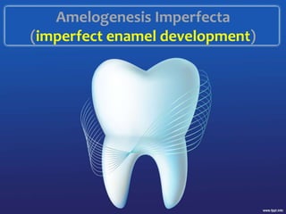
- 1. Amelogenesis Imperfecta (imperfect enamel development)
- 2. Outline • Introduction • Classification • Etiology • Clinical diagnostic features & pictures • Radiological diagnostics • Syndromic associations
- 3. Introduction • Genomic condition affecting dental enamel • May be associated with morphologic or biochemical changes elsewhere in the body and may or may not be associated with systemic disorder • Prevalence varies from 1:700 to 1:14,000 • Teeth affected may be discolored, sensitive or prone to disintegration • AI exists in isolation or associated with other abnormalities in syndromes
- 4. • It may show autosomal dominant, autosomal recessive, sex-linked and sporadic inheritance patterns • Diagnosis is based on the family history, pedigree plotting and meticulous clinical observation • Hereditary enamel dysplasia • Hereditary brown enamel • Hereditary brown opalescent teeth
- 6. Etiology • Normal enamel is 96% large, well organised hydroxyapatite crystals • 4% organic(90% amelogenins) • 2.5mm thick • Formed 4um/day • The final crystal structure is controlled in ameloblasts via molecular interaction amongst matrix proteins. • Mutations in genes regulating any of these proteins would lead to AI
- 7. The matrix proteins • Amelogenin Xp • Ameloblastin 4q • Tuftelin 1q • Amelotin 4q • Enamelin 4q • DSPP 4q • Kallikrein 4 19q • MMP 20 11q • Osterix
- 8. Formation • Elaboration of matrix proteins= which are latter degraded by MMP20(enamelysin) and KLK4 which in turn are later resorbed by mature ameloblasts • Mineralization • Maturation, ie elongation and organization of crystal rods
- 9. Classification (WITKOP)- • 5 genes are responsible for the various types oF AI • AMELX ENAM MMP20 (enamelysin) KLK4 DLX3
- 10. Classification
- 11. • Hypoplastic generalized, localized, AD smooth, XD smooth, enamel agenesis patterns • Hypocalcified AR and AD patterns • Hypomatured pigmented, X-linked, snow capped patterns(“Denture in White Paint” sign) • Hypomatured/hypoplastic
- 12. Hypoplastic AI • Inadequate matrix deposition • Teeth appear like crown preparations • Any available matrix is appropriately mineralized and matured • Patterns: Generalised, Localised, AD smooth, XD smooth, Rough, Enamel agenesis Generalized; Buccal pinpoint pits across teeth in rows/columns +/- stained. Normal intervening enamel Localised; Horizontal row of pits, linear depressions or one large area of hypoplasia surrounded by hypocalcification. Loves middle 1/3 of buccal surface. Only one dentition may be affected(esp deciduous). And some or all teeth. AR variant is more severe( all teeth in all dentitions) AD smooth pattern Smooth, thin hard glossy surface. AOB + spontaneous resorption of unerupted teeth
- 13. hypoplastic
- 14. XD smooth Lyonization(takes place @ approx. 16th wk embryonic life). Males= diffuse thin smooth glossy enamel and almost all exhibit AOB Female = alternating vertical furrows of hypoplastic enamel with normal enamel. Banding is evident on radiographs unlike the x-linked hypomaturation. X- linked Rough Thin hard rough enamel. Denser enamel and so less attrition than the smooth type. AOB + resorption of unerupted teeth common Enamel agenesis (complete lack of enamel formation) Teeth are shape and color (yellow-brown) of dentin. Dentin is rough. There’s lack of eruption usually + resorption. AOB is frequent.
- 15. Hypocalcified AI • No mineralization at all and hence no maturation. • Hyporadiodense than dentin • But normal matrix amount • So normal shaped teeth on eruption but its soft and easily lost so may look like hypoplastic variant • The cervical portion is better calcified bcos it isn’t lost from occlusal wear and stays longer to be mineralized by salivary ions F- Ca2+ PO4 - • Intact interproximal contacts. • Typical yellow-brown-black discoloration. • AOB + Unerupted teeth (It is suggested that frequency of AOB and AI is caused by genetically determined anomaly of craniofacial development rather than due to local factors influencing alveolar growth.) • 2 types: AD and AR • AR more severe than AD but AD more common
- 17. Hypomaturation type • Mineralization begins but maturation into dense crystal structure fails leading to mottling, opaque white to brownish yellow discoloration. • Soft chipping enamel, similar dentin radiodensity • Mostly due to mutations in MMP20 and KLK4 genes( most cases AR bcos most enzyme def are AR) • Severe forms mimic hypocalcified type • Patterns= Pigmented, x-linked, snow-capped patterns. • Pigmented- enamel is soft too and chips. • Mottled agar-brown color (mottling means marked with spots or smears of diff color) • Most cases AR
- 18. X-linked • Lyonization but not as obvious as x linked hypoplastic type so Males=severe mottling Females= vertical bands alternation with normal translucent enamel. Banding is demonstrable by transillumination and not under normal light Snow-capped • Opaque-white enamel on incisal/occlusal 1/3. “denture in white paint sign” • Most cases are X-linked recessive but AD form reported •
- 19. Hypomaturation
- 20. Hypomaturation/hypoplastic AI • Hypomat/hypoplast +/- taurodontism • Hypoplast/hypomat +/- taurodontism • Especially hypertaurodontism • Association with TDO • Mutation in DLX3 is responsible for this AI • AR
- 21. Clinical diagnosis • Yellowish-orange-brown teeth • Sensitivity and poor oral hygiene • Disintegration– pre-eruptive & post eruptive • Soft cheesy enamel • Pseudo-microdontia with spacing • Enamel ridges, grooves & pits • Anterior open bite • Familial pattern • Syndromic • History of consanguinity
- 22. Radiology • Picket fence appearance • Spacing • Thin enamel • Hypo radiodensity of enamel • Skeletal problems
- 23. Hypoplastic type
- 24. Hypomaturation
- 25. hypocalcified
- 26. Radiology
- 28. Looks like • Dental fluorosis • MIH • Dentinogenesis imperfecta • Gunther’s disease • Tetracycline hypoplasia/discoloration
- 29. Syndromic associations • Trichodentoosseous syndrome • Cone rod dystrophy • Kohlshutter-Tonz syndrome • Platyspondyly with AI • AI with nephrocalcinosis • Ameloonychohypohidrotic syndrome • Brachyolmia and AI • Morquio-Brailsford syndrome •