Cell as basic unit of life ppt 88 slides
•Transferir como PPTX, PDF•
107 gostaram•48,760 visualizações

This Power point presentation describes Cell as basic unit of life. The slides provide information about Discovery of cell,cell theory,number,size,shape and cell types .Differentiates prokaryotic and eukaryotic cell types and point out major differences in plant and animal cell and also about structure and function of cell organelles

Recomendados
Mais conteúdo relacionado
Mais procurados
Mais procurados (20)
Semelhante a Cell as basic unit of life ppt 88 slides
Semelhante a Cell as basic unit of life ppt 88 slides (20)
Mais de ICHHA PURAK
Mais de ICHHA PURAK (20)
Último
Último (20)
Cell as basic unit of life ppt 88 slides
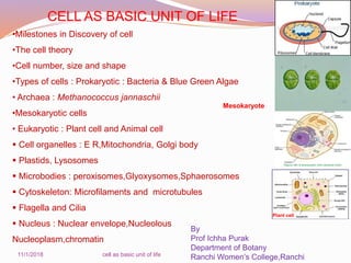
- 1. CELL AS BASIC UNIT OF LIFE •Milestones in Discovery of cell •The cell theory •Cell number, size and shape •Types of cells : Prokaryotic : Bacteria & Blue Green Algae • Archaea : Methanococcus jannaschii •Mesokaryotic cells • Eukaryotic : Plant cell and Animal cell Cell organelles : E R,Mitochondria, Golgi body Plastids, Lysosomes Microbodies : peroxisomes,Glyoxysomes,Sphaerosomes Cytoskeleton: Microfilaments and microtubules Flagella and Cilia Nucleus : Nuclear envelope,Nucleolous Nucleoplasm,chromatin By Prof Ichha Purak Department of Botany Ranchi Women’s College,Ranchi11/1/2018 cell as basic unit of life Mesokaryote Plant cell
- 2. The cell is the smallest unit of living matter. The cell is the smallest unit having all the properties of life. All organisms are composed of structural and functional units of life called cells. The body of some organisms like bacteria, protozoans and some algae is made up of a single cell ( Unicellular organisms) while the body of fungi, plants and animals are composed of many cells (Multicellular organisms) Cells vary in size and structure as they are specialized to perform different functions. But the basic components of the cell are common to all cells.
- 3. MILESTONES IN DISCOVERY OF CELL Robert Hooke (1665), an English scientist, discovered and coined the term cell while examining a thin slice of cork under a self-designed compound microscope. The term cell was derived from a Latin word cellular (meaning little room or chamber). In 1672, Antony Van Leeuwenhoek observed bacteria, sperms and red blood corpuscles, all of which were cells. In 1831, Robert Brown, an Englishman observed that all cells had a centrally positioned body which he termed the nucleus. The crude microscope employed by Robert Hook(1665) Thin slice of cork showing chambers/cells
- 4. WHAT IS A CELL ? ( DEFINITION OF CELL ) Cell is the structural and functional unit of living organisms, enclosing a bit of protoplast consisting of numerous micro and macro organic and inorganic molecules, dissolved or suspended in colloidal or watery matrix. The molecules are busy in reactions with release or uptake of energy. The protoplasm of the cell is limited by a membrane. All types of cells include three major components 1.Cell membrane 2.Cytoplasm and 3. DNA naked in prokaryotes (Bacteria,Archaea and BGA) and covered by a membrane in all other organisms. Mature Plant cells have in their cytoplasm large vacuole containing non-living inclusions like crystals, pigments etc. The bacteria have neither organelles nor a well formed nucleus.
- 5. THE CELL THEORY The cell theory was jointly put forward by Schleiden and Schwann (1839) in their paper “Microscopic investigations on the similarity of structure and growth in animals and plants” : As follows 1) All organisms are composed of one or more cells . 2) Cells are the basic living units within organisms and the chemical reactions of life take place within cells. 3) All cells arise from pre existing cells by division and are not formed de novo Theodor Schwan M J Schleiden In 1855, a German medical doctor named Rudolph Virchow observed, under the microscope, cells dividing. He reasoned that all cells come from other pre-existing cells by cell division
- 6. Modern cell theory includes following features 1. The cell is the fundamental unit of structure and function in living organisms. 2. New cells arise from pre-existing cells through division. All new cells contain the same amount and degree of genetic information as contained in the parent cell 3. Energy flow occurs within cells. 4. Cells contain hereditary information (DNA) which is passed from cell to cell during cell division. 5. All cells are basically the same in chemical composition in organisms of similar species. Haeckel (1866) established that nucleus stores and transmits hereditary traits. Cell theory was modified accordingly and was renamed as cell doctrine or cell principle Ernst Haeckel (1834-1919)
- 7. 6. All known living things are made up of one or more cells. 7. Some organisms are made up of only one cell and are known as unicellular organisms. All life begins as a single cell. 8. Others are multicellular, composed of a number of cells. 9. The activity of an organism depends on the total activity of independent cells. 10.Depending upon specific requirement, the cells get modified, e.g. elongated in muscle and nerve cells, loss of nucleus in RBCs or cytoplasm in outer skin cells. Modern cell theory includes following features continued
- 8. NUMBER OF CELLS Some organisms are unicellular i.e. bacteria, yeast, Chlamydomonas, diatoms, Cosmarium etc whereas most of organisms as plants and animals are multicellular. Human body is built of about one trillion cells. In case of unicellular forms single cell is capable of independent existence and to perform all the essential functions of life In green alga Pandorina, the coenobium is having a fixed number of cells (8/16/32 ) . Colonies like Volvox ,Nostoc etc have many cells enclosed by mucilaginous sheath A multicellular organism is made of many cells Different cells become specialized for performing different functions. Cells are grouped into tissues, tissues into organs and organs into organ systems. Organ systems together form organism NUMBER, SIZE AND SHAPE OF CELL Cell Tissue Organ Organ system Organism
- 9. Figure : 1 Living Organisms may be unicellular /multicelluar Cosmarium Spirogyra starfish Pedistrum Volvox E coli Nostoc Chara Chlorella Paramaecium Pinnularia Plant
- 10. SIZE OF CELL Generally cell size ranges between 0.2µm-20 µm Smallest cell : PPLO : Mycoplasma gallisepticum :0.1 µm Bacteria : Unicellular : About 10 times smaller than Animal cell Escheritia coli : Rod shaped Bacteria : 1-2 micron long Protozoa : Amoeba proteus : 220–760 µm Generally plant cells are larger than animal cell Generally Animal cell range from 10µm to 30 µm & plant cell 10µm to 100µm Exceptions : Ostrich egg cell : 75 mm long Acetabularia a unicellular green alga is about 10 cms in length Bast fibre (phloem fibre ) of some plants (Ramie) are about 50-55 cm long A giraffe's nerve cell can reach about two meters Cell volume The volume of a cell is fairly constant for a particular cell type and is independent of the size of organism. The difference in the total mass of the organ or organism depends on the number ,not on the volume of the cells
- 11. Which Cell Type is Larger? 11 Plant cell Animal cell Bacteria> > > Generally plant cells are larger than animal cell Animal cells are larger than bacterial cell
- 12. 12 CELL SIZE Typical cells range from 5 – 50 micrometers (microns) in diameter copyright cmassengale
- 13. WHY ARE CELLS SO SMALL ? In order to survive, cells must constantly interact with their surrounding environment and can grow only to a certain size . If the cell grows beyond a certain limit , enough material will not be able to cross the membrane to accommodate with increased cellular volume. The reason cells can grow only to a certain size has to do with their surface area to volume ratio. Here, surface area is the area of the outside of the cell, called the plasma membrane. The volume is how much space is inside the cell. The ratio is the surface area divided by the volume. If the surface area to volume ratio is small, the cell is very big. When this happens, the cell must divide into smaller cells with favourable surface area/volume ratios. If the ratio is big, the surface area is greater than the volume then the cell is small.
- 14. There is great variability in cell shape i.e. spherical, polygonal, disc like, cuboidal, columnar,spindle like or fibre like. Generally cells are spherical but in multicellular forms due to pressure become polyhedral . In plants as cells have cell wall appear hexagonal or polygonal . Cells some times change shape because of function as Amoeba and leucocytes . While animal cells tend to have irregular shapes, plant cells are typically rectangular or cube shaped. CELL SHAPE
- 15. Chlamydomonas Paramaecium Amoeba Nerve cells Muscle cells Acetabularia Caulerpa Epithelial cells Onion peel cells Pinnularia E. coli Red blood cells Figure- 1a Cells of different shapes
- 16. TYPE OF CELLS Dougherty (1957) classified cells into prokaryotic (Pro meaning primitive, karyon meaning nucleus) and eukaryotic (Eu meaning true, Karyon meaning nucleus) types on the basis of structural organization of their nucleus. The cells which possess a primitive type of nucleus devoid of nuclear membrane are the prokaryotic cells. On the other hand, eukaryotic cells are those which possess a true, well organized nucleus having typical chromosomes and nuclear membrane. Dodge et al (1966) proposed a third type (Mesokaryotic) which can be placed in between prokaryotic and eukaryotic cells.
- 17. PROKARYOTIC CELLS There is primitive type of nucleus which is not bounded by nuclear membrane as true nucleus and is termed as nucloid There are no membrane bound cell orgalleles, infolding of plasma membrane called mesosomes take their function The nuclear material lies diffused in central part of the cell, the genetic material consists of DNA, but it is not associated with proteins (Histones). Prokaryotic cells are generally smaller than eukaryotic cells. The cells of bacteria, Archaea, blue green algae, Mycoplasma, rickettsiae are included in this type
- 18. EUKARYOTIC CELLS Eukaryotic: In this type of cell organization, there is definite or true nucleus surrounded by definite nuclear membrane. The genetic material consists of DNA complexed with histone proteins to form well developed chromatin/ chromosomes. Nucleus has nucleolous. There are different membrane bound cell organelles such as mitochondria, Endoplasmic Reticulum, Golgi body ,plastids etc for different functions. Plastids are presents in the plants cell only. This types of cells are in general larger and more organized than prokaryotic cells Most of the plants and animal cells fall under this category except Blue Green Algae(BGA).
- 19. MESOKARYOTIC CELLS In some organisms ( the Dinoflagellates ) the genetic material is surrounded by a nuclear membrane ( Eukaryotic character) but histone protein is not associated with DNA ( prokaryotic character) The nucleus is larger in size and has been named as mesokaryon by Dodge (1966). These types of cells which are intermediate between prokaryotic and eukaryotic are called mesokaryotic cells. In these types of cells, the mitotic spindle is not formed. The cells of certain algae like Gymnodinium and Peridinium are the example of such type.
- 20. Some cells are even smaller than prokaryotic cell Smallest known cell is of Mycoplasma gallisepticum (PPLO-Pleuro Pneumonia like organism- a bacterium prokaryote ) Size – 0.1 µm -0.3 to 0.5 µm . The size of this cell may not be more than 1000 to 5000 times that of a hydrogen atom. W. V. Iterson (1969) placed PPLOs with bacteria in the group Mycoplasmata. Novikoff and Holtzman (1970) have excluded them from bacteria and considered them as simplest prokaryotic cells. OTHER CELL TYPES
- 21. 1. Absence of cell wall and mesosomes 2. Selectively permeable plasma membrane (75A⁰ in thickness ), composed of lipoproteins 3.The cytoplasm has ribosomes and enzymes required for protein synthesis and ATP metabolism 4. Nucleus is absent and instead there occurs double stranded DNA molecule of circular or fibrillar type. 5.It does not need a host cell (can live freely) STRUCTURE OF PPLO
- 22. Character Prokaryotic cell Eukaryotic cell Cell compartments No compartments Distinct compartments in the cell. i.e. the cytoplasm and the nucleus Cell wall Present, composed of amino sugars and muramic acid Absent in animal cell, present in plant cell, composed of cellulose Plasma membrane Present, core of lipid bilayer with proteins embedded as mosaic Present,lipid bilayer, with embedded proteins Cell organelles Absent, Enzymatic functions are carried by infoldings of plasma membrane- mesosomes Different cell organelles present for different functions ,bound by extensions of plasma membrane Respiratory system Mesosome(part of plasma membrane) Mitochondria Capsule If present , composed of mucopolysaccharides Absent Photosynthetic apparatus and chlorophyll Photosynthetic pigments chlorophylls and carotenes on grana lamellae not enclosed by membrane, no plastid Pigments present on lamellar system, enclosed by membrane in cytoplasm. plastids present in plant cells Organization of the nucleus Nucleus not distinct , It is in the form of nuclear zone (Nucloid) Distinct nucleus Table-1 Difference between Prokaryotic and Eukaryotic cell
- 23. Character Prokaryotic cell Eukaryotic cell Nuclear membrane Absent Well formed nuclear membrane Nucleoplasm Undifferentiated from cytoplasm Denser and differentiated from cytoplasm by nuclear membrane Hereditory material Single naked strand of DNA / RNA Circular in shape.DNA not linked with Histone proteins More than one strand of DNA & Histone protein inside nuclear membrane as chromosomes Nucleolous Absent Present Flagellum Single,simple composed of protein flagellin No 9+2 fibrillar pattern complex 9+2 fibrillar pattern composed of tubulin and other proteins Cell division Amitosis (Binary fisson) spindle not formed Mitosis or meiosis Spindle formed Ribosomes 70S (30S+50S ) 80 S ( 40S+60S) Cytoplasmic movements Rare or absent Often occurs Vacuoles Absent Present Lysosome Absent Present Growth Can grow both under aerobic and anaerobic conditions Can grow only under aerobic condition Size 1-10 µm 10-100 µm Table-1 Difference between Prokaryotic and Eukaryotic cell
- 24. STRUCTURE OF A PROKARYOTIC CELL : BACTERIAL CELL Average size- 1.25 µm. The smallest bacterium is Dialister pneumosintes which measures about 0.15 µ to 0.3µ in length and the largest bacterium is Spirillum volutons which measures about 13 µ to 15µ in length. A typical bacterial cell consists of three parts (a) outer coverings, (b) cytoplasm and its (c) constituents. Body of the bacteria is basically prokaryotic type. Outer covering in the bacteria is made up of three layers namely: (i) Capsule (ii) Cell wall & (iii) Plasma membrane 11/1/2018 Cell as basic unit of life 24
- 25. Figure - 2 :Structure of Prokaryotic cell (Bacteria)
- 26. Capsule - Most bacterial cells have a slimy capsule outside the cell wall, composed of polysaccharides. which lies exterior to the cell wall . It acts as a protective covering and is of uniform thickness. Cell wall is about 10 µm thick, consisting of carbohydrates,lipids,proteins, certain inorganic salts along with specific amino acid diaminopimalic acid and a glucose derivative muramic acid (has oligopeptide and polysaccharide) On staining properties of cell wall bacteria are of two types Gram positive (+) and Gram Negative (- ) Appendix Gram staining
- 27. Contents of Gram Stain Dye : Crystal violet stain Mordant : Gram iodine Decolourising agent : Ethyl Alcohol Counter stain : Safarnin PROTOCOL Slide with bacterial smear dipped in Crystal violet stain for 1 minute Washed by water Slide dipped in Gram iodine (Mordant) solution Washed by water Slide dipped in Absolute Ethyl Alcohol Gram Positive Bacterial Slide retains the stain: not washed out: Bacteria take Violet colour Gram Negative Bacterial Slide donot retain the stain : washed out Slide dipped in Counter stain Safarnin : Gram Negative Bacteria take pink colour
- 28. The differences in cell wall composition of Gram positive and Gram negative bacteria accounts for the Gram staining differences. Gram positive cell wall contain thick layer of peptidoglycan with numerous teichoic acid cross linking (Figure- 2a) which resists the decolourization with ethanol Gram-Positive Bacteria have thicker cell walls with less fatty substances (lipids) than Gram-Negative bacteria. (Figure- 2b). When washed with solvent, the cell pores close becoming less permeable and are able to retain the stain, in this case, purple. Gram-Negative Bacteria have thinner cell walls with more lipids. The solvent dissolves the lipids, which combined with thinner cell walls, washes out or decolorizes the stain. In order to see the decolorized bacteria, a counter stain (Safranin ) is added which gives pink colour to Gram-Negative cells.
- 29. 1a 1b Figure-2a and 2b Cell wall components of Gram positive and Gram negative Bacteria
- 30. Hans Christian Gram(1884) developed the technique to stain bacterial cells . The Gram stain allows one to distinguish between Gram-positive and Gram- negative bacteria on the basis of differential staining with a crystal violet-iodine complex and a safranin counter stain. The cell walls of Gram-positive organisms retain this complex after treatment with alcohol and appear purple, whereas gram-negative organisms decolorize following such treatment and appear pink on treatment with counterstain safranin.
- 31. Plasma membrane is present inner to cell wall, composed of lipoproteins. usually gets modified as mesosomes/ desmosomes(chondriods)which is associated with various functions as Electron Transport and DNA replication. Figure - 3 Diagrammatic representation of fluid mosaic model of plasma membrane
- 32. Gram +ve bacteria Gram –ve bacteria I Gram +ve bacteria remain coloured ( purple ) even after washing with absolute alcohol Gram –ve bacteria do not retain colour when washed with absolute alcohol Ii The wall is single layered .Outer membrane is absent The wall is two layered.Outer membrane is present Iii The thickness of the wall is 20-80nm The thickness of wall is 8-12nm Iv The lipid content of the wall is quite low The lipid content of the wall is 20-30% V Murein or mucopeptide content is 60-90% Murein or mucopeptide content is 10% Vi Techoic acid is present in cell wall Techoic acid is not present in cell wall Vii Porins are absent Porins or hydrophilic channels occur in outer membrane Viii Example: Staphylococcus,Streptococcus, Pneumococcus, Bacillus,Mycobacterium,Strptomyces Example :Salmonella,E coli, Pseudomonas,E coli, Rhizobium ,Azotobacter Table No-2 Difference between Gram +ve and Gram –ve bacteria
- 33. Flagella Generally rod shaped bacteria possess one or more flagella made up of single fibril (100-200A⁰ thick) Flagellin protein Pilli (Fimbriae) are stout hair like structures, extra cellular outgrowths, meant for attachment to host for infection or during conjugation ,made up of helically arranged subunits of pilin protein Cytoplasm Dense, colloidal, contain granules of glycogen, protein, fats, poly β hydroxyl butyric acid, volutin granules and some polymers of glucose . Some photosynthetic bacteria possess chromatophores with bacteriochlorophyll and bacterioviridin pigments.
- 34. In cytoplasm ribosomes (70S) are freely present. During protein synthesis many ribosomes may attach to same messenger RNA one after one forming polyribosome. In bacteria polyribosomes are present free in the cytoplasm whereas in eukaryotes , polyribosomes are attached to the surface of rough endoplasmic reticulum. Prokaryotic Ribosomes contain slightly more RNA than Eukaryotic ribosomes.
- 35. Nuclear material In the interior of cytoplasm, there is light nuclear region or nucleoid, consisting of single circular DNA molecule ( E coli- 1 nm) (Bacterial chromosome or genophore) . DNA molecule is folded or packed in the nuclear region and is not separated from rest of cytoplasm by nuclear membrane. In addition to nucleoid, bacterial cell may have extra chromosomal circular DNA called plasmid, which is self replicating unit. Plasmids are of 3 types F (Fertility),R (Resistance) and Col (colicin) produce toxic material. Plasmids can be isolated and can be used as tools in Recombinant DNA Technology to insert foreign genes resulting in chimeric plasmids
- 36. Carl Woese (1977) mentioned that prokaryotes can be divided into EuBacteria and Archaebacteria. However subsequently it was shown that this group Archaea is distinct from bacteria and Eukaryotes. Today, these groups are considered to form two out of three domains of life. The third domain (Eukarya) includes all eukaryotes, such as plants, animals, and fungi. The living cells are therefore now divided into following three domains : Bacteria, Archaea and Eukarya This classification came into existence after complete genome sequencing of Archaeon Methanococcus jannaschii by Bult et al 1996. Nucleotide sequence determined were compared with that of bacteria and Eukaryotes . It was found that some of its genes resemble prokaryotes and other to Eukaryote and some unique genes
- 37. Table No 3 Distribution of prokaryotic,Eukaryotic and Novel unique genes in Archaea Eukaryotic genes Prokaryotic genes Novel unique genes 1. DNA replication 2. Transcription 3. Translation 4. Histone genes 1. transport of inorganic ions (Na+,K+) across wall 2. Cell surface structure 1. only one DNA polymerase 2. Several tRNA Aminoacyl synthetase enzymes missing 3. Numerous inteins are known that are inserted into proteins & then are removed Protein splicing elements (Inteins) are protein sequences that catalyze their own removal from a longer protein, splicing together the two surrounding protein halves (exteins) in the process
- 38. Table No - 4 Comparison of Archaea, Bacteria and Eukarya
- 39. Holger Jannasch (1982) discovered Methanococcus jannaschii from a hot spot of deep sea ( 3 km deep pacific ocean ) as Methane producing organism Following are some of the novel features of this Archaean, to create new Domain Archaea 1. 56% of the genes of Methanococcus are new to science. 2. It lives in temperature ranging from 48⁰C →94⁰C and pressure 200 atm 3.It is killed by oxygen 4.It is autotrouph using CO2, N2 and Hydrogen 5.Produces methane and is a methanogen 6. Some of its genes resemble prokaryotes and other to Eukaryotes It has also been shown that Archaeal system is more likely a Eukaryotic system. In fact scientists now believe that common ancestor of all living creatures was a thermophile, which branched off more than 3.6 billion years ago into 3 major life forms Prokaryotes(bacteria) ,Archaea and Eukarya.
- 40. Figure- 5 Evolution of three major life forms ( Prokarya,Archaea and Eukarya) from a common ancestor (a thermophile/Archaeon} Common ancestor
- 41. ANOTHER PROKARYOTE : CYANOBACTERIA (BLUE GREEN ALGAE) Flagella and cilia are absent, have capacity of movement by gliding. Figure-6 Generalized structure of cell of Blue Green Algae (under electron microscope) Examples are Nostoc,Anabaena,Oscillitoria, GleocapsaTrichodesmium,Spirulina, Microcystis
- 42. Blue green algae are found in diverse habitats as fresh water, terrestrial, marine and hot springs. STRUCTURE OF BLUE GREEN ALGAL CELL (CYANOBACTERIA) BGA may be unicellular, filamentous or colonial. Mucilaginous sheath is always present around the cell/cells. Cyanobacteria have primitive( prokaryotic) cell organization. Cell wall consists of cellulose, pectic substances, lipoproteins, lipopolysaccharides and mucoproteins. Plasmamembrane Lipoproteinous,invaginates as mesosomes
- 43. contains chlorophyll, carotenes, myxoxanthophyll, phycocyanin and The cytoplasm can be distinguished into central colourless region-centroplasm and an outer blue green pigmented region chromoplasm phycoerythrin along with oil drops, glycogen and few spherical bodies called cyanophycean granules evenly distributed in chromoplasm located as small granular chromophores (cyanoplasts) Both blue and red pigments are collectively called as phycobilins, which are covalently bound with proteins forming phycobilisomes (cyanosomes) Each cyanosome measures about 400A⁰ in diameter and contain about 10 subunits. Photosynthetic pigments chlorophyll and cartenoids are present on flattened disc, lamella are arranged concentrically in chromoplasm.
- 44. Eukaryotic cell is bounded by plasma membrane which encloses a bit of protoplast. The major components of the cell are (1) cell membrane, (2) cytoplasm, and (3) nucleus. In eukaryotes, a complete set of genetic instructions is found on the DNA molecules, which exist as multiple linear structures called chromosomes that are confined within the nucleus. Eukaryotic cells also contain other membrane bound organelles within their cytoplasm (the region between the nucleus and the plasma membrane). These subcellular structures vary tremendously in structure and function. STRUCTURE OF EUKARYOTIC CELL
- 45. Plant cells have special features as cell wall, plasmodesmata, large vacuole and plastids Animal cells possess Centrosome and centriole Common features Plasma membrane Cytoplasm Endoplasmic reticulum Golgi body Mitochondria Lysosome Microbodies Nucleus In animal cell, outer most boundary is cell membrane, plant cell have additional outermost covering out side plasma membrane, this is cell wall. Because of cell wall plant cells has definite shape, animal cells have no definite shape. Figure :-7 A typical plant cell and A typical animal cell
- 46. S N Plant cell Animal Cell 1 Cellulosic cell wall present around cell membrane Cell wall is absent 2 Vacuoles are usually large Generally vacuoles are absent and if present, are usually small 3 Plastids are present. Except Euglena ,no animal has Plastids 4 Golgi body present in the form of many units known as dictyosomes. Single highly complex and prominent Golgi apparatus . 5 Centrosome and Centriole absent Centrosome and Centriole present Table No- 5 Difference between plant cell and animal cell
- 47. Plant cells differ in several key features from cells of other eukaryotic organisms A cell wall composed of cellulose and hemicellulose, pectin and in many cases lignin, is secreted by the protoplast on the outside of the cell membrane. This contrasts with the cell walls of fungi (which are made of chitin), and of bacteria, which are made of peptidoglycan. Cell wall is rigid, transparent, permeable ,having pits or pores. Cell wall protects the protoplast against loss of water, excessive heat and foreign attacks and provides mechanical strength to cell Cell wall is differentiated into 3 layers a) Primary wall – Cellulosic, delicate, 1-3µm thick. first deposition product of protoplasm, parenchyma, merismatic cells, unicellular forms have only primary wall.
- 48. b) Secondary wall is more than 4 µm thick, consists of cellulose, hemicelluloses and lignin. It is not deposited uniformly leave unthickened areas(pits) . Through pits fine cytoplasmic strands known as plasmodesmata pass from one cell to another cell and connect endoplasmic reticulum and cytoplasm of two neighbouring cells. This is specialized cell-cell communication through cell junctions or pits. Besides the cell junctions, some protein molecules are there in between the cells of a tissue for binding cells together. These protein molecules are called Cell Adhesion Molecules (CAMs). c) Middle lamella – is the cementing layer present between two cells, consisting Ca and Mg pectinate and cellulose. It is sticky gel like, swells with water and join two cells
- 49. In mature plant cell a large central cavity or vacuole enclosed by delicate membrane (tonoplast) is present .Vacuole is filled with sap and help in excretion of waste and excess water. Cell sap has many organic and inorganic substances Young plant cells are completely filled by cytoplasm ( Cytosol) ,having many small vacuoles and nucleus in the centre. These small vacuoles later on fuse to form a single large vacuole, pushing aside cytoplasm and nucleus towards cell wall (Figure-8 ) Figure : - 8 Young and mature plant cell
- 50. THE CYTOPLASM AND THE CELL ORGANELLES Cytoplasm is the protoplasm outside nucleus . Cytoplasm has two parts 1) Cytosol 2) Cell organalles Cytosol is jelly like, optically clear, homogenous, colloidal or viscous fluid in which granules and fibrils are suspended . It is the living part of the cell, most reactions take place in cytosol. Chemical composition of cytosol Water- 90% proteins -7% carbohydrates – 1.5% Lipids and inorganic ions – 1.5%
- 51. Plasma membrane is present between cell wall and cytoplasm. It is delicate, semi permeable – permeability depends on physiological state of cell and size and nature of molecules . Plasma membrane regulates flow of water and other organic and inorganic molecules through it. Fluid Mosaic Model SJ Singer and G L Nicolson(1972) proposed the fluid mosaic model for describing the structure and thermodynamics of plasma ( cell ) membranes a) Bilayer of phospholipids form core of plasma membrane b) Proteins are embedded randomly on peripheral or integral portion of bilayer c) Bilayer has fluid consistency ( not rigid ) due to number of unsaturated fatty acids in phospholipids
- 52. Figure : 9 Structural detail of Plasma membrane according to Fluid Mosaic Model
- 53. By courtsey
- 54. Cell organelles of cytoplasm are related with different functions Those that trap and release energy e.g. Plastids and mitochondria Those that are secretory or are involved in synthesis and transport e.g Endoplasmic reticulum,ribosomes and Golgi apparatus The organelles related with motility – Flagella and Cilia Suicidal bags – Lysosome Microbodies – Peroxisomes,Glyoxysome and Sphaerosomes Related with cell division Centrosome and centriole Cytoskeleton- Microfilaments and Microtubules Nucleus which controls all activities of the cell, and carries the hereditary material
- 55. Plastids – Largest cell organelle present only in plant cell (Except BGA and Fungi). Plastids are not found in animal cell. Plastids are of three types a) Leucoplast – Colourless plastids, Store starch , present in cells of underground parts such as tuber, roots b) Chromoplast – Coloured plastids (red, orange, yellow or violet) due to presence of carotene, xanthophylls and Anthocyanin, are present in petals of flowers and fruits c) Chloroplast – green plastids , present in mesophyll cells of leaves and green stems contains pigments as chlorophylls and carotenoid for light harvesting and photosynthesis Chloroplast is enveloped by double membranous sac. It encloses proteinaceous matrix ( stroma). Lamellar structure run in matrix, it is denser at certain places due to pigments (grana) stacked in a group of many.
- 56. The photosynthetic pigments are chlorophylls and carotenoids. The stroma of chloroplast has DNA, RNA , ribosomes, fat globules, starch and a number of enzymes.(Figure- 10) Main function of chloroplast is photosynthesis Figure :- 10 Structure of Chloroplast
- 57. Mitochondria (Chondriosome) Power house of the cell Mitochondria are .0.5 – 1.0 µm long, appear spherical or rod shaped under compound microscope . Particles present on inner membrane are oxysomes( Respiratory particles/F1 particles ) . These particles play important role in electron transport. Matrix of mitochondria has enzymes for TCA ( Krebs) cycle, ATP synthesis, protein and fat synthesis and contains DNA, RNA and Ribosomes. (Figure- 11) Mitochondria appears to be surrounded by two membranes. Inner membrane is folded inwards deep in matrix, even upto opposite side forming cristae. Intermembranous space is filled with a liquid. Inner membrane has large surface area than outer membrane .
- 58. Figure : 11 Structure of Mitochondria Drawn By Prof Ichha Purak Mitochondria contain 70% enzymes of the cell and is mainly connected with respiration which gives energy so is termed as power house of cell. Mitochondia possess in total 70 enzymes and 14 coenzymes.
- 59. In 1970, American biologist, Lynn Margulis, provided evidence that some organelles within cells were at one time free living cells themselves Supporting evidence included organelles with their own DNA Both chloroplast and mitochondria are semi autonomous as they have DNA ( genetic material), RNA and Ribosomes ( Protein synthesizing machinery) are considered as endosymbionts ( Intracellular parasites)
- 60. Endoplasmic Reticulum (E R ) Porter et al (1945) reported and described structure of Endoplasmic Reticulum for the first time Plasma membrane is folded ( extended) inwards into the cytoplasm in the form of double membranous system of thousands of tiny tubules ( branched/unbranched ) This membranous system extends from plasma membrane upto nuclear membrane or plasma membrane to cytoplasm or nuclear membrane to cytoplasm . Endoplasmic reticulum keeps on changing Space enclosed by tubules is cisternae, two cisternae are connected by inter cisternal connection. On sides of cisternae spherical bodies are present. These are known as vesicles. Endoplasmic reticulum is of two types Smooth E R - no particles present on its surface Rough E R - On surface of Endoplasmic Reticulum , Ribosomes and Microsomes ( Particles ) are present ( Figure -12 )
- 61. Figure : -12 ENDOPLASMIC RETICULUM & RIBOSOMES
- 62. Functions of Endoplasmic Reticulum Mechanical support and act as endoskeleton(cytoskeleton) of the cell 1) Divides cytoplasm into a number of compartments for different enzymatic biochemical reactions 2) Cisternae (Channels) provide space for transport of metabolities from cytoplasm to nucleus and in between two cells through pits. 3) Help in synthesis of sterols and phospholipids 4) Excretion of metabolites outsides Plasma membrane 5) Holds ribosomes for synthesis of secretory proteins
- 63. Ribosomes RNA and Protein non membranous particles , present free in cytoplasm or attached to outer surface of nuclear membrane or on Endoplasmic Reticulum. Ribosomes are also present in chloroplast and mitochondria. Ribosomes are site for protein synthesis Ribosomes present free in cytoplasm or attached to E R are 80S particles having two subunits 40S and 60S,whereas ribosomes present in chloroplast and mitochondria are 70S having two subunits 30S and 50S.(Figure-12 ) Svedberg unit- Components of cell after centrifugation, sediment or settle down at different speeds depending upon their mass and size. The coefficient of sedimentation is represented in Svedberg unit and depicted as S.
- 64. Golgi bodies (Apparatus) Dictyosome / Lipochondria discovered by Camillo Golgi (1898) in animal cell Stacks of flat sac like structures – cisternae ( 4-10 ), surrounded on sides , above and below by swollen structures the vesicles. Margins of cisternae are curved to cup or bowl like appearance. Structure of golgi apparatus continuously changes, some discs appear to grow others shrink. Some cisternae are formed on Cis( forming ) face and some are destroyed on trans face ( Maturity). Cis face lies towards nucleus and Endoplasmic Reticulun , trans face lies towards Plasma Membrane . On cis face small vesicles fuse and add to Golgi Body while vesicles are destroyed from trans face .(Figure-13 )
- 65. In plant cell, groups of cisternae, are attached to other similar groups with the help of Inter cisternal tubules. Each group is reffered as dictyosome, collectively form a golgi complex. Figure :- 13 Structure of Golgi body Functions of Golgi body Helps in secretion of polysaccharides, enzymes, proteins etc, cell plate formation during cell division, vesicles separate and in cell plate region fuse with each other and storage and secretion of cellular products.
- 66. Lysosomes are called “suicidal bags” as enzymes contained in them can digest the cell’s own material (can perform intracellular digestion). Lysosomes are about 0.2-0.8 µ in diameter, generally present in animal cells and some lower plants as fungi. Lysosome is sac enclosed by unit membrane. It encloses various digestive enzymes, which when released break down various organelles, help in digestion of food particles and extracellular substances ( Bacteria , Virus) Lysosomes enable the cell to process bulk material taken by endocytosis ( along with water current ) Lysosome contains 30 different types of hydrolytic enzymes break down cell organelles and cell itself.(Figure-14 )
- 67. Centrosome and centriole It is generally present in animal cell, but in some algae, fungi, moss and fern, the cells have centriole, near the nucleus. It has a pair of dots ( granules) surrounded by transparent cytoplasm centrospheres, whole structure is known as centrosome . centrioles have astral rays and are functional in cell division in formation of spindle. Figure :-15 Structure of centriole and centrosome
- 68. Cilia and flagella (the organelles for mobility) Cilia and Flagella: - Cilia and Flagella are hair-like micro-tubular organelles projecting from the cell surface into the extracellular medium and are concerned with cell motility. i) Some unicellular organisms like Paramaecium and Euglena swim in water with the help of cilia and flagella respectively. (ii) In multicellular organism some living tissues (epithelial tissues) have cilia. They beat and create a current in the fluid in order to move in a given direction e.g. in the wind pipe (trachea) to push out the mucus and dust particles. iii) Reproductive cells as spores and gametes bear one, two or many cilia or flagella for movement. Human sperm Isogametes Cladophora Chlamydomonas
- 69. (iv) Cilia beat like tiny oars or pedals (as in a boat) and flagella bring about whip like lashing movement. (iv) Both are made up of contractile protein tubulin in the form of microtubules. (v) The arrangement of the microtubules is 9 + 2, that is, two central microtubules surrounded by nine peripheral ones. (vi)The cilia and flagella appear to arise from the basal bodies. (vii) Cilia are shorter and more numerous on cells Flagella are longer and fewer (usually 1-3) on cells C P
- 70. Microbodies are spherical bodies 0.5-1.5 µm, single membrane bound organelles having granular interior of proteins. Some of these bodies are associated with chloroplast, mitochondria, E R and golgi bodies. On the basis of enzymes present and tissue in which microbodies are present, they are classified as a) peroxisomes – Discovered by Tolbert (1969) . Found both in plant and animal cells. They are found in leaves of higher plants, are present closely associated with chloroplast. They takes part in photorespiration in plant cells along with chloroplast ant mitochondria. They also bring about fat metabolism in cells. They participate in oxidation of substrates resulting in the formation of hydrogen peroxide
- 71. b) Glyoxysomes – Discovered by Beevers ( 1961), found in plant cell, present in cotyledons and endosperm of seeds are also surrounded by unit membrane and possess the enzymes required for conversion of fats into carbohydrates during germination of seeds, the enzymes are aconitase, citrate synthetase, glycolate oxidase, malate dehydrogenase, catalase and lipase etc. Glyoxysomes have not been reported in animal cells. c) Sphaerosomes – spherical (0.5- 2.0 µm) bodies bounded by half of unit membrane, contain fatty materials, act as centres for synthesis and storage of fats. Cell inclusions – Life less substances in the cell called ergastic matters. These may be stored food ( Strach, oil, proteins) or waste materials deposited as crystals, Calcium carbonate, Calcium oxalate, gums, mucilage. Latex, tannin, nector (help in fertilization)
- 72. Microfilaments and Microtubules (Cytoskeleton) The cytoskeleton is a structure that helps cells maintain their shape and internal organization, and it also provides mechanical support that enables cells to carry out essential functions like division and movement and is located near membrane Microfilaments are solid structures (5-7nm in length) rods made up of a proteins called actin , generally present in muscle cell of animals. Microtubules are the largest type of filament, with a diameter of about 25 nanometers (nm), and they are composed of a protein called tubulin Intermediate filaments, as their name suggests, are mid-sized, with a diameter of about 10 nm. Unlike actin filaments and microtubules, intermediate filaments are constructed from a number of different subunit proteins.
- 73. Nucleus : the hereditary organelle. It is the largest cell organelle. It is present in Eukaryotic cells but not present in prokaryotic ( Bacteria & BGA) cells- In these cells nuclear material is not bounded by nuclear membrane. The eukaryotic cells may be uninucleate (higher plants) or multinucleate ( Algae and Fungi ). Multinucleate cells may be syncytium or coenocytes Nucleus is spherical and denser organelle in the cell. It is bounded by double layered porous nuclear membrane. Nucleus always remains surrounded by cytoplasm and never touches cell wall or vacuole. Chemically nucleus consists of approximately 72% protein, 20% DNA, 5% RNA and 3% Lipids.
- 74. Figure :- 16 Structure of Nucleus .The outermost boundary of the nucleus is the nuclear envelope. Nuclear envelope consists of two phospholipid bilayers (membranes)—an outer membrane and an inner membrane (credit: modification of work by NIGMS, NIH) Figure -17 Structure of Nucleus
- 75. Nuclear membrane is double membrane of lipo-proteinous nature like plasma membrane. It separates the nucleoplasm from cytoplasm. Outer nuclear membrane continues with ER through pores and also has ribosomes attached on the outer surface which make the outer membrane rough. Nuclear envelope consists of two phospholipid bilayers (membranes)—an outer membrane and an inner membrane ,in contrast to the plasma membrane, which consists of only one phospholipid bilayer.( Figure -16) Structually nucleus consists of nuclear membrane, having nuclear pores, nucleoplasm, chromatin (karyolymph) and nucleolous. The pores allow the transport of large molecules in and out of nucleus Nucleoplasm consists of two parts a) A structure less phase the nuclear sap which is made up of phospholipids, proteins and enzymes b) Structural phase consisting of chromatin network and nucleolous (Figure-17)
- 76. Chromatin network changes with stages of cell division, form thread like structure known as chromosomes on which genes are located in linear fashion. On staining with feulgen stain chromosomes show bands of dark stained regions known as heterochromatin is more condensed and is genetically inactive and light stained regions known as euchromatin is less condensed and is genetically active. Chromatin contains DNA, bound to basic proteins( Histones) and small amount of RNA Nucleolous – interphase nucleus may have 1-2 or many nucleoli. The nucleoli are organized by the activities of certain specific areas of some chromosomes. Those chromosomes which take part in formation of nucleolous are known as Nucleolar organizer (N O ) chromosomes.
- 77. The main contents of Nucleolous are RNA and proteins ( diffuse from cytoplasm), rRNA and proteins pack to form ribosomal precursors. Nucleolous is ribosome producing machine. Nucleoous is not bounded by a membrane. In chromatin network of some nuclei, here and there deeply stainable groupings or lumps are present knowns as chromocentres, these are formed due to close coiling of chromonemata. Nucleolous functions for synthesis of RNA and as store house of RNA The cell cannot survive without nucleus. It controls the metabolic activities of the cell and also carries hereditary material from one generation to next and helps in division of the cell, therefore it is known as control centre of cell.
- 78. REFERENCES Bult C J I et al (1996 )Complete genome sequence of the methanogenic archaeon,methanococcus jannaschii. Science : 273(5278) : 1058-73 Cooper, T. G., and Beevers, H. (1969) Mitochondria and Glyoxysomes from Castor Bean Endosperm. Enzyme Constituents and Catalytic Capacity J. Biol. Chem. 244, 3507-3513 D ODGE , J. D. (1965). Chromosome structure in the dinoflagellates and the problem of mesokaryotic cells.Excerpta Medica, International Congress Series 91: 339–345 Doughtery, E. C. (1957). Neologism needed for structures of primitive organisms. 1. Types of nuclei. J. Protozool. 4:14 Gram, H.C. (1884). "Über die isolierte Färbung der Schizomyceten in Schnitt- und Trockenpräparaten". Fortschritte der Medizin (in German). 2: 185–189. Haeckel, E. (1866). Generelle Morphologie der Organismen, Allgemeine Anatomie der Organismen Vol. I, Reimer, Berlin Jannasch HJ, Wirsen CO, Taylor CD (1982) Dark sea Bacteria. Isolation in the absence of decomposition . science 216: 1315-1317 Mazzarello, Paolo (1999). "Camillo Golgi's Scientific Biography". Journal of the History of the Neurosciences. 8 (2): 121–131. Porter KR, Claude A, Fullam EF (1945). “ A study of Tissue Culture Cells by Electron Microscopy”.J Exp Med. 81 (3) : 233-246.
- 79. Schleiden, M. J. (1839). “ Beitrage Zur Phytogenesis”. Archiv für Anatomie, Physiologie und wissenschaftliche Medicin: 137–176. Schwann, Theodor (1839). Microscopical Researches into the Accordance in the Structure and Growth of Animals and Plants. Berlin Singer SJ and Nicolson GL ( 1972). "The fluid mosaic model of the structure of cell membranes". Science. 175 (4023): 720–731. Tolbert NE, Oeser A, Yamazaki RK, Hageman RH, Kisaki T (1969) A survey of plants for leaf peroxisomes. Plant Physiol 44: 135–147 Woese, C. R., and G. E. Fox. (1977). Phylogenetic structure of the prokaryotic domain: the primary kingdoms. Proc. Natl. Acad. Sci. USA 74:5088-5090. Books referred 1.The Cell A molecular approach By G M Coopoer and R E Hausmen 6th edition 2.Cell and Molecular biology By P K Gupta: Rastogi Publications 3. Cell Biology (Cytology,Biomolecules and Molecular Biology) By Verma PS and Agarwal V K ) S Chand & Company Pvt Ltd. 4. Biocyclopaedia Is online for students and researchers in Biology biocyclopedia.com 5. Cell Biology by Dr S P Singh and Dr B S Tomar Rastogi Publication,Meerut,U P ,India
- 80. Questions/Problems Select correct choice for following MCQs on cell structure and function 1. Which is differentially permeable a) Cell wall b) Middle lamella c) Cell membrane d) cuticle 2. Which one of the following cell organelle remains enveloped by single membrane a) Mitochondria b) Plastid c) Ribosome d) Lysosome 3. Which one of these give mechanical support to cell ? a) Cilia b) Lysosome c) Endoplasmic reticulum d) Golgi bodies 4. The unit for measurement of sedimentation of ribosome is a) Svedberg(S) b) Angstrom (A⁰) c) Micron (µ) d) Nanometre (nm) 5. Extranuclear DNA is found in a) Lysosome b) Golgi bodies c) Mitochondria d) None of these 6. The ribosome of 70S sediment coefficient breaks into 2 subunits a) 50S and 20S b) 50S and 40S c) 50S and 30S d) 60S and 40S 7. Lysosomes are called suicide bags because of a) Phagocytic enzymes b) Hydrolytic enzymes c) Proteolytic enzymes d) Respiratory enzymes 8. A cluster of ribosomes held together on a messenger RNA is called a) Mesosome b) Dictyosome c) Polysome d) Microsome
- 81. 9. The oxysomes are made of a) Glycoproteins b) Lipoproteins c) Phosphoproteins d) None of these 10. Mitochondria are called power house of the cell because these a) contain enzymes b) Release ATP c) contain ribosomes d) Contain RNA and DNA 11)Which is not a membrane bound structure ? a) Centriole b) Nucleus c) E R d) None of These 12) Non-living substance of cells are collectively known as a) Cytoplasm b) Plasmagel c) Mesoplasm d) Deutoplasm 13) Centromere is also called a) Secondary constriction b) Primary constriction c) Satellite body d) Locus 14) Plastids contain a) Cristae b) microtubules c) Porous membrane d) thylakoids 15) Which of the following is semi-autonomous ? a) Lysosome b) Sphaerosome C) peroxisome d) mitochondria 16) Sphaerosomes are associated with a) Protein metabolism b) Liptd metabolism c) Nitrogen metabolism d) Glycolate metabolism 17) Smallest cells so far observed are of a) Bacteria b) virus c) PPLO d) yeast
- 82. 18) Which is exoskeleton of plant cell ? a) Endoplasmic reticulum b) Golgi bodies c) Plasma membrane d) cell wall 19) ‘Fluid Mosaic Model’of plasma membrane was given by a) Du Praw b) Danieli c) Robertson d) Singer and Nicolson 20) Semi-autonomous nature of chloroplast is due to a) DNA b) DNA and RNA polymerase c) Ribosomes d) All of the above 21) Ribosomes of chloroplast are of the type a) 60S b) 70S c) 80S d) 120S 22) Ribosomes are attached to endoplasmic Reticulum through a) r-RNA b) t-RNA c) Hydrophobic interaction d) Ribophorins 23) Largest cell organelle is a) Golgi bodies b) Peroxisome c) Chloroplast d) Mitochondria 24) Which one of the following is known as store house of RNA ? a) Nucleus b) Chromosome c) Nucleolous d) None of the above 25) The longest plant cell is a) Fibre b) tracheid c) Vessel d) companion cell
- 83. 26. Single envelope system is characteristic of a) Prokaryotic cell b) Eukaryotic cell c) None d) Both 27. Cell boundary of animal cell is a) Cell wall b) Plasma membrane c) None of these d) Both 28. The granular structure found attached with endoplasmic reticulum are a) Ribosomes b) Lysosomes c) Desmosome d) Kinetosome 29. The power house of the cell a) Golgi complex b) Chloroplast c) Mitochondria d) Lysosome 30. Prokaryotes and eukaryotes have the common a) Mitotic apparatus b) Histone c) Genetic code d) Mitochondria 31.What is the name of fluid part of the cytoplasm of the Eukaryotic cells ? a)Protoplasm b)Cytokine c) Cytoplasmic inclusions d) Matrix 32.Which of the following is not available in animal cells? a) Plastid b)Mitochondria c)Lysosomes d)Centrosomes 33. Which part of the cells are concerned with respiration? a)Ribosome b)Golgi body c) Mitochondria d) Nucleus 34.Which part of the cell contain hydrolytic enzyme? a)Flagella b)Centrosome c)Ribosome d) Lysosomes 35. Microfilaments are made of: a) RNA b)Protein c)DNA d) Enzyme Answers : Note : Options in red colour are correct choice
- 84. Fill in the blanks of the following 1) The fluid-mosaic model of plasma membrane was propsed by ---------------------- Sanger and Nicolson ii) Most abundant lipid in plasma membrane is ---------------------------------phospholipid iii) Golgi complex plays a major role in ---------------------------post translational modification of proteins iv) --------------------------- organelle is enclosed by single membrane lysosome v) colchicines treated cells are arrested in ---------------------------.spindle formation vi) Plant cell wall contain --------------------.cellulose vii) ------------------------ is known as power house of the cell Mitochondria viii) Peroxisome contains ---------------------- enzyme. Oxidative enzymes ix) Histone proteins are enriched in ---------------- amino acid Histidine/Lysine/Arginine(Basic amino acids) x) Kinetochore is the attachment site for ---------------------------. Spindle fibres
- 85. Short Answers Questions 1Apart from nucleus which two other cell organelles have their independent DNA ? 2.Summarize important diagnostic features of prokaryotes 3.Write a short notes on PPLOs 4.Write short note on prokaryotic cell 5.Give the structure of eukaryotic cell 6. Describe the factors which control the shape of the cells . 7. What are the conditions which set upper limit in cell size? 8. Differentiate between cell wall and cell membrane. 9.Draw Singer and Nicholson’s model of cell membrane. 10.Draw structure of mitochondria or chloroplast as seen by electron microscope. 11. List functions of mitochondria or chloroplast. 12. Name the self – duplicating cell organelles? Why are they called so? 13. Differentiate between functions of ER, ribosomes and Golgi bodies. 14. Most organelles are membrane – bound. What is the advantage of such arrangement. 15. Differentiate between the structure and function of centriole and cilia/flagella.
- 86. Long Answer Questions 1.Describe the structure of a prokaryotic cell 2.Describe generalised structure of bacterial cell 3.Describe the structure of a cyanobacterial cell 4.Give electron microscopic structure of PPLO cell. 5. Give a well labelled diagram of a eukaryotic animal cell. 6. Define the cell. 7. Describe the structure of eukaryotic cell. 8. Describe the general shape,size and number of cells in living organisms 8.Describe the detailed structure of a plant cell with the help of labelled diiagram. 9. Describe the structure of eukaryotic cell . Enumerate the differences in plant cell and animal cell. 10. What are differences between prokaryotic and eukaryotic cells ?
- 87. Justify the statement that mitochondrion is semi-autonomous cell organelle Mention components of prokaryotic cell Give an account of different phases of cell cycle Write short note on nuclear envelop and nucleolous Give an account of structure and function of peroxisome Explain structure and function of smooth and rough Endoplasmic reticulum. Explain the structure of flagella and cilia with examples Describe the structure of cell membrane. Describe the structure of typical animal cell. Mention the differences between plant cell and animal cell . Describe different kinds of plastids. Describe the structure of chloroplast as observed under electron microscope. Describe different types of ribosomes ? Mention their role in protein synthesis Describe briefly the cell theory mentioning contribution of important scientists . Describe the structure and function of plant cell wall Defferentiate between prokaryotic cell structure and eukaryotic cell structure with labeled diagrams Explain structural features of mitochondria Why lysosome is known as suicidal bag of the cell ? Briefly describe Fluid mosaic model of plasma membrane with a suitable diagram. Discuss about the structure and function of Golgi complex. Explain the cytoskeletal system for eukaryotic cell. Discuss about the following a) Nucleosome model b) euchromatin and heterochromatin . Describe signal molecule and their role in cellular signaling. Answer the following questions