Chronic pyogenic osteomyelitis
•Transferir como PPTX, PDF•
1 gostou•361 visualizações

Chronic pyogenic osteomyelitis- etiology, pathogenesis,

Recomendados
Mais conteúdo relacionado
Mais procurados
Mais procurados (20)
Semelhante a Chronic pyogenic osteomyelitis
Semelhante a Chronic pyogenic osteomyelitis (20)
Último
Último (20)
Chronic pyogenic osteomyelitis
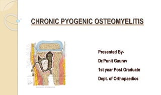
- 1. Presented By- Dr.Punit Gaurav 1st year Post Graduate Dept. of Orthopaedics
- 2. TOPICS TO BE COVERED • Introduction • Causes • Pathogenesis • Pathology • Roentgenology • Diagnosis • Differential Diagnosis
- 3. INTRODUCTION Nelaton (1834) : coined osteomyelitis Definition: “ A severe,persistent and incapacitating infection of bone and bone marrow ”. Chronic osteomyelitis is often defined as the presence of ongoing bone infection for longer than 3 weeks in the presence of devitalized bone.
- 4. Osteomyelitis (osteo- derived from the Greek word osteon, meaning bone, myelo- meaning meaning marrow, and -itis meaning inflammation) simply means an infection of the infection of the bone or bone marrow. marrow. Infection mainly involves - Marrow spaces - Haversian canals - Subperiosteal Spaces
- 5. Bone is involved secondarily. Hallmark of chronic osteomyelitis is infected dead bone within a compromised soft-tissue envelope. Infected foci within the bone are surrounded by sclerotic, relatively avascular bone covered by a thickened periosteum and scarred muscle and subcutaneous tissue.
- 6. This avascular envelope of scar tissue leaves systemic antibiotics essentially ineffective. Secondary infections are common, and sinus track cultures usually do not correlate with cultures obtained at bone biopsy.
- 7. Multiple organisms may grow from cultures taken from sinus tracks and from open biopsy specimens of surrounding soft tissue and bone.
- 8. Osteomyelitis occurs when an adequate number of a sufficiently virulent organism overcomes the host's natural defenses (inflammatory and immune responses) and establishes a focus of infection, for example; local skeletal factors also play a role in the development of infection
- 9. Relative absence of phagocytic cells in the metaphyses of bones in children may explain why acute hematogenous osteomyelitis is more common in this location.
- 10. Factors responsible for chronicity Local factors: Cavity, Sequestrum, Sinus, Foreign body, Degree of bone necrosis. General: Nutritional status of the involved tissues, vascular disease, DM, low immunity. Organism: Virulence. Treatment: Appropriateness and compliance. Risk factors: Penetrating trauma, prosthesis, Animal bite.
- 11. ETIOLOGY
- 12. ORGANISMS CAUSING POSSIBLE PROBLEM 1)Staphylococcus aureus Pressure ulcer, penetrating wound, open fracture, orthopedic surgery, vascular insufficiency disorder. 2)Staphylococcus epidermidis Indwelling prosthetic device. 3)Streptococcus viridans Abscessed tooth, gingival disease. 4)Escherichia coli Urinary tract infection.
- 13. 5)Mycobacterium tuberculosis Tuberculosis 6)Neisseria gonorrhoeae Gonorrhea 7)Pseudomonas sp. Puncture wounds, intravenous drugs 8)Salmonella sp. Sickle cell disease 9)Fungi, mycobacteria Immunocompromised host
- 14. According To Age: 1)Infants < 1yr : Gr. B streptococci, S. aureus, E.coli 2) 1-16 yrs : S. aureus, S. pyogens, H.influenzae 3) > 16 yrs : S.aureus, S epidermidis, Gram –ve Bacilli
- 15. WHY STAPHYLOCOCCUS MOST COMMON? S.aureus and S.epidermis elements of normal skin flora S.aureus increased affinity for host proteins (traumatised bone) Enzymes (coagulase, surface factor A) hampers hosts immune response . Inactive “L” forms dormant for years “Biofilm” (polysaccharide “slime” layer) increases bacterial adherence to any substrate . Large variety of adhesive proteins and glycoproteins mediate binding with bone components.
- 16. PATHOGENESIS
- 17. BACTERIAL RESISTANCE BY BIOFILM Most infections encountered in orthopaedics are related to BIOFILM(A coherent cluster of bacterial cells imbedded in a matrix—which are more tolerant to most anti-microbials and the host defence than planktonic bacterial cells forming bacteria.)
- 18. First, the bacteria need to find an inert surface (e.g., implant or dead tissue). Implants or dead tissue that have been integrated by the host with some type of surface are not inert and will resist colonization. Then, the colonization process will continue until mature colonies are formed. Once mature, the colonies can change based on environmental signals or signals between colonies
- 20. Hematogenous spread usually involves the metaphysis of long bones in children or the vertebral bodies in adults Direct inoculation of microorganisms into bone penetrating injuries and surgical contamination are most common causes Contiguous focus of infection seen in patients with severe vascular disease. Microorganism s in bone Osteomyelitis
- 21. Whatever may be the inciting cause the bacteria reaches the metaphysis of rapidly growing bone & provokes an inflammatory response. Why metaphysis is involved? 1. Infected embolus is trapped in U-shaped small end arteries located predominantly in metaphyseal region 2. Relative lack of phagocytosis activity in metaphyseal region 3. Highly vascularised region ---minor trauma— hemorrhage ----locus minoris resistantae---excellent culture medium.
- 22. Sharp hairpin turns Flow becomes considerably slower and more turbulent. Relatively fewer phagocytic cells than physis and metaphysis.
- 23. EVOLUTION Described by Trueta in 1959. 3 stages- 1) Stage I : Boil in the Bone with pain which is severe,constant,with tenderness.
- 24. Stage II- The signs and symptoms become more marked and the general symptoms of inlmmation appear.
- 25. Stage III- The inflammation spreads outward and produces subperiosteal inflammatory collection- so called sub periosteal abscess.
- 26. These are end-artery branches of the nutrient artery Acute inflammatory response due to infection Tissue necrosis, breakdown of bone Obstruction Avascular necrosis of bone Chronic osteomyelitis
- 28. THE INFLAMMATORY RESPONSE TO OSTEOMYELITIS: Prostaglandin-E production has been shown to be five to thirty fold higher in infected bone than in normal bone. - postulated to be responsible for bone resorption and sequestrum formation. Effective phagocytosis is defense in patients with osteomyelitis Intramedullary oxygen tensions important for phagocytic function - oxygen tensions of <30 mm Hg impair normal phagocytic function
- 29. In any infection of bone , there is an attempt at repair, that if incomplete, it results in chronic persistence of infection. This repair is accomplised by hyperemia of the surrounding tissue , which effects the decalcification of the bone. Granulation tissue forms and carries in osteoclasts n osteoblasts.
- 30. Necrotic cancellous bone is readily absorbed and replaced by new bone. Dead cortex is gradually absorbed about its surface and is detached from living bone to form a sequestrum.(this requires several months).
- 31. PATHOLOGY The most common site is lower femoral metaphysis. Other sites - upper tibial -upper femoral -upper humeral metaphyis
- 32. Pathologic features of chronic osteomyelitis SEQUESTRUM – is a piece of dead bone ,surrounded by infected granulation tissue trying to “eat” the sequestrum away. Appears pale having smooth inner surface and a rough outer.
- 33. Different types of SEQUESTRA TYPES DISEASE TUBULAR PYOGENIC RING EXTERNAL FIXATORS BLACK ACTINOMYCOSIS CORALLIFORM PERTHE'S DISEASE COKE TUBERCULOSIS SANDY TUBERCULOSIS FEATHERY SYPHILIS
- 34. When SEQUESTRUM IS COMPLETE, it lies in the free cavity and is LESS attacked by granulation tissue and is absorbed more slowly. Meanwhile , the surrounding living bone attempts to wall off the infection by forming a thick , dense wall , the INVOLUCRUM. INVOLUCRUM is the dense sclerotic bone overlying the sequestrum.
- 35. An Involucrum usually has multiple openings , the cloacae , through which exudate , bone debris , and sequestra find exit and pass through sinus tracts to the surface. CONSTANT DESTRUCTION of neighboring soft tissue leads to THIN skin which is easily traumatised , skin epithelium grows inwards to line the sinus tract.
- 37. In chronic osteomyelitis of long standing ,multiple cavities and sequestra exist throughout the bone. The shaft becomes thickened , irregular and deformed.
- 40. Factors affecting physiological class Systemic Factors Local Factors Malnutrition Renal,Livere failure Alcohol abuse Immune deficiency Chronic Hypoxia Malignancy Diabetes Mellitus Extremes of ages Steroid therapy Tobacco abuse Chronic Lymphedema Venous stasis Major vessel compromise Arteritis Extensive scarring Radtion fibrosis
- 42. USE OF THIS CLASSIFICATION - To decide whether treatment should be 1) Simple or Complex 2) Curative or Palliative 3) Limb sparing or Ablative
- 43. CLINICAL PICTURE DURING THE PERIOD OF INACTIVITY: - Usually no symptoms - Skin over the focus is dusky, thin, scarred, poorly nourished - Break in the skin causes ulceration that heals slowly - Muscles are scarred & leads to contractures of the adjacent joints.
- 44. DURING ACUTE EXACERBATION - Aching pain worsening at night, overlying soft tissue becomes edematous, warm redddened & tender - Patient is febrile - As infection progresses, sinus may open up & drain extruding small sequestrum at intervals
- 45. - Intervals between flare ups may be months or years. - Flare ups may be due to poor general condition & lowered resistance. - Recurrent toxemia will eventually cause debilitary & sometimes fatal.
- 46. DIAGNOSIS The diagnosis is based on Clinical , Laboratory and Imaging studies. The “GOLD STANDARD” is to obtain a biopsy specimen for histological and microbiological evaluation of the infected bone.
- 47. CLINICAL Physical examination should be focused on integrity of skin and soft tissue . Determination of area of tenderness. Assessing bone stability. And evaluation of neuro vascular status of the limb.
- 48. LABORATORY Lab studies generally are nonspecific and give no indication for severity of the infection. ESR and C- Reactive protein are elevated in most patients. But WBC’S elevated in only 35%.
- 49. The white blood cell count will show a marked leucocytosis as high as 20,000 or more. Peak elevation of the ESR occurs at 3 to 5 days after infection and returns to normal approximately 3 weeks after treatment is begun. CRP increases within 6 hours of infection, reaches a peak elevation 2 days after infection, and returns to normal within 1 week after adequate treatment has begun.
- 50. CULTURAL STUDIES Key to the successful management of osteomyelitis is the isolation of the involved pathogens before the initiation of antibiotics. Cultures of superficial wounds or sinus tracks should not be relied on because they have been shown to be poor indicators of deep infection and usually are polymicrobial.
- 51. The preferred specimen in most bacterial and yeast infections is aspirated fluid (joint or purulent fluid). A deep wound biopsy or a curetted specimen after leaning the wound is acceptable.
- 52. RADIOLOGY
- 53. Multiple imaging technique are available to evaluate chronic osteomyelitis ,however no technique can absolutely confirm or exclude presence of osteomyelitis. Imaging should be done to confirm the diagnosis and prepare for surgery. Initial plain radiographs to be performed it yields valuable information.
- 54. PLAIN RADIOGRAPH Earliest changes are swelling of the soft tissue, periosteal thickening and/or elevation, and focal osteopenia. More diagnostic lytic changes are delayed and are associated with subacute and chronic osteomyelitis.
- 55. USG May detect a subperiosteal collection of fluid in Early stages of osteomyelitis. To establish if joint effusion is present. To localise needle aspiration.
- 56. Sinography Sinography can be preformed if a sinus track is present. Roentgenograms made in two planes after injection of radiopaque liquid into sinus. Helpful in locating focus of infection in chronic osteomyelitis. A valuable adjunct to surgical planning
- 57. RADIONUCLEOTIDE SCAN Isotopic bone scanning is more useful in acute osteomyelitis than chronic osteomyelitis. Most common is 99mTc phosphate, which can detect osteomyelitis.
- 58. Three-phase bone scan consists of (1)Flow phase- shows blood flow (2)Immediate or equilibrium - shows relative flow and distribution of radio isotope into extracellular matrix (3) Delayed phase-shows osteoblastic activity Osteomyelitis shows increase uptake in all three phase
- 59. MRI MRI has very high sensitivity and specificity for the diagnosis of osteomyelitis. Classic findings of osteomyelitis on MRI are a decrease in the normally high marrow signal on T1 images and a normal or increased signal on T2 images.
- 60. May reveal a well defined rim of high signal intensity surrounding the focus of active disease (RIM SIGN). The reported abnormal images reflect an increase in water content, resulting from edema in the marrow cavity.
- 61. Marrow fat is replaced by edema and cellular infiltrates that are lower in signal than fat on T1 images and higher in signal than fat on T2 and STIR image.
- 62. CT SCAN CT provides excellent definition of cortical bone and a fair evaluation of the surrounding soft tissues and is especially useful in identifying sequestra.
- 63. DIFFERENTIAL DIAGNOSIS TUBERCULOSIS OF BONE- -Thin watery discharge -Undermining Bluish discolouration, -Diaphyseal involvement more common -H/o Pulmonary TB -Often Multifocal SOFT TISSUE INFECTION -Absence of bony changes
- 64. EWING SARCOMA -Radiological D/D -Acute presentation -Biopsy is diagnostic OSTEONECROSIS OSTEOID OSTEOMA HISTIOCYTIC EOSINOPHILIC GRANULOMA GARRE'S OSTEOMYELITIS
- 65. REFERENCES CAMPBELL’S OPERATIVE ORTHOPAEDICS,14th edition Apley and Solomon’s System of Orthopaedics and Trauma 10th Edition Kulkarni's Textbook Of Orthopedics And Trauma 3rd edition Turek’s Orthopaedics Principles and Their Applications 7th edition.